. 1997 Dec;8(12):2617-29.
doi: 10.1091/mbc.8.12.2617.

On the role of myosin-II in cytokinesis: division of Dictyostelium cells under adhesive and nonadhesive conditions

Affiliations
Free PMC article

On the role of myosin-II in cytokinesis: division of Dictyostelium cells under adhesive and nonadhesive conditions

J H Zang et al. Mol Biol Cell. 1997 Dec.
Free PMC article

Abstract

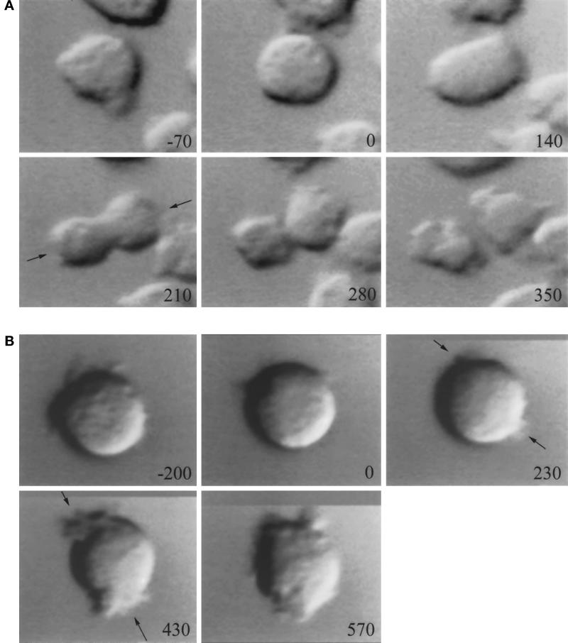
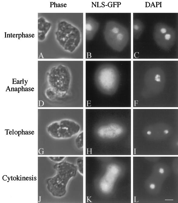
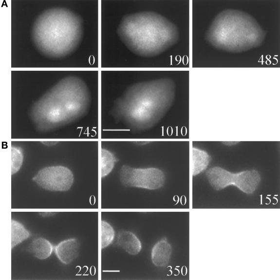
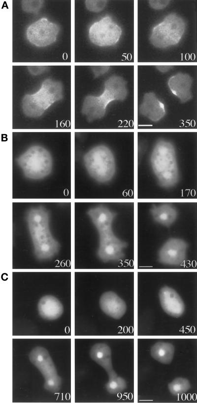
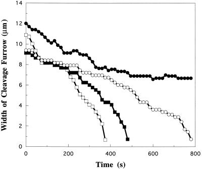
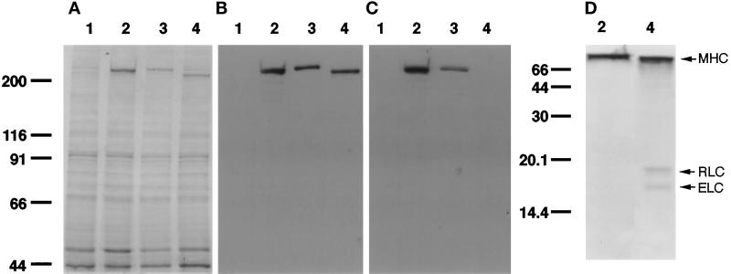
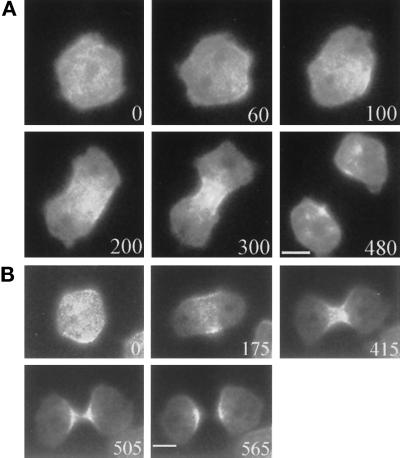
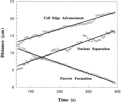
We have investigated the role of myosin in cytokinesis in Dictyostelium cells by examining cells under both adhesive and nonadhesive conditions. On an adhesive surface, both wild-type and myosin-null cells undergo the normal processes of mitotic rounding, cell elongation, polar ruffling, furrow ingression, and separation of daughter cells. When cells are denied adhesion through culturing in suspension or on a hydrophobic surface, wild-type cells undergo these same processes. However, cells lacking myosin round up and polar ruffle, but fail to elongate, furrow, or divide. These differences show that cell division can be driven by two mechanisms that we term Cytokinesis A, which requires myosin, and Cytokinesis B, which is cell adhesion dependent. We have used these approaches to examine cells expressing a myosin whose two light chain-binding sites were deleted (DeltaBLCBS-myosin). Although this myosin is a slower motor than wild-type myosin and has constitutively high activity due to the abolition of regulation by light-chain phosphorylation, cells expressing DeltaBLCBS-myosin were previously shown to divide in suspension (Uyeda et al., 1996). However, we suspected their behavior during cytokinesis to be different from wild-type cells given the large alteration in their myosin. Surprisingly, DeltaBLCBS-myosin undergoes relatively normal spatial and temporal changes in localization during mitosis. Furthermore, the rate of furrow progression in cells expressing a DeltaBLCBS-myosin is similar to that in wild-type cells.


Figures

Figure 1
Figure 1
Dicytostelium myosin-null cells fail to form furrows during cytokinesis in suspension as shown by sequential differential interference contrast images. In these images, time 0 indicates the start of rounding and quiescence. (A) Cells expressing wild-type myosin can round up, elongate, furrow, and separate. Times of the panels are −70, 0, 140, 210, 280, and 350 s. Ruffling of the polar regions, as indicated by black arrows, coincided with the furrowing (210 s). (B) Myosin-null cells cannot divide in suspension. Times of the panels are −200, 0, 230, 430, and 570 s. They can round up, but they fail to elongate and furrow. However, like wild-type cells, they are able to polar ruffle, as indicated by arrows, at about the same time as wild-type cells (230 s).
Figure 2
Figure 2
NLS-GFP is localized to the nucleus, except during early mitosis. Myosin-null cells expressing NLS-GFP were fixed and stained with DAPI. The left column shows phase-contrast images, the center column shows NLS-GFP localization, and the right column shows DAPI staining. (A–C) Interphase cells. (D–F) An early anaphase cell. (G–I) A telophase cell. (J–L) A cell undergoing cytokinesis. Bar, 5 μm.
Figure 3
Figure 3
Myosin-null cells cannot divide on a hydrophobic surface. (A) A myosin-null cell expressing NLS-GFP fails to form a furrow on such a surface. Times of the panels are 0, 190, 485, 745, and 1010 s. (B) A cell expressing GFP-myosin divides successfully on a hydrophobic surface. Times of the panels are 0, 90, 155, 220, and 350 s. Bar, 5 μm.
Figure 4
Figure 4
Myosin-null cells are able to divide on an adhesive surface, such as glass. (A) Cells expressing GFP-myosin are able to divide on a glass surface successfully. Times of the panels are 0, 50, 100, 160, 220, and 350 s. (B) Myosin-null cells can undergo mitosis-coupled division with morphologies similar to cells expressing functional myosin. Times of the panels are 0, 60, 170, 260, 350, and 430 s. (C) However, sometimes the cleavage furrow is not centered, resulting in unequal daughter cells. Times of the panels are 0, 200, 450, 710, 950, and 1000 s. Bar, 5 μm.
Figure 5
Figure 5
The rate of furrowing for myosin-null cells dividing on a glass surface is not constant. The width of the cleavage furrow vs. time is presented for four representative cells. One cell (closed circles) failed to divide into two.
Figure 6
Figure 6
Expression of GFP-ΔBLCBS-myosin in comparison to that of myosin and GFP-myosin. (A–C) Whole cell lysates were run on 7.5% SDS-PAGE gels. Lane 1, myosin null cells; lane 2, cells expressing GFP-ΔBLCBS-myosin; lane 3, cells expressing GFP-myosin; lane 4, cells expressing wild-type myosin. (A) Coomassie-stained gel. (B) Immunoblot probed with antimyosin antibody. (C) Immunoblot probed with anti-GFP antibody. (D) Neither light chain binds to GFP-ΔBLCBS-myosin. Purified GFP-ΔBLCBS-myosin protein and wild-type myosin were run on a 15% SDS-PAGE gel and subsequently Coomassie stained to visualize the light chains. MHC, myosin heavy chain; ELC, essential light chain; RLC, regulatory light chain.
Figure 7
Figure 7
Cells expressing GFP-ΔBLCBS-myosin are able to divide successfully on an adhesive surface (A) or on the hydrophobic surface (B). The number in the lower right corner indicates the progression of division in seconds. Bar, 5 μm.
Figure 8
Figure 8
Analysis of rate of cleavage furrow constriction, nuclear separation, and cell edge advancement for GFP-ΔBLCBS-myosin cells. Cleavage furrow constriction in cells expressing GFP-ΔBLCBS-myosin is linear.

References

    1. Bradford MM. A rapid and sensitive method for the quantitation of microgram quantities of protein utilizing the principle of protein-dye binding. Anal Biochem. 1976;72:248–254. - PubMed
    1. Burton K, Taylor DL. Traction forces of cytokinesis measured with optically modified elastic substrata. Nature. 1997;385:450–454. - PubMed
    1. Cramer LP, Mitchison TJ. Investigation of the mechanism of retraction of cell margin and rearward flow of nodules during mitotic cell rounding. Mol Biol Cell. 1997;8:109–119. - PMC - PubMed
    1. de Hostos EL, Rehfuess C, Bradtke B, Waddell DR, Albrecht R, Murphy J, Gerisch G. Dictyostelium mutants lacking the cytoskeletal protein coronin are defective in cytokinesis and cell motility. J Cell Biol. 1993;120:163–73. - PMC - PubMed
    1. De Lozanne A, Spudich JA. Disruption of the Dictyostelium myosin heavy chain gene by homologous recombination. Science. 1987;236:1086–1091. - PubMed

Publication types

LinkOut - more resources