. 1997 Dec;8(12):2647-57.
doi: 10.1091/mbc.8.12.2647.

Defining extracellular integrin alpha-chain sites that affect cell adhesion and adhesion strengthening without altering soluble ligand binding

Affiliations
Free PMC article

Defining extracellular integrin alpha-chain sites that affect cell adhesion and adhesion strengthening without altering soluble ligand binding

C Pujades et al. Mol Biol Cell. 1997 Dec.
Free PMC article

Abstract

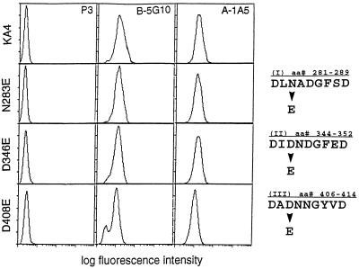
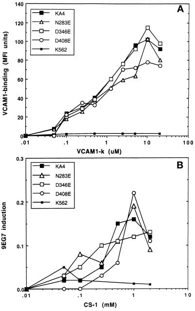
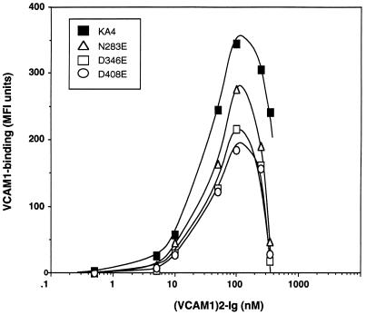
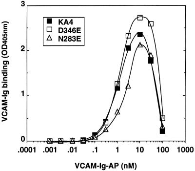
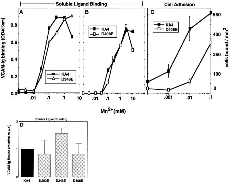
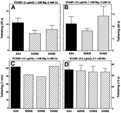
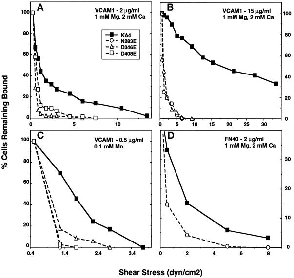
It was previously shown that mutations of integrin alpha4 chain sites, within putative EF-hand-type divalent cation-binding domains, each caused a marked reduction in alpha4beta1-dependent cell adhesion. Some reports have suggested that alpha-chain "EF-hand" sites may interact directly with ligands. However, we show here that mutations of three different alpha4 "EF-hand" sites each had no effect on binding of soluble monovalent or bivalent vascular cell adhesion molecule 1 whether measured indirectly or directly. Furthermore, these mutations had minimal effect on alpha4beta1-dependent cell tethering to vascular cell adhesion molecule 1 under shear. However, EF-hand mutants did show severe impairments in cellular resistance to detachment under shear flow. Thus, mutation of integrin alpha4 "EF-hand-like" sites may impair 1) static cell adhesion and 2) adhesion strengthening under shear flow by a mechanism that does not involve alterations of initial ligand binding.


Figures

Figure 1
Figure 1
Flow cytometric analysis of α4 and β1 present in K562 cells. Transfected K562 cells were stained using the control antibody P3, anti-α4 MAb B-5G10, and anti-β1 MAb A-1A5, and cell surface levels were determined by flow cytometry. The sequences of the α4 EF hand-like domains containing each mutation are indicated.
Figure 2
Figure 2
Indirect analysis of soluble monovalent ligand binding. Cells were tested for binding by VCAM-1-k (A) or CS1 peptide (B). For VCAM-1-k binding, cells were incubated for 30 min at 4°C, with increasing amounts of VCAM-1-k, in the presence of 1 mM MnCl2, and then stained with FITC-antimouse κ chain antibody and analyzed by flow cytometry. For each experiment, VCAM-k binding in the presence of 5 mM EDTA was used to obtain the background signal (typically <5–10% relative to the maximal signal), which was then subtracted. For this experiment, fluorescence intensity (MFI units) due to VCAM-1-k binding were corrected by a factor of 1.3 for the D346E and D408E mutants, because the surface level of wild-type α4 was 1.3-fold greater than that of the mutants. The level of CS1 peptide binding was determined indirectly by measuring induction of the 9EG7 epitope, as described in MATERIALS AND METHODS. The 9EG7 epitope expression results are presented as a fraction of the total β1 subunit (measured using mAb 13).
Figure 3
Figure 3
Indirect bivalent (VCAM-1)2-Ig binding as a function of ligand concentration. Cells were incubated with increasing amounts of (VCAM-1)2-Ig in the presence of 2mM MnCl2. After washing, the cells were incubated with FITC-antihuman IgG and analyzed using a FACscan. Prior to incubation with the ligand, nonspecific binding was blocked with anti-Fc receptor antibodies and human serum. All experiments were done at 4°C and with 0.02% sodium azide to minimize receptor and/or ligand internalization. Background binding, determined in the presence of 5 mM EDTA, was subtracted (and was typically < 10 MFI units). At the time of this experiment, differences among wild-type and mutant α4 levels were not more than twofold.
Figure 4
Figure 4
Direct bivalent binding of (VCAM-1)2-Ig-AP to α4β1. Cells were incubated with increasing amounts of (VCAM-1)2-Ig-AP in the presence of 2mM MnCl2. After washing, cells were incubated with AP substrate and color development was determined at OD 405 nm. Background binding to mock-transfected K562 cells (typically <0.1 OD units) was subtracted. At the time of this experiment, differences among cell surface levels of α4 wild-type, D346E, and N283E were not more than twofold.
Figure 5
Figure 5
Mn2+ effects on binding of (VCAM-1)2-Ig-AP. Cells were incubated with 4 nM (VCAM-1)2-Ig-AP, and α4-specific binding was determined as described in the legend to Figure 4. (A and B). Wild-type α4 was synthesized at comparable levels to D408E (A) and D346E (B), and in two separate experiments binding was determined over a range of MnCl2 concentrations. (C) Cell adhesion to VCAM-1, coated at 2 μg/ml, was measured for KA4 and D408E cells at various MnCl2 concentrations. (D) Binding of (VCAM-1)2-Ig-AP was carried out in 0.1 mM MnCl2. Results were normalized relative to wild-type α4 binding (given an arbitrary value of 1.0). Each point represents the mean of two to three experiments. Typical OD405 values for specific (VCAM-1)2-Ig-AP binding ranged from 0.5 to 1.0. Also, data in D were adjusted for differences in α4 surface levels, which varied from each other by not more than 1.5-fold.
Figure 6
Figure 6
Tethering of K562 transfectants on rsVCAM-1. Into a parallel plate flow chamber, 2–5 × 10⁵ cells were perfused at low wall shear stress (0.30–0.45 dyn/cm2) and tethering to rsVCAM-1 was analyzed. The flow chamber was coated the indicated concentrations of rsVCAM. Experiments were carried out in 1 mM Mg2+, 2 mM Ca2+ (A–C), or 0.1 mM Mn2+ (D). Tethering events were accumulated for 30 s (A and B), 1 min (C), or 15 s (D).
Figure 7
Figure 7
Shear resistance of K562 transfectants on rsVCAM-1 or FN40. Using initial conditions as in Figure 6, A, B, and D, an average of ∼50 cells was allowed to accumulate in shear flow for 15 to 30 s on rsVCAM-1 coated at the indicated levels (A–C). Then cells were subjected to increasing shear stresses in small increments at 10-s intervals. After each 10-s interval, the percentage of cells remaining bound was determined at each shear. In D, cells were allowed to accumulate in static conditions for 2 min on FN40 (coated at 2 μg/ml). Then resistance to detachment was determined as for A–C. The values indicated in the detachment profiles (A, B, and D) are the mean of three to four experiments. Results in C are representative of four different experiments.

References

    1. Akiyama SK, Yamada SS, Chen W-T, Yamada KM. Analysis of fibronectin receptor function with monoclonal antibodies: roles in cell adhesion, migration, matrix assembly, and cytoskeletal organization. J Cell Biol. 1989;109:863–875. - PMC - PubMed
    1. Alon R, Hammer DA, Springer TA. Lifetime of the P-selectin-carbohydrate bond and its response to tensile force in hydrodynamic flow. Nature. 1995a;374:539–542. - PubMed
    1. Alon R, Kassner PD, Carr MW, Finger EB, Hemler ME, Springer TA. The integrin VLA-4 supports tethering and rolling in flow on VCAM-1. J Cell Biol. 1995b;128:1243–1253. - PMC - PubMed
    1. Babu A, Su H, Ryu Y, Gulati J. Determination of residue specificity in the EF-hand of troponin C for Ca2+ coordination, by genetic engineering. J Biol Chem. 1992;267:15469–15474. - PubMed
    1. Bajt ML, Goodman T, McGuire SL. β2 (CD18) mutations abolish ligand recognition by I domain integrins LFA-1 (αLβ2, CD11a/CD18) and MAC-1 (αMβ2, CD11b/CD18) J Biol Chem. 1995;270:94–98. - PubMed

Publication types

MeSH terms

LinkOut - more resources