. 1997 Dec;8(12):2693-705.
doi: 10.1091/mbc.8.12.2693.

Identification of a second myosin-II in Schizosaccharomyces pombe: Myp2p is conditionally required for cytokinesis

Affiliations
Free PMC article

Identification of a second myosin-II in Schizosaccharomyces pombe: Myp2p is conditionally required for cytokinesis

M Bezanilla et al. Mol Biol Cell. 1997 Dec.
Free PMC article

Abstract

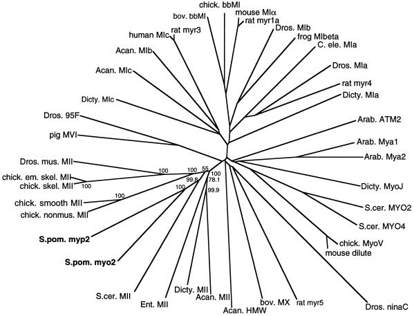
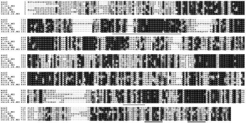
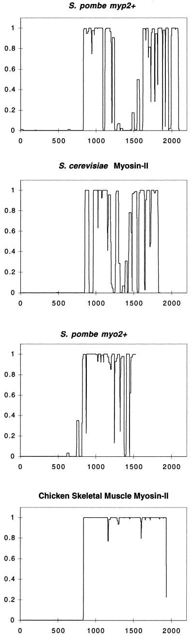
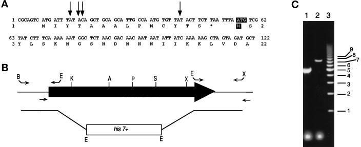
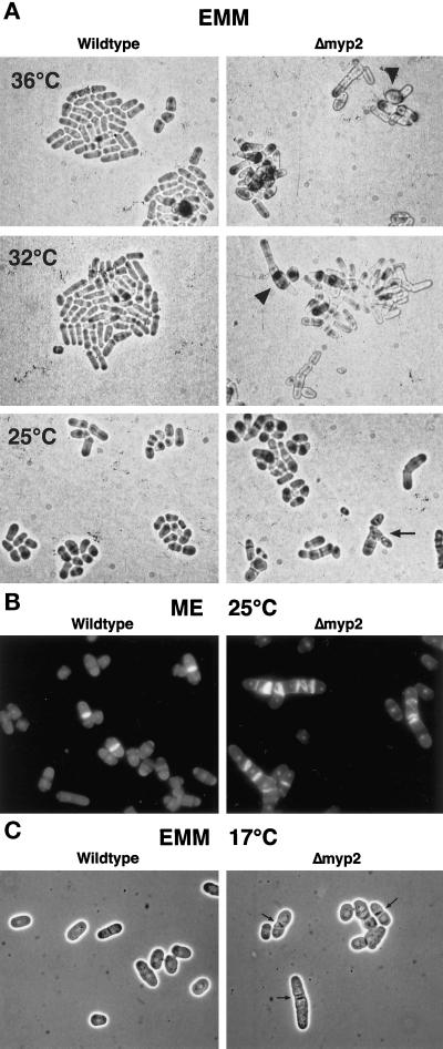
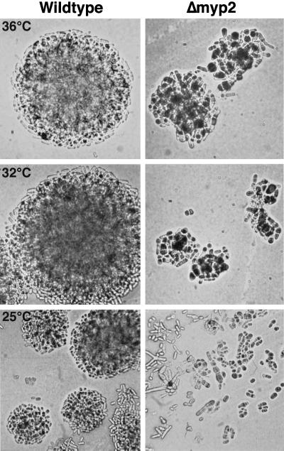
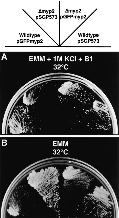
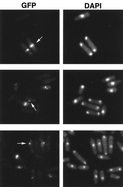
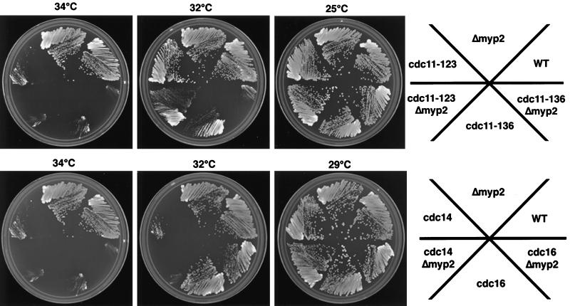
As in many eukaryotic cells, fission yeast cytokinesis depends on the assembly of an actin ring. We cloned myp2(+), a myosin-II in Schizosaccharomyces pombe, conditionally required for cytokinesis. myp2(+), the second myosin-II identified in S. pombe, does not completely overlap in function with myo2(+). The catalytic domain of Myp2p is highly homologous to known myosin-IIs, and phylogenetic analysis places Myp2p in the myosin-II family. The Myp2p sequence contains well-conserved ATP- and actin-binding motifs, as well as two IQ motifs. However, the tail sequence is unusual, since it is predicted to form two long coiled-coils separated by a stretch of sequence containing 19 prolines. Disruption of myp2(+) is not lethal but under nutrient limiting conditions cells lacking myp2(+) function are multiseptated, elongated, and branched, indicative of a defect in cytokinesis. The presence of salt enhances these morphological defects. Additionally, Deltamyp2 cells are cold sensitive in high salt, failing to form colonies at 17 degrees C. Thus, myp2(+) is required under conditions of stress, possibly linking extracellular growth conditions to efficient cytokinesis and cell growth. GFP-Myp2p localizes to a ring in the middle of late mitotic cells, consistent with a role in cytokinesis. Additionally, we constructed double mutants of Deltamyp2 with temperature-sensitive mutant strains defective in cytokinesis. We observed synthetic lethal interactions between Deltamyp2 and three alleles of cdc11ts, as well as more modest synthetic interactions with cdc14ts and cdc16ts, implicating myp2(+) function for efficient cytokinesis under normal conditions.


Figures

Figure 1
Figure 1
Phylogenetic analysis of myp2+. An unrooted phylogenetic tree was generated from an alignment of catalytic domains of myosins from several families using Clustal W (Thompson et al., 1994). The numbers indicate the bootstrapping value in percentage of 1000 bootstrapping trials for the type II myosin subfamily. In this analysis, the lengths of the branches joining two proteins are proportional to the percentage of amino acid sequence divergence between the two proteins. The myosin-catalytic domain sequences do not include the light chain-binding region, since they were defined as ending 18 residues after the conserved threonine in the TKVFF sequence. The accession numbers for the sequences are given in MATERIALS AND METHODS. The abbreviations used are as follows: Acan., Acanthamoeba; Arab., Arabidopsis; bov., bovine; chick., chicken; em., embryonic; skel., skeletal; mus., muscle; nonmus., nonmuscle; Dicty., Dictyostelium; Dros., Drosophila; C. ele., C. elegans; S. cer., S. cerevisiae; S. pom., S. pombe.
Figure 2
Figure 2
Sequence alignment of myp2+ with six myosin-IIs from other organisms. The catalytic domain of myp2+ was aligned with myosin-IIs from S. pombe, S. cerevisiae, amoebae, and chicken using Clustal W (Thompson et al., 1994). From top to bottom: S. pombe myp2+, S. pombe myo2+, S. cerevisiae myosin-II (MYO1p), Entamoeba myosin-II, Acanthamoeba myosin-II, Dictyostelium myosin-II, and chicken skeletal muscle myosin-II. The sequence accession numbers are given in MATERIALS AND METHODS. The numbers to the left of the amino acid sequence indicate amino acid position. Amino acid identities are shown in white lettering against black, conserved residues are shown in black lettering against gray. The putative ATP-binding motif is underlined. The putative actin-binding site has asterisks underneath. The putative IQ motifs are underlined twice.
Figure 3
Figure 3
Coiled-coil predictions of myosin-IIs from different organisms. The predictions were generated by the Coils program (Lupas et al., 1991) for S. pombe Myp2p, S. cerevisiae myosin-II (MYO1p), S. pombe Myo2p, and chicken skeletal muscle myosin-II. x-axis, amino acid position. y-axis, probability of forming a coiled-coil. A window of 28 amino acids was used to generate the profiles shown.
Figure 4
Figure 4
Cloning 5′ of myp2+ and construction of the myp2-disrupted strain. (A) The DNA sequence for the 5′ region of myp2+ in the pmyp2BS construct is shown along with the deduced amino acid sequence. The arrows indicate the position of the beginning of four separate 5′ RACE PCR products that were sequenced. The putative start codon and methionine are highlighted with white lettering against black. (B) Schematic representation of the genomic locus of myp2+. The shaded arrow represents the open reading frame of 6312 bp. The primers used to amplify the 5′ and 3′ regions of myp2+ that were used to generate the knockout construct are represented above the locus. The restriction sites used to ligate together the knockout construct are indicated at the ends of the primers. B, BamHI; E, EcoRI; X, XhoI. The primers used to amplify the genomic locus from His+ haploids generated from the diploid transformed with the knockout construct are indicated below the locus. (C) Amplification of the genomic locus by colony PCR from His+ haploids. PCR products were separated on an 0.8% agarose gel. Lane 1 represents a homologous recombination event resulting in disruption of the myp2+ locus by his7+. The disrupted locus is 2.5 kb smaller than the wild-type locus. Lane 2 represents a heterologous recombination event in which his7+ has been integrated elsewhere in the genome thus leaving the myp2+ locus intact. Lane 3 is the molecular weight marker with sizes in kilobases indicated to the right.
Figure 5
Figure 5
Phenotype of Δmyp2 strain. (A) Wild-type and Δmyp2 strains on EMM-agar grown at 36°C (top), 32°C (middle), and 25°C (bottom). Cells were taken from YES plates and streaked onto EMM plates. Cells were visualized using a 40× long-working-distance bright-field objective and photographed directly off the plate. Both strains form groups of cells that will eventually form colonies of roughly the same size. Many of Δmyp2 cells have multiple septa, and a few are branched (arrow). Δmyp2 cells are slightly wider than wild-type and are occasionally swollen (arrowheads) at the higher temperatures. (B) Fluorescence micrographs of wild-type and Δmyp2 cells grown on ME agar, removed from the plate, and stained with DAPI and calcofluor. (C) Phase contrast micrographs of wild-type and Δmyp2 cells grown on EMM agar at 17°C smeared on a microscope slide. Δmyp2 cells have very pronounced and misshapen septa (arrows).
Figure 6
Figure 6
Phenotype of Δmyp2 cells grown on YES + 1 M KCl at 36°C (top), 32°C (middle), and 25°C (bottom). Cells were streaked onto YES + 1 M KCl plates form YES plates. Cells were visualized using a 40× long-working-distance bright-field objective and photographed directly off the plate. Δmyp2 cells eventually form colonies at all three temperatures but are slower than wild-type, as apparent from the smaller colonies at 36°C and 32°C.
Figure 7
Figure 7
Complementation by pGFPmyp2 and overexpression of pGFPmyp2. Ura+ transformants of strains carrying the indicated plasmid (pSGP573 or pGFPmyp2) were streaked to (A) EMM-Ura + 1 M KCl + thiamine (B1) (this plate shows colonies after 4 days at 32°C) and to (B) EMM-Ura (this plate shows colonies after 3 days at 32°C).
Figure 8
Figure 8
Localization of GFP-Myp2p in Δmyp2 cells. Δmyp2 cells were grown in liquid with thiamine and harvested during exponential growth. Cells were stained with DAPI and observed immediately after staining. GFP-Myp2p localizes to a ring in the middle of late mitotic cells (arrow, top). Some late mitotic cells only show a bright spot in the middle of the cell (arrow, middle). Cells in interphase, show GFP-Myp2p localizing to a bright dot near the nucleus (arrow, bottom).
Figure 9
Figure 9
Genetic interactions observed with Δmyp2 strain and several temperature-sensitive mutant strains that affect cytokinesis. Cells of the indicated strains were streaked onto YES + phloxin-B plates at the indicated temperatures and then scanned in once the wild-type colonies have formed. In all cases, the double mutants were darker pink than the single mutants indicating that the double mutants are sicker.

References

    1. Adachi Y, Toda T, Niwa O, Yanagida M. Differential expressions of essential and nonessential alpha-tubulin genes in Schizosaccharomyces pombe. Mol Cell Biol. 1986;6:2168–2178. - PMC - PubMed
    1. Altschul SF, Gish W, Miller W, Myers EW, Lipman DJ. Basic local alignment search tool. J Mol Biol. 1990;215:403–410. - PubMed
    1. Apolinario E, Nocero M, Jin M, Hoffman CS. Cloning and manipulation of the Schizosaccharomyces pombe his7+ gene as a new selectable marker for molecular genetic studies. Curr Genet. 1993;24:491–495. - PMC - PubMed
    1. Balasubramanian MK, Helfman DM, Hemmingsen SM. A new tropomyosin essential for cytokinesis in the fission yeast S. pombe. Nature. 1992;360:84–87. - PubMed
    1. Balasubramanian MK, Hirani BR, Burke JD, Gould KL. The Schizosaccharomyces pombe cdc3+ gene encodes a profilin essential for cytokinesis. J Cell Biol. 1994;125:1289–1301. - PMC - PubMed

Publication types

MeSH terms

Associated data