. 1997 Dec 23;94(26):14320-5.
doi: 10.1073/pnas.94.26.14320.

Identification and characterization of a human protein kinase related to budding yeast Cdc7p

Affiliations

Identification and characterization of a human protein kinase related to budding yeast Cdc7p

W Jiang et al. Proc Natl Acad Sci U S A. .

Abstract

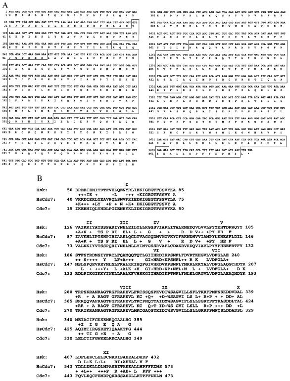
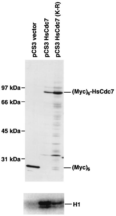
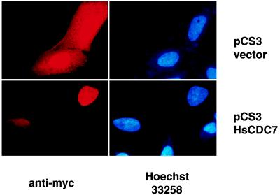
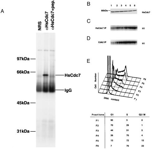
The Cdc7p protein kinase is essential for the G1/S transition and initiation of DNA replication during the cell division cycle in Saccharomyces cerevisiae. Cdc7p appears to be an evolutionarily conserved protein, since a homolog Hsk1 has been isolated from Schizosaccharomyces pombe. Here, we report the isolation of a human cDNA, HsCdc7, whose product is closely related in sequence to Cdc7p and Hsk1. The HsCdc7 cDNA encodes a protein of 574 amino acids with predicted size of 64 kDa. HsCdc7 contains the conserved subdomains common to all protein-serine/threonine kinases and three "kinase inserts" that are characteristic of Cdc7p and Hsk1. Immune complexes of HsCdc7 from cell lysates were able to phosphorylate histone H1 in vitro. Indirect immunofluorescence staining demonstrated that HsCdc7 protein was predominantly localized in the nucleus. Although the expression levels of HsCdc7 appeared to be constant throughout the cell cycle, the protein kinase activity of HsCdc7 increased during S phase of the cell cycle at approximately the same time as that of Cdk2. These results, together with the functions of Cdc7p in yeast, suggest that HsCdc7 may phosphorylate critical substrate(s) that regulate the G1/S phase transition and/or DNA replication in mammalian cells.


Figures

Figure 1
Figure 1
(A) Nucleotide and deduced amino acid (single letter amino acid code) sequences of HsCdc7. HsCdc7 contains an ORF of 574 amino acids and its kinase subdomains are indicated in the boxes. The asterisk represents the stop codon. (B) Multiple alignment of amino acid sequences in HsCdc7, Cdc7p and Hsk1 kinase subdomains (I to XI), which correspond to the boxes in A, using the blast program on the National Center for Biotechnology Information database. Amino acid residues that are identical are indicated by single letters between the sequence lines, and amino acid residues that are similar are indicated by +.
Figure 2
Figure 2
Immunoblot and protein kinase assays of HsCdc7 in 293 cells transfected with pCS3HsCdc7, pCS3HsCdc7 (K-R) and pCS3 control vector plasmids. Lysates from 293 cells transfected with pCS3HsCdc7, pCS3HsCdc7 (K-R) and pCS3 were separated by SDS/PAGE, transferred to Immobilon-P membrane, and then blotted with the 9E10 anti-Myc-tag mAb (Upper). The same cell lysates used for the immunoblotting experiment were immunoprecipitated with 9E10 mAb, and subjected to in vitro kinase reactions using histone H1 as substrate plus [γ-32P]ATP. The products of the kinase reactions were separated by SDS/PAGE prior to autoradiography (Lower) (for details see Materials and Methods).
Figure 3
Figure 3
Immunofluorescence staining in HeLa cells transfected with pCS3HsCdc7 and pCS3 control vector plasmids using 9E10 mAb. HeLa cells were grown on glass coverslips and transiently transfected with the indicated plasmids. After fixation, cells were incubated with the 9E10 mAb. Immunofluorescence staining was performed, and nuclei were visualized by staining with Hoechst dye 33258 as described in Materials and Methods.
Figure 4
Figure 4
The expression levels of HsCdc7 protein and its kinase activity during the cell cycle in 293 cells. (A) Lysates from 293 cells were immunoprecipitated with nonimmune rabbit serum (NRS), HsCdc7 antiserum or HsCdc7 antiserum that had been prebound to HsCdc7 C-terminal peptide. After washing, the immunoprecipitates were subjected to SDS/PAGE, transferred to Immobilon-P membrane and then blotted with HsCdc7 antiserum. (B) Subconfluent 293 cells were fractionated at specific stage of the cell cycle using centrifugal elutriation as described in Materials and Methods. Cell lysates from the indicated fractions (F1–F6) were subjected to SDS/PAGE, transferred to Immobilon-P membrane and then blotted with affinity-purified HsCdc7 antibodies. (C and D) The same cell lysates used in B were immunoprecipitated with HsCdc7 antibodies (C) or Cdk2 antibodies (D), and subjected to in vitro kinase reactions using histone H1 as substrate plus [γ-32P]ATP. The products of the kinase reactions were separated by SDS/PAGE prior to autoradiography. (E) 293 cells from each elutriated fraction as described in B were collected and analyzed for DNA content by flow cytometry. (Lower) The values represent the percentage of cells in the indicated phase(s) of the cell cycle in fractions (F1–F6), determined as described in Materials and Methods.

References

    1. Bell S P, Stillman B. Nature (London) 1992;357:128–134. - PubMed
    1. Diffley J F, Cocker J H. Nature (London) 1992;357:169–172. - PubMed
    1. Li J J, Herskowitz I. Science. 1993;262:1870–1874. - PubMed
    1. Micklem G, Rowley A, Harwood J, Nasmyth K, Diffley J F. Nature (London) 1993;366:87–89. - PubMed
    1. Liang C, Weinreich M, Stillman B. Cell. 1995;81:667–676. - PubMed

Publication types

Associated data