. 1997 Dec 23;94(26):14450-5.
doi: 10.1073/pnas.94.26.14450.

Otogelin: a glycoprotein specific to the acellular membranes of the inner ear

Affiliations

Otogelin: a glycoprotein specific to the acellular membranes of the inner ear

M Cohen-Salmon et al. Proc Natl Acad Sci U S A. .

Abstract

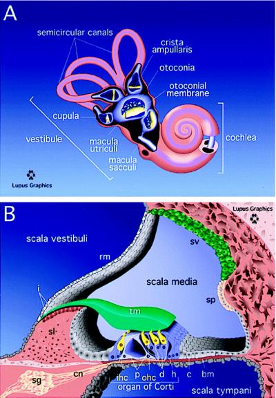
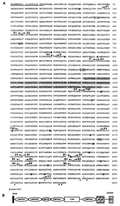
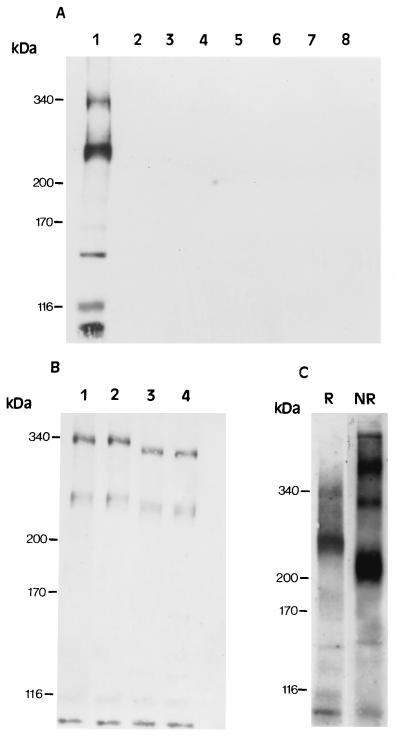
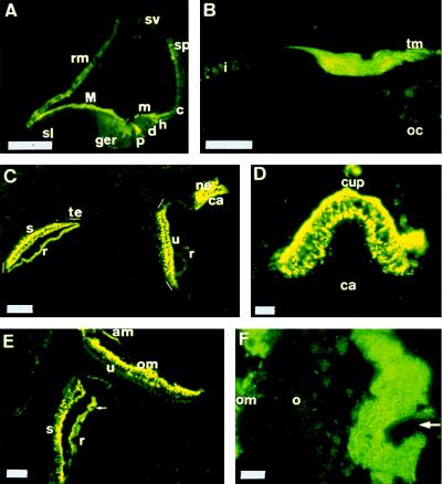
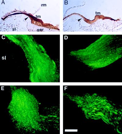
Efforts to identify the specific components of the mammalian inner ear have been hampered by the small number of neuroepithelial cells and the variety of supporting cells. To circumvent these difficulties, we used a PCR-based subtractive method on cDNA from 2-day-old mouse cochlea. A cDNA encoding a predicted 2910-amino acid protein related to mucin has been isolated. Several lines of evidence indicate, however, that this protein does not undergo the O-glycosylation characteristic to mucins. As confirmed by immunocytochemistry and biochemical experiments, this protein is specific to the inner ear. Immunohistofluorescence labeling showed that this protein is a component of all the acellular membranes of the inner ear: i.e., the tectorial membrane of the cochlea, the otoconial and accessory membranes of the utricule and saccule, the cupula of the semicircular canals, and a previously undescribed acellular material covering the otoconia of the saccule. The protein has been named otogelin with reference to its localization. A variety of nonsensory cells located underneath these membranes could be identified as synthesizing otogelin. Finally, this study revealed a maturation process of the tectorial membrane, as evidenced by the progressive organization of otogelin labeling into thick and spaced radial fiber-like structures.


Figures

Figure 1
Figure 1
(A) General organization of the mature inner ear. (B) Cross section of the cochlea. bm, basilar membrane; i, interdental cells; rm, Reissner’s membrane; sv, stria vascularis; sp, spiral prominence; p, pillar cells; d, Deiter’s cells; h, Hensen’s cells; c, Claudius cells; ohc, outer hair cells; ihc, inner hair cells; tm, tectorial membrane; sg, spiral ganglion; cn, cochlear nerve; sl, spiral limbus.
Figure 2
Figure 2
(A) Deduced amino acid sequence of otogelin. The signal peptide is underlined. Arrows delineate the different protein domains. WF D-type domains D1 (residues 135–498), D2 (500–870), D′ (873–971, truncated domain), D3 (972–1450), and D4 (2093–2459) share between each other and with the D domains of WF a sequence similarity of about 50%. The D3 domain contains a unique 103-residue insertion (position 1242–1394), which is shaded. The conserved residues of the multimerization site are shaded. Five WF B-type domains, B1 (2460–2492), B2 (2497–2527), B3 (2532–2563), B4 (2266–2597), B5 (2602–2632) are shown. The TSP domain (1451–2036) contains 13% threonines, 13% serines, and 15% prolines and is devoid of cysteine; CT, carboxyl-terminal end (2825–2910). The potential N-glycosylation sites are underlined and in bold. | indicates the additional 3′ RACE PCR alternatively spliced sequence presented at the end of the sequence. The cysteine involved in the dimerization of WF and TGF-β2 (position 2873) and the conserved glycine (position 2852) are indicated by an asterisk. The cysteines involved in the TGF-β2 knot cystine structure formation are numbered. (B) Schematic representation of the structure of otogelin. The thick bar indicates the predicted signal peptide.
Figure 3
Figure 3
Western blot analysis of otogelin using the 7.8 antiserum. Molecular mass standards (in kDa) are indicated on the left. (A) Tissue distribution of otogelin in the P15 mouse: inner ear (1), eye (2), brain (3), liver (4), intestine (5), kidney (6), spleen (7), and lung (8). (B) Deglycosylation assays of P2 mouse inner ear extracts: control buffer (1), neuraminidase (2), N-glycosidase (3), both enzymes (4). Note that the concentration of the diffuse 240–270-kDa band and the 170- and 150-kDa bands were weaker compared with 15-day-old extracts. (C) P15 mouse cochlear extracts in denaturing reducing (R) and nonreducing (NR) conditions (see Materials and Methods).
Figure 4
Figure 4
Otogelin in the inner ear by immunohistofluorescence using the 7.8 antiserum. (A) Cochlea at P0. (B) Cochlea at P15. (C) Vestibule at P0. (D) Anterior crista ampullaris at P4. (E) Saccule and utricle at P20. (F) Saccule at P20 (laser confocal microscopy). The arrowhead in E and F indicates the staining covering the otoconia (○). ger, greater epithelial ridge; M, major tectorial membrane; m, minor tectorial membrane; s, saccule; u, utricle; ca, crista ampullaris; te, transitory epithelium; ne, neuroepithelium; r, roof; cup, cupula; ca, crista ampullaris; om, otoconial membrane; am, accessory membrane. (C) The OMs and the cupula were lost during tissue preparation. Other abbreviations as before. Scale bars: 60 μm (A), 50 μm (B), 100 μm (C–E), 10 μm (F).
Figure 5
Figure 5
Otogelin in the tectorial membrane. Immunoperoxidase labeling at P4 (A) and P6 (B) with the 7.8 antiserum. Immunohistofluorescence using the 7.8 antiserum and laser confocal microscopy of the outer edge of the spiral limbus area (arrowhead in A and B) at P4, (C), P6 (D), P8 (E), and P20 (F). The tectorial membrane has the same orientation in all of the figures (A–F). Abbreviations as before. Scale bar: 100 μm (A, B), 10 μm (C–F).

References

    1. Hudspeth A J. Nature (London) 1989;341:397–404. - PubMed
    1. Hudspeth A J, Gillepsie P G. Neuron. 1994;12:1–9. - PubMed
    1. Denk W, Holt J R, Sheperd G M G, Corey D P. Neuron. 1995;15:1311–1321. - PubMed
    1. Lim D L, Anniko M. Acta Otolaryngol Suppl. 1985;42:1–69. - PubMed
    1. Tilney L G, Tilney M S, Saunders J S, DeRosier D J. J Cell Biol. 1986;106:355–365.

Publication types

Associated data