. 1997 Dec 23;94(26):14489-94.
doi: 10.1073/pnas.94.26.14489.

Receptors induce chemotaxis by releasing the betagamma subunit of Gi, not by activating Gq or Gs

Affiliations

Receptors induce chemotaxis by releasing the betagamma subunit of Gi, not by activating Gq or Gs

E R Neptune et al. Proc Natl Acad Sci U S A. .

Abstract

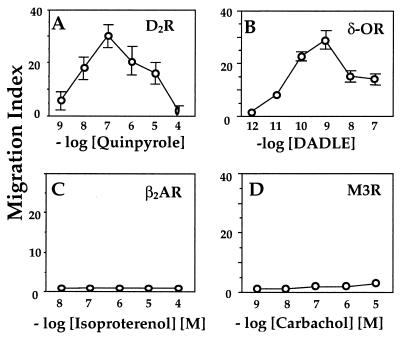
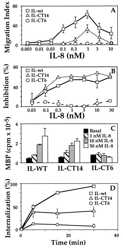
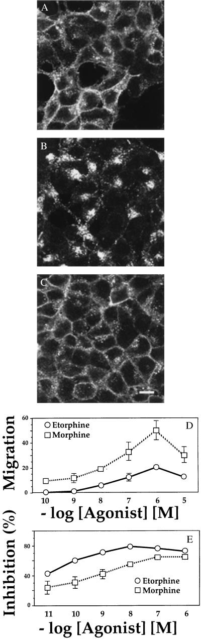
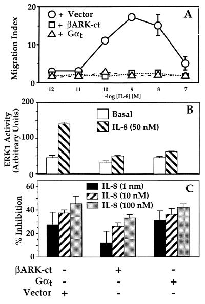
Many chemoattractants cause chemotaxis of leukocytes by stimulating a structurally distinct class of G protein-coupled receptors. To identify receptor functions required for chemotaxis, we studied chemotaxis in HEK293 cells transfected with receptors for nonchemokine ligands or for interleukin 8 (IL-8), a classical chemokine. In gradients of the appropriate agonist, three nonchemokine Gi-coupled receptors (the D2 dopamine receptor and opioid mu and delta receptors) mediated chemotaxis; the beta2-adrenoreceptor and the M3-muscarinic receptor, which couple respectively to Gs and Gq, did not mediate chemotaxis. A mutation deleting 31 C-terminal amino acids from the IL-8 receptor type B quantitatively impaired chemotaxis and agonist-induced receptor internalization, but not inhibition of adenylyl cyclase or stimulation of mitogen-activated protein kinase. To probe the possible relation between receptor internalization and chemotaxis, we used two agonists of the mu-opioid receptor. Morphine and etorphine elicited quantitatively similar chemotaxis, but only etorphine induced receptor internalization. Overexpression of two betagamma sequestering proteins (betaARK-ct and alphat) prevented IL-8 receptor type B-mediated chemotaxis but did not affect inhibition of adenylyl cyclase by IL-8. We conclude that: (i) Nonchemokine Gi-coupled receptors can mediate chemotaxis. (ii) Gi activation is necessary but probably not sufficient for chemotaxis. (iii) Chemotaxis does not require receptor internalization. (iv) Chemotaxis requires the release of free betagamma subunits.


Figures

Figure 1
Figure 1
Chemotaxis of cells expressing nonchemokine receptors. Migration assays were performed in a 48-well Boyden chamber, as described (15) on stable transfectants expressing (A) D2 dopamine receptor, (B) HA-tagged δ opioid receptor, (C) FLAG-tagged β2-adrenoreceptor, and (D) HA-tagged M3-muscarinic receptor. Values represent the mean ± SE of six determinations. Similar results were obtained in three or more independent experiments. In these and all other migration assays, chemokinesis controls (described in Materials and Methods) showed that chemokinesis could not account for >15% of total cell migration (data not shown).
Figure 2
Figure 2
Wild-type and C-terminal truncation mutants of the IL-8 receptor. (A) Migration assays, performed as described in the legend to Fig. 1, on cells expressing C-terminal truncation mutants (IL-CT14 or IL-CT6) or the wild-type IL-8 receptor (15). (B) cAMP accumulation. Cells were incubated with 200 μM forskolin and the indicated concentrations of IL-8. Values represent percent inhibition by IL-8 of the forskolin-stimulated cAMP response, measured as described (17). Forskolin increased cAMP >100-fold over basal. (C) MAPK (ERK) activation. Stable transfectants were transiently transfected with 1 μg HA-ERK1 DNA and after 48 hr ERK activities were assessed as described (13). ERK activation is expressed in PhosphorImager units. (D) Receptor internalization. The percent of surface receptors remaining after various times of exposure to agonist was measured in whole-cell binding assays using [125I]-IL-8. Data shown represents the mean ± SE of triplicate determinations. Results similar to those in A–D were obtained in three or more independent experiments. HEK293 cell lines expressing the wild-type and mutant IL8Rs were obtained from Adit Ben-Baruch (Tel Aviv University) (15).
Figure 3
Figure 3
Responses of cells expressing the μ opioid receptor. (A–C). Immunofluorescence localization. Cells stably expressing a FLAG-tagged μ-opioid receptor were incubated for 1 hr in the absence of ligand (A) or in the presence of 100 nM etorphine (B) or 100 nM morphine (C), then fixed in 4% formaldehyde. The receptor was detected by incubation with the monoclonal antibody M2, followed by fluorescein isothiocyanate-conjugated anti-mouse antibody (16). Specimens were examined by confocal microscopy. Scale bar = 10 μm. (D) Chemotaxis, assessed as in Fig. 1, in response to morphine or etorphine. Similar results were obtained in three separate experiments. Values represent the mean ± SE of six determinations. (E) Inhibition of forskolin-stimulated cAMP accumulation was measured in response to increasing concentrations of morphine or etorphine, as in Fig. 2B. Similar results were obtained in three separate experiments. Bars = mean ± SE of three determinations.
Figure 4
Figure 4
Responses to IL-8 in cells expressing proteins that sequester βγ. (A) Chemotaxis. Migration assays were performed as described in the legend to Fig. 1 on cells expressing the wild-type IL-8 receptor stably cotransfected with either pcDNA1, pRK-βARK-1-(495–689) (βARK-ct), or αt-pcDNA1, each in combination with a plasmid encoding a hygromycin-resistance marker. (B) MAPK (ERK) activation. Double transfectants were analyzed as described in the legend to Fig. 2. ERK1 activity is expressed in PhosphorImager units. Epidermal growth factor caused 4- to 5-fold activation of ERK in each cell line. (C) Inhibition of cAMP accumulation. Transfectants were incubated with 200 μM forskolin and the indicated concentrations of IL-8. Bars = percent inhibition by IL-8 of the forskolin-stimulated cAMP response. In cells treated with forskolin alone, cAMP accumulation over basal values (measured as the ratio of [3H]cAMP to the sum of [3H]cAMP plus [3H]ATP) was 115, 160, or 247 × 10−3 in cells expressing pcDNA1, βARK-ct, or αt. Results similar to those in each panel were obtained in at least two independent experiments. Data shown represent the mean ± SE of triplicate determinations for A and C and the mean and range of duplicate determinations for B.

References

    1. Baggiolini M, Dewald B, Moser B. Adv Immunol. 1994;55:97–179. - PubMed
    1. Murphy P M. Annu Rev Immunol. 1994;12:593–633. - PubMed
    1. Premack B A, Schall T J. Nat Med. 1996;2:1174–1178. - PubMed
    1. Gerard C, Gerard N P. Curr Opin Immunol. 1994;6:140–145. - PubMed
    1. Freedman N J, Lefkowitz R J. Recent Prog Horm Res. 1996;51:319–351. ; discussion, 352–353. - PubMed

Publication types

LinkOut - more resources