

Chemotaxis in a lymphocyte cell line transfected with C-C chemokine receptor 2B: evidence that directed migration is mediated by betagamma dimers released by activation of Galphai-coupled receptors
- PMID: 9405641
- PMCID: PMC25033
- DOI: 10.1073/pnas.94.26.14495
Chemotaxis in a lymphocyte cell line transfected with C-C chemokine receptor 2B: evidence that directed migration is mediated by betagamma dimers released by activation of Galphai-coupled receptors
Abstract
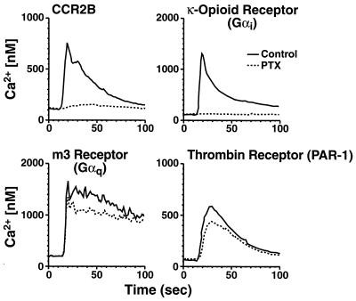
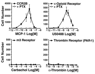
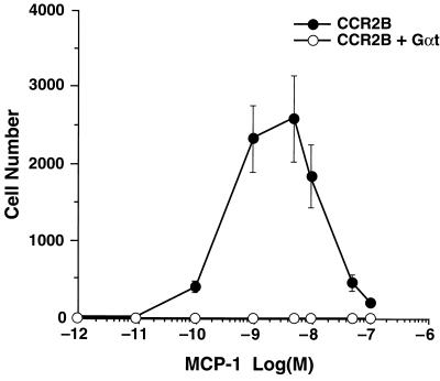
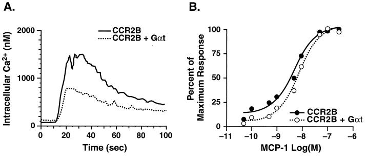
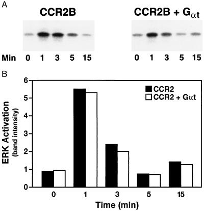
Chemotaxis is mediated by activation of seven-transmembrane domain, G protein-coupled receptors, but the signal transduction pathways leading to chemotaxis are poorly understood. To identify G proteins that signal the directed migration of cells, we stably transfected a lymphocyte cell line (300-19) with G protein-coupled receptors that couple exclusively to Galphaq (the m3 muscarinic receptor), Galphai (the kappa-opioid receptor), and Galphas (the beta-adrenergic receptor), as well as the human thrombin receptor (PAR-1) and the C-C chemokine receptor 2B. Cells expressing receptors that coupled to Galphai, but not to Galphaq or Galphas, migrated in response to a concentration gradient of the appropriate agonist. Overexpression of Galpha transducin, which binds to and inactivates free Gbetagamma dimers, completely blocked chemotaxis although having little or no effect on intracellular calcium mobilization or other measures of cell signaling. The identification of Gbetagamma dimers as a crucial intermediate in the chemotaxis signaling pathway provides further evidence that chemotaxis of mammalian cells has important similarities to polarized responses in yeast. We conclude that chemotaxis is dependent on activation of Galphai and the release of Gbetagamma dimers, and that Galphai-coupled receptors not traditionally associated with chemotaxis can mediate directed migration when they are expressed in hematopoietic cells.
Figures
References
-
- Hugli T E, Gerard C, Kawahara M, Scheetz M E, II, Barton R, Briggs S, Koppel G, Russell S. Mol Cell Biochem. 1981;41:59–66. - PubMed
-
- Yokomizo T, Izumi T, Chang K, Takuwa Y, Shimizu T. Nature (London) 1997;387:620–624. - PubMed
-
- Baggiolini M, Dewald B, Moser B. Annu Rev Immunol. 1997;15:675–705. - PubMed
-
- Hedrick J A, Zlotnik A. Curr Opin Immunol. 1996;8:343–347. - PubMed
Publication types
MeSH terms
Substances
Grants and funding
LinkOut - more resources
Full Text Sources
Other Literature Sources
