

doi: 10.1073/pnas.94.26.14584.
Sequence of the FRA3B common fragile region: implications for the mechanism of FHIT deletion
Affiliations
- PMID: 9405656
- PMCID: PMC25062
- DOI: 10.1073/pnas.94.26.14584
Item in Clipboard
Sequence of the FRA3B common fragile region: implications for the mechanism of FHIT deletion
Proc Natl Acad Sci U S A.
.
Abstract
The hypothesis that chromosomal fragile sites may be "weak links" that result in hot spots for cancer-specific chromosome rearrangements was supported by the discovery that numerous cancer cell homozygous deletions and a familial translocation map within the FHIT gene, which encompasses the common fragile site, FRA3B. Sequence analysis of 276 kb of the FRA3B/FHIT locus and 22 associated cancer cell deletion endpoints shows that this locus is a frequent target of homologous recombination between long interspersed nuclear element sequences resulting in FHIT gene internal deletions, probably as a result of carcinogen-induced damage at FRA3B fragile sites.
Figures
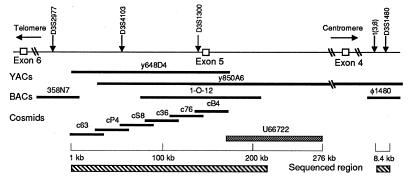
The normal FHIT/FRA3B locus. The top line represents the locus with positions of reported markers and FHIT exons. The long hatched bar represents the 210-kb sequenced region, which overlapped the reported U66722 sequence (solid bar) (17). Genomic YAC, BAC, phage and cosmid clones used in the analysis are shown. The small hatched bar represents the sequence of chromosome 3 at the t(3;8) break.
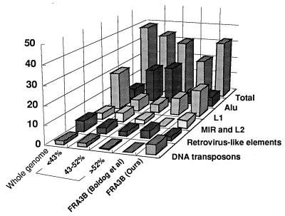
Repeats in the FHIT/FRA3B region. In this modification of Smit’s original figure (36), the whole genome was divided into three groups according to GC content, <43%, 43–52%, and >52%. MIR and L2 elements are combined. The ordinate shows the fraction of the respective sequenced region occupied by specific repetitive classes.
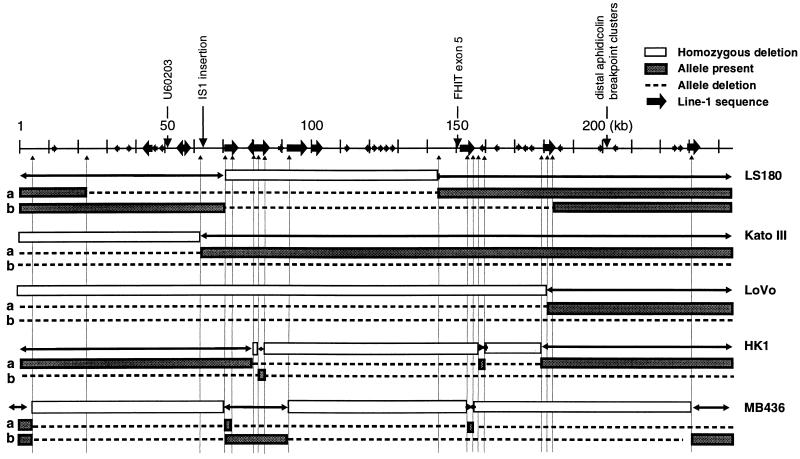
Deletions in cancer cell lines. Depiction of 21 breakpoints (not including the LoVo breakpoint in BAC clone 358N7) with locations relative to L1 elements. L1 sequences of >1 kb, bold arrows; L1 sequences of <1 kb, small arrowheads
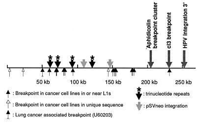
Positions of FRA3B/FHIT rearrangements. Illustration of the 276-kb sequenced region summarizing positions of deletion endpoints, hybrid breaks, plasmid or viral flanking sequences, and trinucleotide repeats.
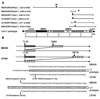
L1 associated breakpoints and sequences. (A) Six L1 sequences in the 210-kb region are aligned to the active LINE-1 (L1.2) prototype sequence (46). These L1 elements showed >80% sequence homology to the prototype over 1,000 bp. Note that all L1s were truncated, retaining only a 3′ portion of the sequence, and most breakpoints were located at the 5′ end of the retained sequence. EN, endonuclease; RT, reverse transcriptase; C, cysteine-rich region. (B) Rearrangements and sequence of breakpoints in MB436 and LS180; note sequence homology to the L1 prototype.
References
-
- Sutherland G R, Richards R I. Curr Opin Genet Dev. 1995;5:323–327. - PubMed
-
- Jones C, Penny L, Mattina T, Yu S, Baker E, Voullaire L, Langdon W Y, Sutherland G R, Richards R I, Tunnacliffe A. Nature (London) 1995;376:145–149. - PubMed
-
- Yu S, Mangelsdorf M, Hewett D, Hobson L, Baker E, Eyre H J, Lapsys N, Paslier D L, Doggett N A, Sutherland G R, Richards R I. Cell. 1997;88:367–374. - PubMed
-
- Warren S T, Zhang F, Licambi G R, Peters J F. Science. 1987;237:420–423. - PubMed
Publication types
MeSH terms
Substances
Associated data
- Actions
- Actions
- Actions
- Actions
- Actions
- Actions
- Actions
- Actions
- Actions
- Actions
Grants and funding
LinkOut - more resources
Full Text Sources
Other Literature Sources
