. 1997 Dec 23;94(26):14683-8.
doi: 10.1073/pnas.94.26.14683.

The anticoagulant activation of antithrombin by heparin

Affiliations

The anticoagulant activation of antithrombin by heparin

L Jin et al. Proc Natl Acad Sci U S A. .

Abstract

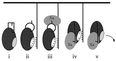
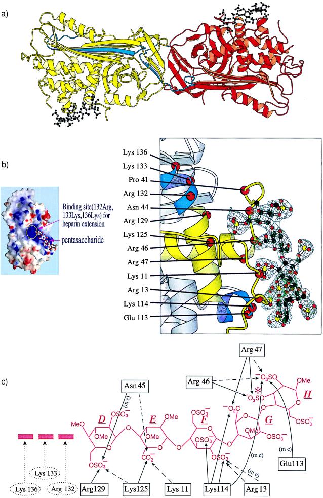
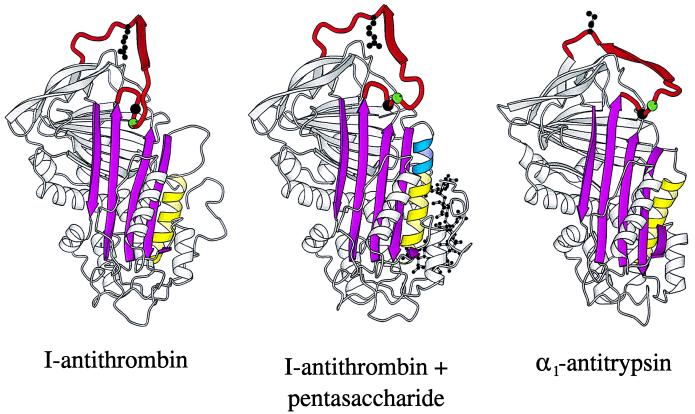
Antithrombin, a plasma serpin, is relatively inactive as an inhibitor of the coagulation proteases until it binds to the heparan side chains that line the microvasculature. The binding specifically occurs to a core pentasaccharide present both in the heparans and in their therapeutic derivative heparin. The accompanying conformational change of antithrombin is revealed in a 2.9-A structure of a dimer of latent and active antithrombins, each in complex with the high-affinity pentasaccharide. Inhibitory activation results from a shift in the main sheet of the molecule from a partially six-stranded to a five-stranded form, with extrusion of the reactive center loop to give a more exposed orientation. There is a tilting and elongation of helix D with the formation of a 2-turn helix P between the C and D helices. Concomitant conformational changes at the heparin binding site explain both the initial tight binding of antithrombin to the heparans and the subsequent release of the antithrombin-protease complex into the circulation. The pentasaccharide binds by hydrogen bonding of its sulfates and carboxylates to Arg-129 and Lys-125 in the D-helix, to Arg-46 and Arg-47 in the A-helix, to Lys-114 and Glu-113 in the P-helix, and to Lys-11 and Arg-13 in a cleft formed by the amino terminus. This clear definition of the binding site will provide a structural basis for developing heparin analogues that are more specific toward their intended target antithrombin and therefore less likely to exhibit side effects.


Figures

Figure 1
Figure 1
Schematic: i, circulating antithrombin; ii, contacts endothelial heparans with induction of high-affinity binding and reactive site loop exposure; iii–iv, complexes with factor Xa followed by loop cleavage and insertion with diminished heparin affinity; and v, the complex is released into the circulation for catabolism by the liver.
Figure 2
Figure 2
(a) Dimer of L-antithrombin (yellow) and I-antithrombin (red) each complexed with the pentasaccharide (black). The reactive site loop of each molecule is in blue, the loop being fully inserted in the A β-sheet in L-antithrombin with the reactive loop of the I-antithrombin replacing the vacated strand site s1C in the L-molecule through its P3–P8 residues (amino-terminal to the P1 reactive center). (b) (Left) Electrostatic surface potential map of antithrombin (red, negative potential; blue, positive potential) with the pentasaccharide outline and showing its extension region including 132-Arg, 133-Lys, and 136-Lys. (Right) A σA-weighted difference map, calculated after omitting the pentasaccharide, is displayed at a contour level of 3σ within volume 4 Å around the omitted atoms. Superimposed on the omitmap are the atoms of the pentasaccharide DEFGH (D above) and a ribbon representation of I-antithrombin. The binding site is in yellow and also includes the P-helix, the lower of the two induced helical segments, in blue. (c) Hydrogen bonding to pentasaccharide DEFGH. Likely bonds in full lines, possible bonds in interrupted lines, (mc) indicates main-chain bonding. Arg-132, Lys-133, and Lys-136 are beyond hydrogen bonding distance from the pentasaccharide but could interact with extended oligosaccharides. The extra sulfate present in the high affinity pentasaccharide (16) is asterisked in saccharide H.
Figure 3
Figure 3
Ribbon diagrams of (from left) I-antithrombin (15), pentasaccharide-complexed I-antithrombin, and α1-antitrypsin (32). The pentasaccharide activation of I-antithrombin is seen to involve a closing of the A-sheet (magenta), an extension (blue) of helix D (yellow), and an expulsion of residues P14 (green sphere) and P15 (black sphere) of the reactive site loop (red). The reactive loop of both antithrombin molecules is constrained by the dimer contact (see Fig. 2a) of the β-pleated P3–P8 (ribboned arrow). An indication of the likely free conformation, with exposure of the P1 reactive center (shown as a ball–stick model), is provided by the optimal inhibitory conformation of the reactive loop present in α1-antitrypsin (32).
Figure 4
Figure 4
Exposure of reactive center arginine by pentasaccharide. Native PAGE at pH 8.0 of a mutant (P1 His) antithrombin (26) in lanes 1–3, and of normal (P1 Arg) antithrombin in lanes 4–6, is shown. In lanes 2 and 5, the antithrombins have been incubated with deiminase; in lanes 3 and 6, incubation with deiminase has been carried out in the presence of the pentasaccharide. The change in mobility consequent to deimination occurs only in lane 6, with the P1 Arg antithrombin plus pentasaccharide.

References

    1. Marcum J A, McKenney J B, Galli S J, Jackman R W, Rosenberg R D. J Clin Invest. 1986;74:341–350. - PMC - PubMed
    1. Thunberg L, Backstrom G, Lindahl U. Carbohydr Res. 1982;100:393–397. - PubMed
    1. Choay J, Petitou M, Lormeau J C, Sinay P, Casu B, Gatti G. Biochem Biophys Res Commun. 1983;116:492–499. - PubMed
    1. Lane D A, Denton J, Flynn A M, Thunberg L, Lindahl U. Biochem J. 1984;218:725–732. - PMC - PubMed
    1. Olson S T, Björk I, Sheffer R, Craig P A, Shore J D, Choay J. J Biol Chem. 1992;267:12528–12538. - PubMed

Publication types

Associated data