A system for functional analysis of Ebola virus glycoprotein
- PMID: 9405687
- PMCID: PMC25111
- DOI: 10.1073/pnas.94.26.14764
A system for functional analysis of Ebola virus glycoprotein
Abstract
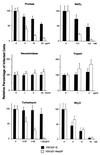
Ebola virus causes hemorrhagic fever in humans and nonhuman primates, resulting in mortality rates of up to 90%. Studies of this virus have been hampered by its extraordinary pathogenicity, which requires biosafety level 4 containment. To circumvent this problem, we developed a novel complementation system for functional analysis of Ebola virus glycoproteins. It relies on a recombinant vesicular stomatitis virus (VSV) that contains the green fluorescent protein gene instead of the receptor-binding G protein gene (VSVDeltaG*). Herein we show that Ebola Reston virus glycoprotein (ResGP) is efficiently incorporated into VSV particles. This recombinant VSV with integrated ResGP (VSVDeltaG*-ResGP) infected primate cells more efficiently than any of the other mammalian or avian cells examined, in a manner consistent with the host range tropism of Ebola virus, whereas VSVDeltaG* complemented with VSV G protein (VSVDeltaG*-G) efficiently infected the majority of the cells tested. We also tested the utility of this system for investigating the cellular receptors for Ebola virus. Chemical modification of cells to alter their surface proteins markedly reduced their susceptibility to VSVDeltaG*-ResGP but not to VSVDeltaG*-G. These findings suggest that cell surface glycoproteins with N-linked oligosaccharide chains contribute to the entry of Ebola viruses, presumably acting as a specific receptor and/or cofactor for virus entry. Thus, our VSV system should be useful for investigating the functions of glycoproteins from highly pathogenic viruses or those incapable of being cultured in vitro.
Figures


References
-
- Peters C J, Sanchez A, Rollin P E, Ksiazek T G, Murphy F A. In: Fields Virology. Fields B N, Knipe D M, Howley P M, editors. Philadelphia: Lippincott-Raven; 1995. pp. 1161–1176.
-
- Feldmann H, Klenk H D, Sanchez A. Arch Virol Suppl. 1993;7:81–100. - PubMed
-
- Feldmann H, Muhlberger E, Randolf A, Will C, Kiley M P, Sanchez A, Klenk H D. Virus Res. 1992;24:1–19. - PubMed
-
- Kiley M P, Regnery R L, Johnson K M. J Gen Virol. 1980;49:333–341. - PubMed
-
- Elliott L H, Kiley M P, McCormick J B. Virology. 1985;147:169–176. - PubMed
Publication types
MeSH terms
Substances
Grants and funding
LinkOut - more resources
Full Text Sources
Other Literature Sources
Medical

