. 1997 Dec 23;94(26):14787-91.
doi: 10.1073/pnas.94.26.14787.

A new mechanism of sound generation in songbirds

Affiliations

A new mechanism of sound generation in songbirds

F Goller et al. Proc Natl Acad Sci U S A. .

Abstract

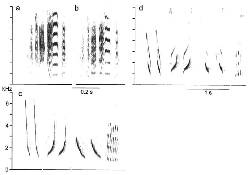
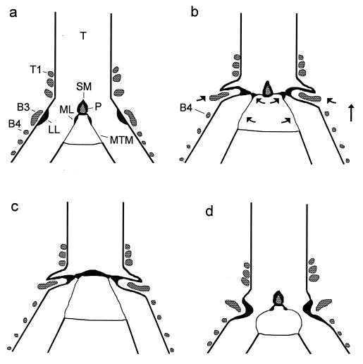
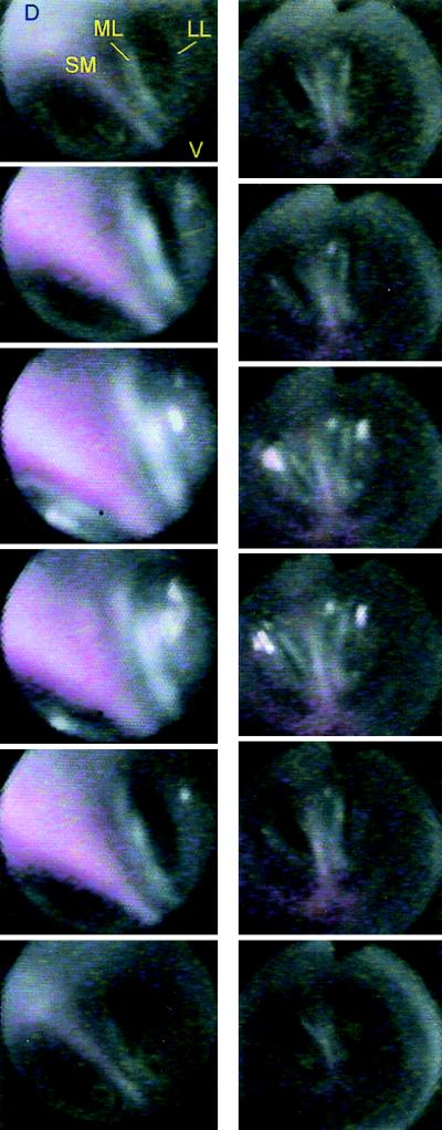
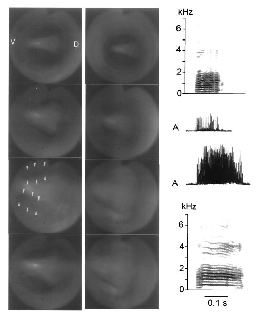
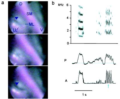
Our current understanding of the sound-generating mechanism in the songbird vocal organ, the syrinx, is based on indirect evidence and theoretical treatments. The classical avian model of sound production postulates that the medial tympaniform membranes (MTM) are the principal sound generators. We tested the role of the MTM in sound generation and studied the songbird syrinx more directly by filming it endoscopically. After we surgically incapacitated the MTM as a vibratory source, zebra finches and cardinals were not only able to vocalize, but sang nearly normal song. This result shows clearly that the MTM are not the principal sound source. The endoscopic images of the intact songbird syrinx during spontaneous and brain stimulation-induced vocalizations illustrate the dynamics of syringeal reconfiguration before phonation and suggest a different model for sound production. Phonation is initiated by rostrad movement and stretching of the syrinx. At the same time, the syrinx is closed through movement of two soft tissue masses, the medial and lateral labia, into the bronchial lumen. Sound production always is accompanied by vibratory motions of both labia, indicating that these vibrations may be the sound source. However, because of the low temporal resolution of the imaging system, the frequency and phase of labial vibrations could not be assessed in relation to that of the generated sound. Nevertheless, in contrast to the previous model, these observations show that both labia contribute to aperture control and strongly suggest that they play an important role as principal sound generators.


Figures

Figure 1
Figure 1
Elimination of the MTM as a vibrating sound source does not mute birds and does not prevent them from producing nearly normal song. The song motif of a zebra finch remains intact after large holes were made into the left and right MTM, fully encompassing both. Song is shown spectrographically before (a) and 1 day after (b) the MTM were incapacitated as sound sources. Similarly, when both MTM were fully torn in a cardinal, preoperatively recorded syllable types (c) were sung nearly intact 1 day after the surgical removal of the MTM (d). In both species small changes in the acoustic fine structure of song, especially in harmonic emphasis, may be caused by the loss of the MTM, but surgical effects, such as fluid build-up and the sealing of the interclavicular air sac membrane with tissue adhesive, cannot be ruled out as possible causes.
Figure 2
Figure 2
(a) Configuration of the syrinx during quiet breathing in a frontal section (schematic). B3, B4, third and fourth bronchial ring; P, ossified pessulus; T, trachea; T1, first tracheal ring. (b) In the phonatory position the syrinx is moved rostrad and the bronchi are stretched (indicated by arrows), which, in concert with activity of the dorsal syringeal muscles, causes both labia to move mediad. LL is pushed into the lumen by B3, which presumably is rotated by dorsal muscle activity and also moved mediad by syringeal stretching. Movement of ML presumably is mediated by a thin, cartilaginous extension from the ventral end of B2 (not illustrated). (c) The dome-shaped phonatory position of the crow syrinx lacking an ossified pessulus. (d) The “classical model” of the phonatory position postulates constriction of the syrinx by the LL and vibrating MTM (redrawn after refs. and 11).
Figure 3
Figure 3
Individual frames taken from video segments (duration about 1 s) illustrate syringeal configuration during the transition from quiet respiratory to phonatory position in brown thrasher (Left, Top to Bottom) and cardinal (Right). The vocal organ is viewed from the top with the angioscope inserted into the trachea. The syrinx moves rostrad (increasing size of syringeal structures because of decreasing distance to angioscope lens and increased brightness of light reflections), and the labia move simultaneously into the bronchial lumen, resulting in bilateral closure. Phonation occurred during frames three and four (Left and Right). Black triangular notch on images (Right, top of frames) stems from an indicator in the angioscope.
Figure 4
Figure 4
During spontaneously emitted calls, the crow syrinx (viewed from top) undergoes a reconfiguration similar to that of the thrasher and the cardinal (frames show transition from quiet respiration to phonation). A soft call (left angioscope column, upper spectrogram, and rectified amplitude waveform, A) is generated with incomplete closure of the ventral portion (arrows) of both syringeal halves. In contrast, during a louder call (right angioscope column, lower spectrogram, and waveform) there is complete bilateral closure and a greater rostrad excursion (distance between angioscope lens and syrinx in frame 1 is greater on Right than that on Left) of the syrinx. High subsyringeal pressure inflates the bronchial septum such that the ML cannot be clearly separated from the SM. The absence of an ossified pessulus in crows (15) probably facilitates this deformation. Because of the longer duration of the loud call, the return of the syrinx into respiratory position is not shown (Right).
Figure 5
Figure 5
Amplitude modulation of a brown thrasher vocalization is clearly visible as a correlated partial opening of the right valve formed by the adducted labia. (a) Illustration of the slight partial abduction (arrowhead) and fully closed position of the right side during one modulation cycle. On the left side the labia remain in close contact throughout the cycle (D, dorsal; V, ventral). (b) Series of representative vocalizations evoked by electrical brain stimulation is shown spectrographically (Upper) and as rectified sound amplitude (A) (Lower) together with the subsyringeal air sac pressure (P, horizontal line indicates ambient pressure). Sounds may be almost unmodulated (Left) or 100% amplitude modulated (Right). The blue arrow marks the modulation cycle illustrated in a.

References

    1. Konishi M. Annu Rev Neurosci. 1985;8:125–170. - PubMed
    1. Nottebohm F. J Comp Physiol A. 1996;179:149–156. - PubMed
    1. Marler P. Am Scientist. 1970;58:669–673. - PubMed
    1. Hauser M D. The Evolution of Communication. Cambridge, MA: MIT; 1996.
    1. Fink B R. The Human Larynx: A Functional Study. New York: Raven; 1975.

Publication types

LinkOut - more resources