. 1997 Dec 23;94(26):14809-14.
doi: 10.1073/pnas.94.26.14809.

In vitro-generated neural precursors participate in mammalian brain development

Affiliations

In vitro-generated neural precursors participate in mammalian brain development

O Brüstle et al. Proc Natl Acad Sci U S A. .

Abstract

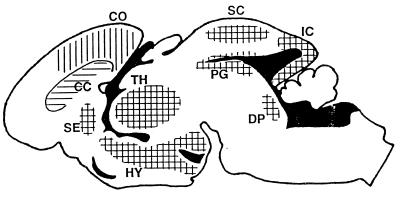
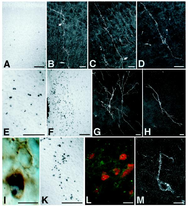
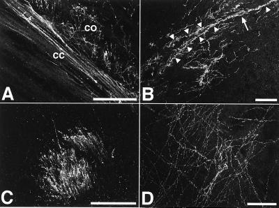
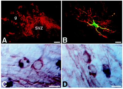
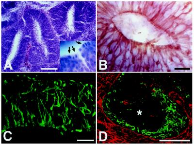
During embryogenesis, pluripotent stem cells segregate into daughter lineages of progressively restricted developmental potential. In vitro, this process has been mimicked by the controlled differentiation of embryonic stem cells into neural precursors. To explore the developmental potential of these cell-culture-derived precursors in vivo, we have implanted them into the ventricles of embryonic rats. The transplanted cells formed intraventricular neuroepithelial structures and migrated in large numbers into the brain tissue. Embryonic-stem-cell-derived neurons, astrocytes, and oligodendrocytes incorporated into telencephalic, diencephalic, and mesencephalic regions and assumed phenotypes indistinguishable from neighboring host cells. These observations indicate that entirely in vitro-generated neural precursors are able to respond to environmental signals guiding cell migration and differentiation and have the potential to reconstitute neuronal and glial lineages in the central nervous system.


Figures

Figure 1
Figure 1
Distribution of ES-cell-derived neural precursors after implantation into the telencephalic vesicle of E16–E18 rats. The schematic represents a midsagittal section through the brain of a newborn recipient. After leaving the ventricular system (solid areas), neurons (vertical lines) and astrocytes (horizontal lines) occupy overlying territories. Donor-derived neurons integrate preferentially into gray matter regions exhibiting neurogenesis until or beyond the time of implantation. ES-cell-derived astrocytes also incorporate into white matter regions such as the corpus callosum (CC). CO, cortex; DP, dorsal pontine area; HY, hypothalamus; IC, inferior colliculus; PG, periaqueductal gray; SC, superior colliculus; SE, septum; TH, thalamus. Donor-derived neurons and astrocytes were also detected in hippocampus (Fig. 3B), olfactory bulb (Fig. 4 A and B), and striatum (Fig. 2E).
Figure 2
Figure 2
ES-cell-derived neural precursors injected into the telencephalic vesicle of fetal rats incorporate individually into a variety of host brain regions and differentiate into neurons. Donor cells are identified by in situ hybridization using a digoxigenin-labeled probe to mouse satellite DNA (A, E, F, and I–L). Immunofluorescence detection of the mouse-specific antigen M6 and confocal laser microscopy were used to reconstruct individual neuronal profiles (B–D, G, H, and M). (A–D) Six days after injection into the telencephalic vesicle of an E17 rat, donor cells have left the ventricle and incorporated into the host cortex. The ES-cell-derived neurons show prominent apical dendrites and basal axons entering the corpus callosum, a morphology appropriate for cortical projection neurons (arrows: perikaryon). Note the characteristic pyramidal morphology in D. (E) Incorporated donor cells in the striatum of a 2-week-old rat transplanted at E18. (F–H) Donor-derived cells in the host hypothalamus. In contrast to cortex, neurons incorporating into the diencephalon frequently exhibited multipolar morphologies (G and H). (I) Host- and donor-derived neurons in the septum of a newborn rat. Both neurons show expression of microtubule-associated protein 2; the donor-derived cell is identified by in situ hybridization. (K–M) Incorporated cells in the host thalamus of newborn (K) and 2-week-old rats (L and M). In L, ES-cell-derived neurons are visualized by fluorescence in situ hybridization (green dots) and subsequent immunofluorescence analysis with an antibody to the nuclear neuronal antigen NeuN (red). Note the mature neuronal phenotype of the integrated cells with the presence of dendritic spines (M). [Bars = 100 μm (A, E, F, and K), 20 μm (B–D, G, H, L, and M), and 10 μm (I).]
Figure 3
Figure 3
Extensive axonal innervation of the host brain. The ES-cell-derived neurons generated a dense axonal network within the recipient brains. Abundant M6-positive axons were found at all levels in both gray and white matter. (A) Donor-derived axons in corpus callosum (cc) and deep layer cortex (co) of a 2-week-old recipient. (B) Axonal innervation of the hippocampal stratum oriens. The M6 immunofluorescence also depicts the perikaryon (arrow) and dendrites (arrowheads) of a large horizontal neuron in the upper stratum oriens. The morphology of this cell is very similar to the outline of Golgi-impregnated horizontal neurons in this area (20). (C) Abundant donor-derived axons in a striatal fiber tract of 2-week-old recipient brain. (D) ES-cell-derived axons in the thalamus of a newborn recipient transplanted at E17. (Bars = 50 μm.)
Figure 4
Figure 4
Incorporation of ES-cell-derived glia. (A and B) ES-cell-derived astrocytes have migrated into the granular layer (g) of the olfactory bulb of a 2-week-old host. Cells are visualized with an antibody to the mouse-specific antigen M2 (red). SVZ, olfactory subventricular zone. An individual astrocyte, double labeled with an antibody to GFAP (green), is shown in B. (C and D) ES-cell-derived oligodendrocytes in the rostral (C) and caudal (D) corpus callosum of a 2-week-old host brain. The donor cells, identified by DNA in situ hybridization (black), are morphologically indistinguishable from adjacent host oligodendrocytes. Host- and donor-derived oligodendrocytes exhibit equivalent immunoreactivity to CNPase (red). [Bars = 100 μm (A) and 10 μm (B–D).]
Figure 5
Figure 5
Generation of neuroepithelial formations. (A and B) Eight days after intrauterine transplantation, the donor cells have generated numerous neural tube-like structures within the host ventricle. Like the developing neural tube, these structures exhibit high mitotic activity at the luminal surface (A) (hematoxylin/eosin; arrows in Inset indicate mitotic figures) and strong expression of the intermediate filament nestin (B). (C and D) Neuroepithelial formation in the ventricle of a 2-week-old animal transplanted at E18. The formation contains abundant radially oriented nestin-positive processes (C). As in the early neuroepithelium, there is an inside-out gradient of differentiation with neuronal markers being expressed at the periphery of the formation (D) (green, tyrosine hydroxylase; red, M6; ∗, center of formation). [Bars = 100 μm (A and D) and 20 μm (B and C).]

References

    1. Evans M J, Kaufman M H. Nature (London) 1981;292:154–156. - PubMed
    1. Martin G R. Proc Natl Acad Sci USA. 1981;78:7634–7638. - PMC - PubMed
    1. Palacios R, Golunski E, Samaridis J. Proc Natl Acad Sci USA. 1995;92:7530–7534. - PMC - PubMed
    1. Bain G, Kitchens D, Yao M, Huettner J E, Gottlieb D I. Dev Biol. 1995;168:342–357. - PubMed
    1. Fraichard A, Chassande O, Bilbaut G, Dehay C, Savatier P, Samarut J. J Cell Sci. 1995;108:3181–3188. - PubMed