. 1997 Dec 23;94(26):14894-9.
doi: 10.1073/pnas.94.26.14894.

Apparent mtDNA heteroplasmy in Alzheimer's disease patients and in normals due to PCR amplification of nucleus-embedded mtDNA pseudogenes

Affiliations

Apparent mtDNA heteroplasmy in Alzheimer's disease patients and in normals due to PCR amplification of nucleus-embedded mtDNA pseudogenes

M Hirano et al. Proc Natl Acad Sci U S A. .

Abstract

In an unprecedented finding, Davis et al. [Davis, R. E., Miller, S., Herrnstadt, C., Ghosh, S. S., Fahy, E., Shinobu, L. A., Galasko, D., Thal, L. J., Beal, M. F., Howell, N. & Parker, W. D., Jr. (1997) Proc. Natl. Acad. Sci. USA 94, 4526-4531] used an unusual DNA isolation method to show that healthy adults harbor a specific population of mutated mitochondrial cytochrome c oxidase (COX) genes that coexist with normal mtDNAs. They reported that this heteroplasmic population was present at a level of 10-15% in the blood of normal individuals and at a significantly higher level (20-30%) in patients with sporadic Alzheimer's disease. We provide compelling evidence that the DNA isolation method employed resulted in the coamplification of authentic mtDNA-encoded COX genes together with highly similar COX-like sequences embedded in nuclear DNA ("mtDNA pseudogenes"). We conclude that the observed heteroplasmy is an artifact.


Figures

Figure 1
Figure 1
PCR amplification of ρ+ and ρo DNA and of DNA from the supernatant (S) and the pelleted cell debris (P) after 10 min of boiling of the buffy coat of an AD patient (subject 1) and a control (subject 2), using primers corresponding to the three COX genes. The predicted sizes of the PCR products, in bp, are at right. M, DNA Mass Ladder markers (GIBCO/BRL) with sizes, in bp, at left.
Figure 2
Figure 2
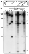
PCR/RFLP analysis of the COX II region. (A) Schematic of the HpaII digestion pattern of the COX II region amplified from ρ+- and ρo-derived DNA by using the COX II primers (P1 and P2). The HpaII sites (interior vertical lines), the location of the two HpaII polymorphisms, and the predicted sizes of the HpaII-digested fragments, in bp, are shown. (B) Autoradiogram of HpaII-digested 0.9-kb PCR products of ρ+ and ρo DNA and of total cellular DNA (T), DNA from the supernatant (S), and DNA from the pelleted cell debris (P) after 10 min of boiling of the buffy coat from the blood of an AD patient (subject 7) and a control (subject 8), by using primers corresponding to the COX II gene. Sizes of expected fragments, in bp, are at right. U, uncut PCR product from ρ+ DNA. Other notations are as in Fig. 1. The photo is a composite of two exposures of the same gel.
Figure 3
Figure 3
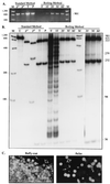
Time course of boiling. (A) The buffy coat pellet from subject 1 was resuspended in water, boiled for the indicated times, amplified with the COX II primers, and electrophoresed through an agarose gel; equal amounts of PCR product were loaded in each lane. DNA from ρ+ and ρo cells, and the total cellular DNA (T) and pelleted buffy-coat DNA after 10 min of boiling (P), all isolated by the standard method, are also shown. (B) The samples shown in A were digested with HpaII and electrophoresed through a nondenaturing polyacrylamide gel. Note the decline of the 270-bp and 94-bp fragments at the later time points (more clearly visible in the original autoradiogram, and in the darker exposure of the 20-min, 30-min, and 60-min results shown in the last three lanes). Other notations are as in Fig. 2. (C) Micrographs of resuspended cells from the buffy coat prior to boiling (the cells were clumped and difficult to disaggregate) and of the resuspended pellet after 10 min of boiling, after staining with MitoTracker. Note the mixed population of lymphocytes (large arrows) and platelets (small arrows). (×400.)
Figure 4
Figure 4
(A) Southern blot hybridization analysis of DNA from subject 7. Total cellular DNA (Tot.), and the DNA from the supernatant (Sup.) and the pelleted cell debris (Pel.) after 10 min of boiling of the buffy coat were digested with PvuII, electrophoresed through agarose, transferred to a nylon membrane, and hybridized with probes to reveal mtDNA and 18S rDNA (predicted sizes, in kb, at right). The blot was overexposed to reveal the nDNA signal in the pellet. (B) PCR amplification of the samples shown in A, as well as of DNA from ρ+ and ρo cells, using primers to amplify the mtDNA-encoded COX II gene (C) and the nDNA-encoded PGK gene (P). Note the strong signal for PGK compared with that for COX II in the supernatant samples. Predicted sizes of the PCR products, in bp, are at right.
Figure 5
Figure 5
Mixing experiment. DNA from ρ+ and ρo cells were mixed in the indicated proportions [reported as the percentage of ρ+ DNA present in the mixture, i.e., ρ+/(ρ+ + ρo) × 100], amplified with the COX II primers (see agarose gel shown in Inset), and the 901-bp fragment was digested with HpaII, and electrophoresed through a nondenaturing polyacrylamide gel. Lane numbers at bottom correspond to those in the Inset. Other notations are as in Fig. 2.

Similar articles

Cited by

References

    1. Goate A, Chartier-Harlin M-C, Mullan M, Brown J, Crawford F, et al. Nature (London) 1991;349:704–706. - PubMed
    1. Sherrington R, Rogaev E I, Liang Y, Rogaeva E A, Levesque G, et al. Nature (London) 1995;375:754–760. - PubMed
    1. Levy-Lahad E, Bird T D. Ann Neurol. 1996;40:829–840. - PubMed
    1. Levy-Lahad E, Wasco W, Poorkaj P, Romano D M, Oshima J, Pettingell W H, Yu C-E, Jondro P D, Schmidt S D, Wand K, Crowley A C, Fu Y H, Fuenette SY, Galas D, Nemens E, Wijsmann E M, Bird TD, Schellenberg GD, Tanzi R E. Science. 1995;269:973–977. - PubMed
    1. Stritmatter W J, Saunders A M, Schmechel D, Pericak-Vance M, Enghild J, Salvensen G S, Roses A D. Proc Natl Acad Sci USA. 1993;90:1977–1981. - PMC - PubMed

Publication types

Substances