. 1997 Dec 15;11(24):3471-81.
doi: 10.1101/gad.11.24.3471.

DNA damage induces phosphorylation of the amino terminus of p53

Affiliations

DNA damage induces phosphorylation of the amino terminus of p53

J D Siliciano et al. Genes Dev. .

Abstract

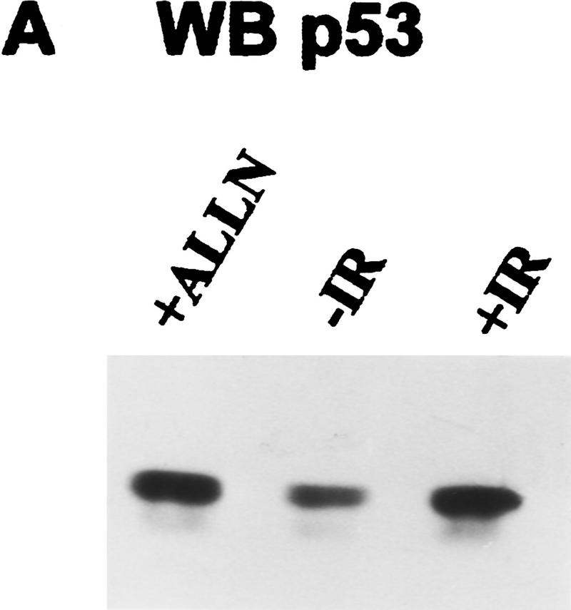
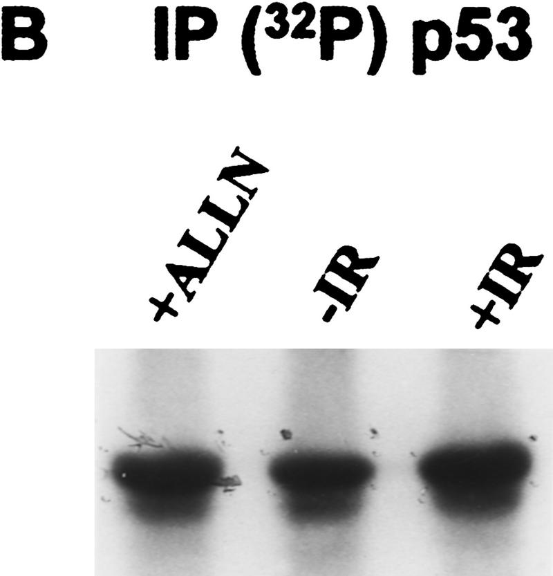
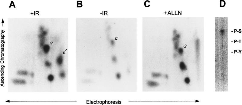
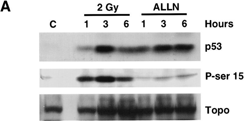
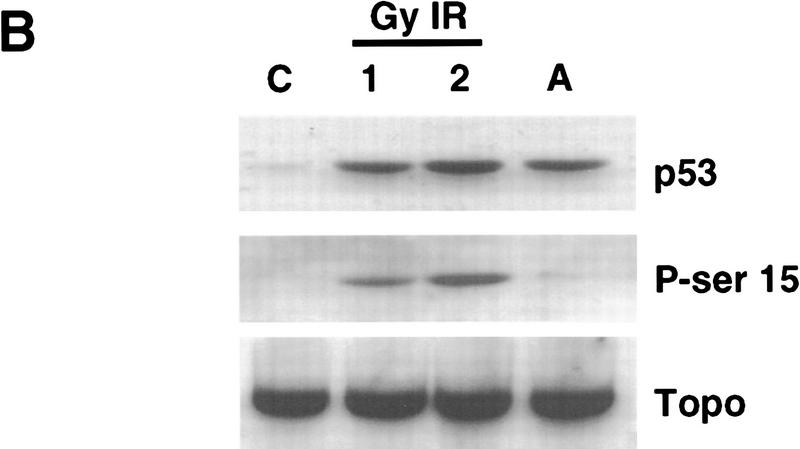
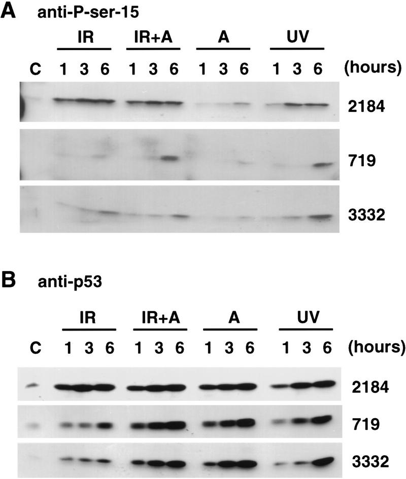
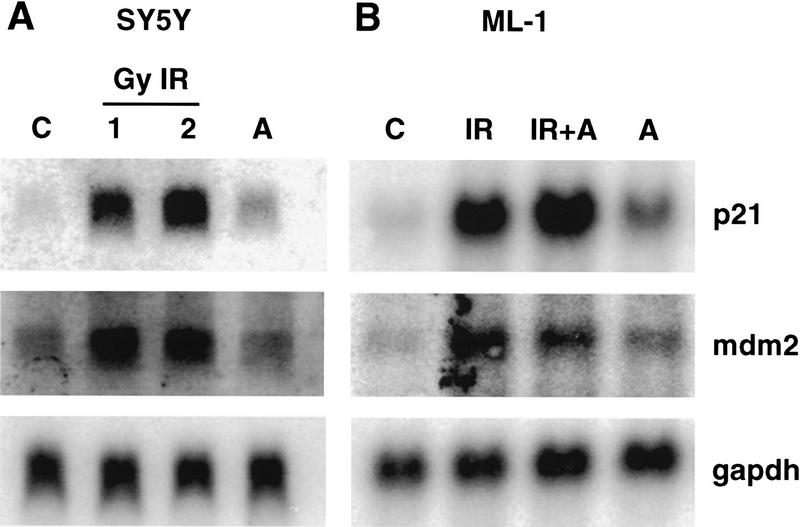
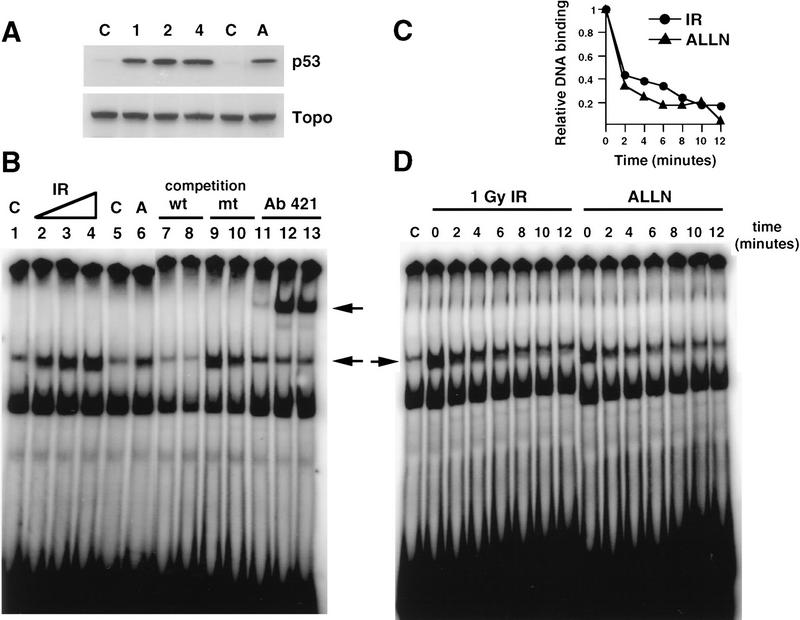
Data are presented demonstrating that DNA damage leads to specific post-translational modifications of p53 protein. Using two-dimensional peptide mapping of in vivo radiolabeled p53 tryptic phosphopeptides, recombinant truncated p53 protein, and synthetic p53 tryptic peptides, a unique p53 phosphopeptide was identified after exposure of ML-1 cells to ionizing irradiation. This peptide represents the first 24 amino acids of p53 and contains three phosphorylated serine residues. A specific p53 phosphopeptide antibody identified serine-15 as one of the two serines in p53 that becomes phosphorylated following DNA damage induced by either ionizing irradiation (IR) or ultraviolet (UV) irradiation in multiple cell types. IR-induced phosphorylation of p53 does not affect the kinetics of p53 binding to or dissociating from DNA as assessed by electrophoretic mobility-shift assays. However, p53 phosphorylation induced by DNA damage correlates with enhanced transcription of downstream p53 target genes. Low levels of phosphoserine-15 p53 are detectable within 6 hr after IR in AT cells, whereas lymphoblasts from normal individuals exhibit this modification within 1 hr. In contrast, phosphorylation of p53 on serine-15 is similar in normal and AT cells after UV irradiation. Our results indicate that p53 is phosphorylated in response to DNA damage, that this de novo phosphorylation may be involved in the subsequent induction and activation of p53, and that although ATM affects the kinetics of p53 phosphorylation after IR, it is not absolutely required for phosphorylation of p53 on serine-15.


Figures

Figure 1
Figure 1
Immunodetection of p53 protein in ML-1 cells. (A) Western blot analysis of ML-1 lysates. ML-1 cells were untreated (−IR) or treated with either 20 μm of ALLN (+ALLN) or 2 Gy irradiation (+IR). Lysates (50 μg) from each sample were resolved by 10% SDS-PAGE and then electrophoretically transferred to nitrocellulose. p53 was detected by immunoblotting. (B) Immunoprecipitation of p53. 32P-Labeled ML-1 extracts prepared from untreated (−IR), irradiated (+IR), or ALLN-treated (+ALLN) cells were immunoprecipitated with anti-p53 antibodies. Immunoprecipitates were resolved by 10% SDS-PAGE and electrophoretically transferred to a PVDF membrane. Radiolabeled p53 was detected by autoradiography. 32P-Labeled p53 from ALLN- and IR-treated cells had approximately twice as many counts per minute as 32P-labeled p53 from unirradiated cells when the isolated bands were counted in a scintillation counter.
Figure 1
Figure 1
Immunodetection of p53 protein in ML-1 cells. (A) Western blot analysis of ML-1 lysates. ML-1 cells were untreated (−IR) or treated with either 20 μm of ALLN (+ALLN) or 2 Gy irradiation (+IR). Lysates (50 μg) from each sample were resolved by 10% SDS-PAGE and then electrophoretically transferred to nitrocellulose. p53 was detected by immunoblotting. (B) Immunoprecipitation of p53. 32P-Labeled ML-1 extracts prepared from untreated (−IR), irradiated (+IR), or ALLN-treated (+ALLN) cells were immunoprecipitated with anti-p53 antibodies. Immunoprecipitates were resolved by 10% SDS-PAGE and electrophoretically transferred to a PVDF membrane. Radiolabeled p53 was detected by autoradiography. 32P-Labeled p53 from ALLN- and IR-treated cells had approximately twice as many counts per minute as 32P-labeled p53 from unirradiated cells when the isolated bands were counted in a scintillation counter.
Figure 2
Figure 2
IR induces phosphorylation of two serines within the first 24 amino acids of p53 in vivo. p53 was immunoprecipitated from 32P-labeled ML-1 cells that were either given 2 Gy irradiation (A), untreated (B), or treated with 20 μm of ALLN (C). Proteins were resolved by 10% SDS-PAGE and electrophoretically transferred to PVDF membrane. Radiolabeled p53 was cut from the membrane and digested with TPCK–trypsin. Radiolabeled peptides were resolved by electrophoresis at pH 3.5 in the first dimension followed by ascending chromatography in the second dimension. The unique, irradiation-induced p53 phosphopeptide (A, black arrow) was eluted from the cellulose and subjected to phosphoamino acid analysis. The position of the unlabeled phosphoamino acid markers are indicated (D). A singly phosphorylated p53 synthetic peptide corresponding to amino acids 1–24 (Ac 1–24; serine-15 P) comigrated with an in vivo peptide in all three maps (A–C, open arrow). The unique, IR-induced phosphopeptide comigrated with a synthetic, triply phosphorylated p53 peptide comprising amino acids 1–24 (Ac 1–24; serine-9 P, serine-15 P, serine-20 P) (A, black arrow).
Figure 3
Figure 3
Identification of serine-15 as one of the two sites phosphorylated within p53 in response to IR. (A) Western blot analysis of ML-1 lysates. ML-1 cells were either untreated (control, C) or treated with 2 Gy irradiation or 20 μm of ALLN. Cells were harvested at 1, 3, and 6 hr after treatment. Lysates (50 μg of protein in each lane) were resolved by 10% SDS-PAGE. After electrophoretic transfer, the nitrocellulose was immunoblotted with either anti-p53 ( Ab-6, top), anti-phosphoserine-15 p53 (middle), or anti-topoisomerase (bottom). (B) Western blot analysis of SY5Y lysates. SY5Y cells were either untreated (control, C), treated with 1 or 2 Gy IR, or treated with 20 μm of ALLN. Cells were harvested 3 hr after treatment. After biochemical fractionation, 30 μg of nuclear lysate from each sample was resolved by 10% SDS-PAGE and then electrophoretically transferred to nitrocellose. The nitrocellulose was immunoblotted with either anti-p53 (Ab-6, top), anti-phosphoserine-15 p53 (middle), or anti-topoisomerase (bottom).
Figure 3
Figure 3
Identification of serine-15 as one of the two sites phosphorylated within p53 in response to IR. (A) Western blot analysis of ML-1 lysates. ML-1 cells were either untreated (control, C) or treated with 2 Gy irradiation or 20 μm of ALLN. Cells were harvested at 1, 3, and 6 hr after treatment. Lysates (50 μg of protein in each lane) were resolved by 10% SDS-PAGE. After electrophoretic transfer, the nitrocellulose was immunoblotted with either anti-p53 ( Ab-6, top), anti-phosphoserine-15 p53 (middle), or anti-topoisomerase (bottom). (B) Western blot analysis of SY5Y lysates. SY5Y cells were either untreated (control, C), treated with 1 or 2 Gy IR, or treated with 20 μm of ALLN. Cells were harvested 3 hr after treatment. After biochemical fractionation, 30 μg of nuclear lysate from each sample was resolved by 10% SDS-PAGE and then electrophoretically transferred to nitrocellose. The nitrocellulose was immunoblotted with either anti-p53 (Ab-6, top), anti-phosphoserine-15 p53 (middle), or anti-topoisomerase (bottom).
Figure 4
Figure 4
IR and UV irradiation induce phosphorylation of p53 on serine-15 in both normal and AT lymphoblasts. (A) Western blot analysis of normal and AT lymphoblasts with anti-phosphoserine-15 p53 peptide antibody. Normal lymphoblasts (2184) and AT lymphoblasts (719 and 3332) were untreated (control, C) or given either 5 Gy IR (IR), 5 Gy IR + 20 μm of ALLN (IR + A), 20 μm ALLN (A), or 10 J/m2 of UV irradiation (UV). Cells were harvested at 1, 3, and 6 hr after treatment. Lysate (50 μg) from each sample was resolved by 10% SDS-PAGE, followed by electrophoretic transfer to nitrocellulose. The nitrocellulose was immunoblotted with anti-phosphoserine-15 p53 peptide antibody. Phosphoserine-15 p53 was detected by ECL. (B) Western blot analysis of normal and AT lymphoblasts with anti-p53. The identical lysates from the above experiment were immunoblotted with anti-p53 antibody (Ab-6). p53 protein was detected with the ECL reagent.
Figure 5
Figure 5
Increased transcription of p21waf1 and mdm2 correlates with IR-induced post-translational modification of p53. (A) Northern blot analysis of SY5Y RNA. Total RNA isolated from SY5Y cells that had been either untreated (control, C) or treated with 1 or 2 Gy IR or treated with 20 μm ALLN (A) was assayed for hybridization to 32P-labeled p21waf1 cDNA (top), 32P-labeled mdm2 cDNA (middle), or 32P-labeled GAPDH cDNA (bottom) coding sequence probes. (B) Northern blot analysis of ML-1 RNA. Total RNA was isolated from ML-1 cells that were either untreated (C) or treated with 5 Gy IR (+IR), 5 Gy IR + 20 μm ALLN (IR + A), or 20 μm ALLN (A). The nitrocellulose was sequentially assayed for hybridization to p21, mdm2, and gadph-labeled cDNA probes (top, middle, and bottom, respectively).
Figure 6
Figure 6
The ability of p53 to bind to or dissociate from a p21waf1 oligonucleotide is not affected by p53 phosphorylation induced by IR. (A) Western blot analysis of SY5Y lysates. Immunoblot analysis with anti-p53 (Ab-6, top) and anti-topoisomerase (bottom) of SY5Y lysates prepared from untreated cells (C), cells treated with 1, 2, or 4 Gy IR, or cells treated with 20 μm ALLN (A). (B) EMSA with SY5Y lysates and a p21waf1 probe. EMSAs of a 32P-labeled p21waf1 oligonucleotide probe were performed with nuclear lysates prepared from either untreated SY5Y cells (lanes 1,5,11), cells given 1 Gy (lane 2), 2 Gy (lanes 3,7,9,12), or 4 Gy (lane 4) irradiation, or cells treated with 20 μm ALLN (lanes 6,8,10,13). Specificity of binding of p53 to the labeled probe was confirmed by competition with either 40-fold excess unlabeled wild-type probe (lanes 7,8) or with excess unlabeled mutant probe (lanes 9,10). Specific binding of p53 to the labeled probe is indicated by the lower arrow. Antibody 421 supershifted this complex (lanes 11–13, upper arrow). (C) Quantitation of rate of p53 dissociation from 32P-labeled p21waf1 oligonucleotide probe. Nuclear lysates were prepared from irradiated (•) or ALLN-treated (▴) SY5Y cells. Binding of p53 to radiolabeled probe was quantitated on a PhosphorImager. (D) Analysis of the dissociation rate of p53 from p21waf1 oligonucleotide probe. SY5Y nuclear lysates prepared from unirradiated (C), irradiated (1Gy IR), or ALLN-treated cells were incubated with radiolabeled p21wafl for 20 min. Excess unlabeled wild-type probe was added. Aliquots of the reaction were applied to a progressively running native gel at various times (0, 2, 4, 6, 8, 10, and 12 min) after the addition of unlabeled wild-type competitor. The gel was dried down and visualized on a PhosphorImager.

References

    1. Adler V, Pincus MR, Minamoto T, Fuchs SY, Bluth MJ, Brandt-Rauf PW, Friedman FK, Robinson RC, Chen JM, Wang XW, Harris CC, Ronai Z. Conformation-dependent phosphorylation of p53. Proc Natl Acad Sci. 1997;94:1686–1691. - PMC - PubMed
    1. Baudier J, Delphin C, Grunwald D, Khochbin S, Lawrence JJ. Characterization of the tumor suppressor protein p53 as a protein kinase C substrate and a S100b-binding protein. Proc Natl Acad Sci. 1992;89:11627–11631. - PMC - PubMed
    1. Bischoff JR, Friedman PN, Marshak DR, Prives C, Beach D. Human p53 is phosphorylated by p60-cdc2 and cyclin B-cdc2. Proc Natl Acad Sci. 1990;87:4766–4770. - PMC - PubMed
    1. Bogue MA, Zhu C, Aguilar-Cordova E, Donehower LA, Roth DB. p53 is required for both radiation-induced differentiation and rescue of V(D)J rearrangement in scid mouse thymocytes. Genes & Dev. 1996;10:553–565. - PubMed
    1. Boyle WJ, van der Geer P, Hunter T. Phosphopeptide mapping and phosphoamino acid analysis by two-dimensional separation on thin-layer cellulose plates. Methods Enzymol. 1991;201:110–149. - PubMed

Publication types

MeSH terms