. 1997 Dec 29;139(7):1663-75.
doi: 10.1083/jcb.139.7.1663.

The Tim54p-Tim22p complex mediates insertion of proteins into the mitochondrial inner membrane

Affiliations

The Tim54p-Tim22p complex mediates insertion of proteins into the mitochondrial inner membrane

O Kerscher et al. J Cell Biol. .

Abstract

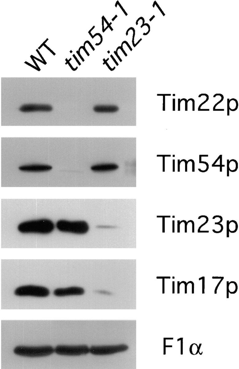
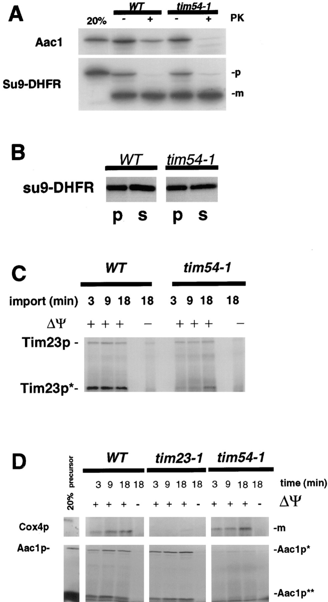
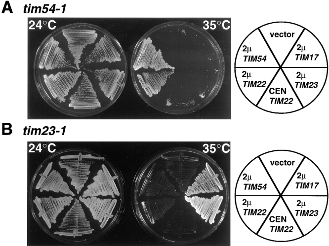
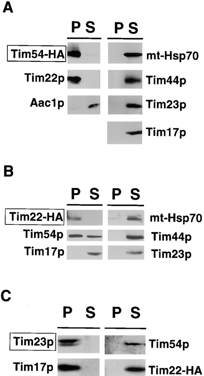
We have identified a new protein, Tim54p, located in the yeast mitochondrial inner membrane. Tim54p is an essential import component, required for the insertion of at least two polytopic proteins into the inner membrane, but not for the translocation of precursors into the matrix. Several observations suggest that Tim54p and Tim22p are part of a protein complex in the inner membrane distinct from the previously characterized Tim23p-Tim17p complex. First, multiple copies of the TIM22 gene, but not TIM23 or TIM17, suppress the growth defect of a tim54-1 temperature-sensitive mutant. Second, Tim22p can be coprecipitated with Tim54p from detergent-solubilized mitochondria, but Tim54p and Tim22p do not interact with either Tim23p or Tim17p. Finally, the tim54-1 mutation destabilizes the Tim22 protein, but not Tim23p or Tim17p. Our results support the idea that the mitochondrial inner membrane carries two independent import complexes: one required for the translocation of proteins across the inner membrane (Tim23p-Tim17p), and the other required for the insertion of proteins into the inner membrane (Tim54p-Tim22p).


Figures

Figure 1
Figure 1
TIM54 is an essential gene. (A) Restriction endonuclease map of the TIM54 gene. Relevant restriction sites in the cloned TIM54 gene, and their position in basepairs with respect to the amino terminus of the Tim54 protein (position 1) are shown. The arrow represents the TIM54 open reading frame, with the arrowhead indicating the 3′-end of TIM54. The location of the URA3 and LEU2 disruptions, which inactivate TIM54, are also shown. (B) Meiotic products from diploid strain 506, in which one of the two TIM54 genes was replaced by the tim54:: URA3 disruption, were separated by micromanipulation and allowed to grow at 30°C for ten days on YEP medium (Rose et al., 1988) containing 2% glucose. The dissection of eleven tetrads is shown, and the position where the four spores were initially placed is indicated (A–D).
Figure 2
Figure 2
Tim54p is located in the mitochondrial inner membrane. (A) Tim54p colocalizes with mitochondria. Yeast strain 501 containing plasmid pOK27, which expresses the Tim54 protein tagged with the HA epitope (Tim54– HA), were fixed, permeabilized, and incubated with mouse antibodies to the HA epitope, or with rabbit antiserum to the β-subunit of the F1-ATPase (F1β). Cells were then incubated with Texas red–conjugated goat anti–rabbit IgG and fluorescein-linked goat anti–mouse IgG, and examined under the microscope at a magnification of 100. The three images in the left column show the same cell visualized by DIC illumination, or by fluorescence using the fluorescein (Tim54–HA) or rhodamine (F1β) channels. The three images in the right column show a group of eight cells examined by DIC and fluorescence. (B) Tim54p cofractionates with a mitochondrial marker. Strain 494, in which the integrated pOK102 construct expresses the Tim54–HA protein, were grown, converted to spheroplasts and homogenized. The homogenate (Hom) was separated into a mitochondrial pellet (Mito) and a postmitochondrial supernatant (PMS) by centrifugation. Aliquots of homogenate, mitochondria, and PMS representing equivalent numbers of cells were subjected to SDS-PAGE, blotted and proteins decorated with 12CA5 antibodies to the HA epitope (Tim54–HA), the β subunit of the F1-ATPase (F1β), or hexokinase. Immune complexes were visualized by chemiluminescence. (C) Tim54p is an integral membrane protein. 150-μg mitochondria isolated from strain 494 were sonicated, treated with either 0.1 M sodium carbonate, 1.5 M sodium chloride, or no additions (buffer), and centrifuged. Pellets (p) and supernatants (s) were analyzed by immune blotting with antibodies to the HA epitope (Tim54–HA), F1β, a peripheral membrane protein, and Tim23p, an integral membrane protein. (D) Tim54p is located in the inner membrane. Mitochondria from strain 494 were sonicated and the resulting vesicles were loaded onto sucrose step-gradients. After centrifugation, fractions were collected and analyzed by immune blotting with antibodies to the HA epitope (Tim54–HA), the inner membrane protein, F1β, or the outer membrane protein, OM45. Fraction 1 represents the top of the gradient. (E) The carboxyl terminus of Tim54 faces the intermembrane space. Mitochondria isolated from strain 494 were digested with 150 μg/ml trypsin for 20 min on ice, followed by the addition of 2 mg/ml soybean trypsin inhibitor. Mitochondria were reisolated by centrifugation and analyzed by immune blotting with antiserum to Tom70p, F1β, Tim23p, and Tim54p. To expose proteins located in the intermembrane space, the mitochondrial outer membrane was ruptured by osmotic shock (OS), and proteins were digested with 150 μg/ml trypsin as above. (F) Import of Tim54p into mitochondria requires an inner membrane potential. Mitochondria were isolated from wild-type strain D273-10B and incubated with the 35S-labeled Tim54 protein. In one sample, the inner membrane potential was dissipated (− Δψ) by the addition of 50 μM carbonyl cyanide m-chlorphenylhydrazone and 1 μM valinomycin prior to the import reaction. After 15 min at 30°C, imports were stopped by incubation at 0°C. Mitochondria were treated with 50-μg/ml proteinase K for 20 min at 0°C, isolated by centrifugation, and solubilized in SDS-sample buffer. Proteins were separated on SDS-polyacrylamide gels, and the radiolabeled Tim54p was identified by fluorography. (G) The bulk of the Tim54 protein faces the intermembrane space. Tim54p was imported into wild-type mitochondria in the presence (+ Δψ) or absence (− Δψ) of inner membrane potential as described above. After import, the mitochondrial outer membrane was disrupted by osmotic shock, and the resulting mitoplasts were digested with 50 μg/ml proteinase K for 20 min. Mitoplasts were recovered by centrifugation and subjected to SDS-PAGE and fluorography. The full-length Tim54 protein (Tim54p) and a fragment of Tim54p (Tim54p*) protected from protease digestion are indicated.
Figure 2
Figure 2
Tim54p is located in the mitochondrial inner membrane. (A) Tim54p colocalizes with mitochondria. Yeast strain 501 containing plasmid pOK27, which expresses the Tim54 protein tagged with the HA epitope (Tim54– HA), were fixed, permeabilized, and incubated with mouse antibodies to the HA epitope, or with rabbit antiserum to the β-subunit of the F1-ATPase (F1β). Cells were then incubated with Texas red–conjugated goat anti–rabbit IgG and fluorescein-linked goat anti–mouse IgG, and examined under the microscope at a magnification of 100. The three images in the left column show the same cell visualized by DIC illumination, or by fluorescence using the fluorescein (Tim54–HA) or rhodamine (F1β) channels. The three images in the right column show a group of eight cells examined by DIC and fluorescence. (B) Tim54p cofractionates with a mitochondrial marker. Strain 494, in which the integrated pOK102 construct expresses the Tim54–HA protein, were grown, converted to spheroplasts and homogenized. The homogenate (Hom) was separated into a mitochondrial pellet (Mito) and a postmitochondrial supernatant (PMS) by centrifugation. Aliquots of homogenate, mitochondria, and PMS representing equivalent numbers of cells were subjected to SDS-PAGE, blotted and proteins decorated with 12CA5 antibodies to the HA epitope (Tim54–HA), the β subunit of the F1-ATPase (F1β), or hexokinase. Immune complexes were visualized by chemiluminescence. (C) Tim54p is an integral membrane protein. 150-μg mitochondria isolated from strain 494 were sonicated, treated with either 0.1 M sodium carbonate, 1.5 M sodium chloride, or no additions (buffer), and centrifuged. Pellets (p) and supernatants (s) were analyzed by immune blotting with antibodies to the HA epitope (Tim54–HA), F1β, a peripheral membrane protein, and Tim23p, an integral membrane protein. (D) Tim54p is located in the inner membrane. Mitochondria from strain 494 were sonicated and the resulting vesicles were loaded onto sucrose step-gradients. After centrifugation, fractions were collected and analyzed by immune blotting with antibodies to the HA epitope (Tim54–HA), the inner membrane protein, F1β, or the outer membrane protein, OM45. Fraction 1 represents the top of the gradient. (E) The carboxyl terminus of Tim54 faces the intermembrane space. Mitochondria isolated from strain 494 were digested with 150 μg/ml trypsin for 20 min on ice, followed by the addition of 2 mg/ml soybean trypsin inhibitor. Mitochondria were reisolated by centrifugation and analyzed by immune blotting with antiserum to Tom70p, F1β, Tim23p, and Tim54p. To expose proteins located in the intermembrane space, the mitochondrial outer membrane was ruptured by osmotic shock (OS), and proteins were digested with 150 μg/ml trypsin as above. (F) Import of Tim54p into mitochondria requires an inner membrane potential. Mitochondria were isolated from wild-type strain D273-10B and incubated with the 35S-labeled Tim54 protein. In one sample, the inner membrane potential was dissipated (− Δψ) by the addition of 50 μM carbonyl cyanide m-chlorphenylhydrazone and 1 μM valinomycin prior to the import reaction. After 15 min at 30°C, imports were stopped by incubation at 0°C. Mitochondria were treated with 50-μg/ml proteinase K for 20 min at 0°C, isolated by centrifugation, and solubilized in SDS-sample buffer. Proteins were separated on SDS-polyacrylamide gels, and the radiolabeled Tim54p was identified by fluorography. (G) The bulk of the Tim54 protein faces the intermembrane space. Tim54p was imported into wild-type mitochondria in the presence (+ Δψ) or absence (− Δψ) of inner membrane potential as described above. After import, the mitochondrial outer membrane was disrupted by osmotic shock, and the resulting mitoplasts were digested with 50 μg/ml proteinase K for 20 min. Mitoplasts were recovered by centrifugation and subjected to SDS-PAGE and fluorography. The full-length Tim54 protein (Tim54p) and a fragment of Tim54p (Tim54p*) protected from protease digestion are indicated.
Figure 6
Figure 6
Mitochondria isolated from the tim54-1 mutant contain reduced amounts of both Tim54p and Tim22p. Mitochondria were isolated from wild-type strain 836, tim54-1 strain 835 and tim23-1 strain 201 grown at 24°C. 50 μg of mitochondria were analyzed by SDS-PAGE and immune blotting with antibodies to the α subunit of the F1-ATPase (F1α), Tim54p, Tim23p, Tim22p, Tim17p.
Figure 3
Figure 3
Mitochondria isolated from the tim54-1 mutant is defective in the insertion of proteins into the inner membrane, but not in the import of matrix proteins. (A) Su9-DHFR and Aac1p imports: Mitochondria were isolated from tim54-1 stain 809 and wild-type strain YPH857, and incubated with the 35S-labeled Aac1p or Su9-DHFR proteins. After 20 min at 30°C, import was stopped by the addition of 40 μM valinomycin and incubation at 0°C. An aliquot of the mitochondria was treated with 200 μg/ml proteinase K for 20 min at 0°C (+ PK). Samples were isolated by centrifugation, and pellets were solubilized in SDS-sample buffer. Proteins were separated on SDS-polyacrylamide gels, and the radiolabeled proteins were identified by fluorography. Precursor (p) and mature (m) forms of the Su9-DHFR are indicated. 20% of the precursor added to each import reaction is also shown. (B) Su9-DHFR is imported into the matrix in tim54-1 mitochondria. Mitochondria were isolated from tim54-1 stain 809 and wild-type strain YPH857, incubated with the 35S-labeled Su9-DHFR protein, and treated with proteinase K as described above. Mitochondria were sonicated and then centrifuged at 200,000 g for 45 min. Equal aliquots of the pellet (p) and supernatant (s) fractions were subjected to SDS-PAGE and fluorography. The mature form of Su9-DHFR is shown. (C) Tim23p imports: 35S-labeled Tim23 protein was imported into either wild-type or tim54-1 mitochondria for 3, 9, or 18 min at 30°C. Import was stopped by the addition of valinomycin, and mitochondria were treated with 200 μg/ml trypsin for 20 min at 0°C. After the addition of 1 mg/ml soybean trypsin inhibitor and centrifugation, the outer membrane of the mitochondria was disrupted by resuspending the mitochondrial pellet in 20 mM Hepes-KOH; pH 7.4, and incubation for 20 min at 0°C. Proteins were digested by treatment with 100 μg/ml proteinase K for 20 min 0°C. Samples were then isolated by centrifugation, and analyzed by SDS-PAGE and fluorography. Tim23* indicates the 14-kD protease-protected fragment of Tim23p indicative of correct insertion into the inner membrane. In some reactions, the inner membrane potential (Δψ) was dissipated prior to import by the addition of 40 μM valinomycin. (D) Import of Aac1p and Cox4p into wild-type, tim23-1, and tim54-1 mitochondria: Mitochondria were isolated from wild-type strain 55, tim23-1 strain 201, and tim54-1 strain 809 and incubated with the 35S-labeled Aac1 or Cox4 proteins for the indicated times at 30°C. After import, mitochondria were reisolated by centrifugation, converted to mitoplasts, and treated with protease as described above.
Figure 4
Figure 4
Multiple copies of TIM22 suppress tim54-1, but not tim23-1. (A) tim54-1 ura3 strain 723 was transformed with the following multi-copy, URA3-containing plasmids: 2μ-TIM17 plasmid pTB1, 2μ-TIM23 plasmid pTB2, 2μ-TIM22 plasmid pJH202, 2μ-TIM54 plasmid pOK32, or the empty vector pRS426 (Sikorski and Hieter, 1989). tim54-1 was also transformed with CEN- TIM22 plasmid pJH201, which carries TIM22 on a centromere-containing plasmid. Ura+ transformants were streaked onto YEP medium containing 2% glycerol and ethanol, and incubated at 24° or 35°C for 5 d. (B) tim23-1 ura3 strain 574 was transformed with the same set of plasmids described above. Transformants were streaked onto YPglycerol/ethanol medium and incubated at 24 or 35°C for 5 d.
Figure 5
Figure 5
Tim54p and Tim22p physically interact, but are not part of the Tim23p– Tim17p complex. Mitochondria were isolated from strain 494, in which the integrated pOK102 construct expresses the Tim54–HA protein (A) or tim22::TRP1 strain 800, which expresses Tim22–HA from plasmid pJH102 (B and C), and solubilized in 0.5% digitonin. Extracts were immune precipitated with antibodies against the HA epitope (A and B), or antiserum to the Tim23 protein (C). Immunoprecipitates (P) and supernatants (S) were analyzed by SDS-PAGE and duplicate samples were immune blotted with antibodies to the HA epitope, Tim22p, Aac1p, Tim23p, Tim17p, Tim44p, or mt-Hsp70. A box in the upper left of each figure highlights the protein that was directly immune precipitated in each experiment.
Figure 7
Figure 7
A model diagramming the role of two import complexes in the mitochondrial inner membrane. The pathway of two imported proteins is shown. Precursor proteins destined for the matrix carry a positively charged, amino-terminal presequence (indicated by the wavy line with + +), which is removed by the matrix-localized processing protease. After their import through the outer membrane machinery (TOM complex), these precursors are translocated across the IM through the Tim23p–Tim17p complex. Polytopic IM proteins carry internal targeting information. After their translocation through the outer membrane, the Tim54p–Tim22p complex mediates their insertion into the IM.

Similar articles

Cited by

References

    1. Altschul SF, Gish W, Miller W, Myers EW, Lipman DJ. Basic local alignment search tool. J Mol Biol. 1990;215:403–410. - PubMed
    1. Armstrong KA, Som T, Volkert FC, Rose A, Broach JR. Propagation and expression of genes in yeast using 2-micron circle vectors. Biotechnology. 1989;13:165–192. - PubMed
    1. Bachhawat AK, Suhan J, Jones EW. The yeast homolog of H<β>58, a mouse gene essential for embryogenesis, performs a role in the delivery of proteins to the vacuole. Genes Dev. 1994;8:1379–1387. - PubMed
    1. Bai C, Elledge SJ. Gene identification using the yeast two-hybrid system. Methods Enzymol. 1996;273:331–347. - PubMed
    1. Bauer M, Sirrenberg C, Neupert W, Brunner M. Role of Tim23 as voltage sensor and presequence receptor in protein import into mitochondria. Cell. 1996;87:33–41. - PubMed

Publication types

MeSH terms

Substances