. 1997 Dec 29;139(7):1821-33.
doi: 10.1083/jcb.139.7.1821.

Actin-binding verprolin is a polarity development protein required for the morphogenesis and function of the yeast actin cytoskeleton

Affiliations

Actin-binding verprolin is a polarity development protein required for the morphogenesis and function of the yeast actin cytoskeleton

G Vaduva et al. J Cell Biol. .

Abstract

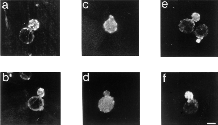
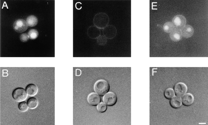
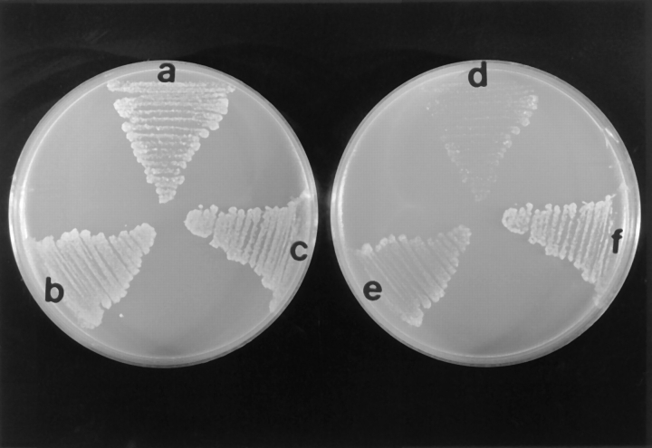
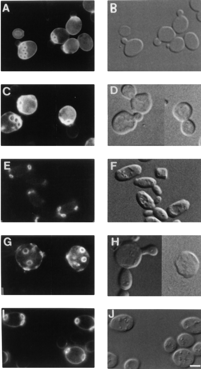
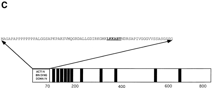
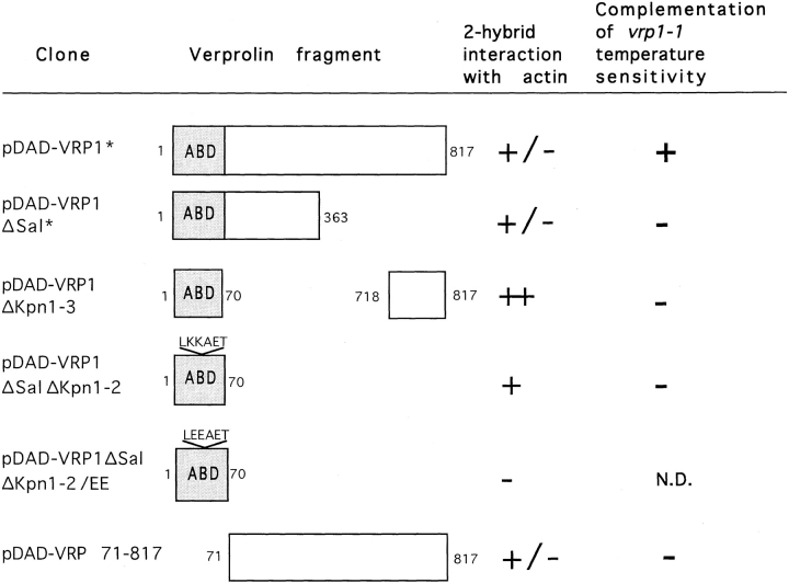
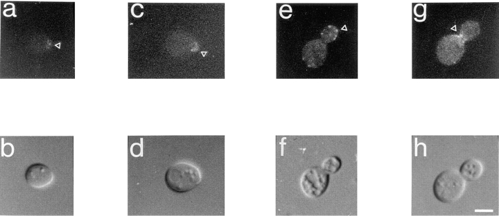
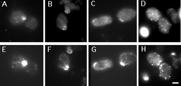
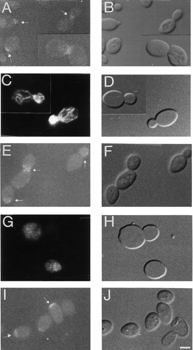
Yeast verprolin, encoded by VRP1, is implicated in cell growth, cytoskeletal organization, endocytosis and mitochondrial protein distribution and function. We show that verprolin is also required for bipolar bud-site selection. Previously we reported that additional actin suppresses the temperature-dependent growth defect caused by a mutation in VRP1. Here we show that additional actin suppresses all known defects caused by vrp1-1 and conclude that the defects relate to an abnormal cytoskeleton. Using the two-hybrid system, we show that verprolin binds actin. An actin-binding domain maps to the LKKAET hexapeptide located in the first 70 amino acids. A similar hexapeptide in other acting-binding proteins was previously shown to be necessary for actin-binding activity. The entire 70- amino acid motif is conserved in novel higher eukaryotic proteins that we predict to be actin-binding, and also in the actin-binding proteins, WASP and N-WASP. Verprolin-GFP in live cells has a cell cycle-dependent distribution similar to the actin cortical cytoskeleton. In fixed cells hemagglutinin-tagged Vrp1p often co-localizes with actin in cortical patches. However, disassembly of the actin cytoskeleton using Latrunculin-A does not alter verprolin's location, indicating that verprolin establishes and maintains its location independent of the actin cytoskeleton. Verprolin is a new member of the actin-binding protein family that serves as a polarity development protein, perhaps by anchoring actin. We speculate that the effects of verprolin upon the actin cytoskeleton might influence mitochondrial protein sorting/function via mRNA distribution.


Figures

Figure 1
Figure 1
Cytoskeletal polarization is restored in vrp1-1 cells (strain TZ33) by two- to fourfold increases in the level of actin. Representative cells of wild-type strain T8.1D bearing an empty plasmid in a and b; vrp1-1 mutant cells (strain TZ33) bearing an empty plasmid in c and d; and vrp1-1 cells plus ACT1 on the centromere-containing plasmid pT3 in e and f. Cells were grown on 2% lactate medium at permissive temperature, fixed as described (Adams and Pringle, 1991) and then stained with rhodamine-phalloidin for 1 h. Bar, 3 μm.
Figure 2
Figure 2
Fluid-phase endocytosis is restored in vrp1-1 cells (strain TZ33) by additional actin. Lucifer yellow uptake in wild-type (strain T8.1D) cells transformed with vector alone in A and B; vrp1-1 mutant cells bearing vector alone (C and D); and vrp1-1 cells bearing ACT1 on pT3 plasmid (E and F). Cells were grown at permissive temperature and then incubated with Lucifer yellow for 2 h. (A, C, and E), FITC channel. In B, D, and F, the same cells visualized under DIC. Bar, 3 μm.
Figure 3
Figure 3
Extra actin partially suppresses the Lys+ phenotype of vrp1-1 cells. The plate in the left is uraleu and the one in the right is uraleulys. Parental T8.1D cells (a and d); vrp1-1 cells (strain TZ33), (c and f); vrp1-1 cells bearing pT3 (encoding actin) (b and e). All strains have Mod5-I,KR6ET gene on a centromere-containing plasmid; the uraleu master plate was first replica plated to uraleulys plate and then to uraleu plate. For all strains, growth was assessed at 23°C.
Figure 4
Figure 4
Budding pattern phenotypes of haploid and diploid vrp1::LEU2 mutant strains and rescue of mutant phenotype by VRP1-GFP. Logarithmic cultures of each strain were grown in selective media at 23°C and then stained with Calcofluor to visualize bud scars. (A) vrp1::LEU2 (strain T65.1D) cells bearing YCpVRP1GFP plasmid show normal axial budding pattern; (C) vrp1::LEU2 (strain T65.1D) cells bearing vector alone show axial pattern; (E) wild-type diploid strain Sc467 bearing an empty plasmid shows bipolar budding pattern; (G) diploid vrp1::LEU2/vrp1:: LEU2 cells bearing vector alone show random budding pattern; (H) morphologically, a large proportion of these cells have multiple buds and shape defects; (I) restoration of bipolar budding in homozygous diploid vrp1::LEU2/vrp1::LEU2 cells by the VRP1-GFP gene on a centromere-containing plasmid. These cells have normal morphology (J). B, D, F, H, J are DIC images of A, C, E, G, I, respectively. Bar, 2.5 μm.
Figure 5
Figure 5
The actin-binding domain of verprolin. (A) Homologies between the first 70 amino acids of verprolin and C. elegans CELR 144.4 protein (“CELR144.4”, GenBank/EMBL/DDBJ accession number U23515), X. laevis proline-rich protein (“Xlpro-rich”, GenBank/EMBL/DDBJ accession number X68249), and rat SH3-domain–binding protein (“RNU25281”, GenBank/EMBL/DDBJ accession number U25281). Alignment was done using BOXSHADE 3.21 program running on a WWW server at ISREC, Switzerland. Asterisks designate the position of the LKKAET hexapeptide. (B) LKKAET-like motifs in various cytoskeletal proteins. (C) Diagram of the localization of the actin-binding domain in verprolin. The LKKAET motif is in bold and underlined. The 11 black bars correspond to potential SH3-binding domains having the general pattern APPL/IP.
Figure 5
Figure 5
The actin-binding domain of verprolin. (A) Homologies between the first 70 amino acids of verprolin and C. elegans CELR 144.4 protein (“CELR144.4”, GenBank/EMBL/DDBJ accession number U23515), X. laevis proline-rich protein (“Xlpro-rich”, GenBank/EMBL/DDBJ accession number X68249), and rat SH3-domain–binding protein (“RNU25281”, GenBank/EMBL/DDBJ accession number U25281). Alignment was done using BOXSHADE 3.21 program running on a WWW server at ISREC, Switzerland. Asterisks designate the position of the LKKAET hexapeptide. (B) LKKAET-like motifs in various cytoskeletal proteins. (C) Diagram of the localization of the actin-binding domain in verprolin. The LKKAET motif is in bold and underlined. The 11 black bars correspond to potential SH3-binding domains having the general pattern APPL/IP.
Figure 6
Figure 6
Mapping of the actin-binding domain of verprolin using two-hybrid analysis. Various DNA fragments encoding truncated Vrp1p were cloned in-frame with the Gal4 DNA-activation domain in vector pDAD and the resultant plasmids were transformed into Y190 strain bearing the pDBD–ACT1 construct encoding actin. Association of verprolin with actin was examined by the qualitative assay for β-galactosidase and by assessing growth on media-lacking histidine in the presence of 100 mM 3-amino-1,2,4-triazole. Numbers represent the length in amino acids. Asterisks designate mitotic instability of pDAD-VRP1 and pDAD-VRP1ΔSal plasmids. (N.D.) not done.
Figure 7
Figure 7
In vivo localization of verprolin-GFP fusion in vrp1::LEU2 disruptant (as shown by arrowheads). (a and c) A verprolin cap marks the site where the potential bud will form. (e) Verprolin localizes to the bud in patches-like structures. (g) Cells preparing for cytokinesis have two rings of verprolin-GFP around the neck. b, d, f, and h are DIC images of a, c, e, and g frames, respectively. Bar, 2 μm.
Figure 8
Figure 8
Co-localization of verprolin-HA and actin. GVY1 cells bearing hemagglutinin-tagged VRP1 on a centromere-containing vector (YCpVRP1-HA) were fixed and stained with 12CA5 antibody against the hemagglutinin tag and then co-stained with Oregon green phalloidin as described in Methods. In A–D representative cells bearing the verprolin-HA fusion are shown. The same cells, visualized in the FITC channel to detect F-actin, are shown in E, F, G, and H. The two proteins co-localize in many of the cortical patches. However, apparently there are actin patches which do not seem to contain verprolin and vice versa. Bar, 1.5 μm.
Figure 9
Figure 9
Localization of verprolin-GFP in the absence or presence of LAT-A in diploid vrp1::LEU2/vrp1::LEU2 cells (strain GVY1) containing YCpVRP1GFP plasmid. (A–H) Cells were grown to logarithmic phase, the culture was then divided in two: half was treated with DMSO alone for 15 min (A–D) and half with LAT-A solubilized in DMSO for 15 min (E–H). (I and J), a separate culture of cells enriched for unbudded cells was treated for 4 h with LAT-A solubilized in DMSO. (A) Localization of verprolin-GFP in the absence of LAT-A shows the polarized localization of verprolin at the presumptive budding site, in small buds, and at the mother-bud neck; (C) An aliquot of the culture used in (A) was fixed and further stained with rhodamine phalloidin to confirm the normal morphology of actin cytoskeletons; (E) Localization of verprolin-GFP in a logarithmic culture after incubation with LAT-A for 15 min. Verprolin-GFP maintains its intracellular localization at the mother-bud neck or at the tip of buds. (G) An aliquot of the culture used in E was fixed and stained with rhodamine phalloidin. Actin cytoskeletons of these cells are disassembled. (I) Localization of verprolin-GFP in the presence of LAT-A (4 h) in cells exiting G0. The verprolin-GFP signal is present at the presumptive budding site while the actin cytoskeletons of these cells are virtually absent (not shown). B, D, F, H, and J are DIC images of A, C, E, G, and I, respectively. Bar, 2 μm.
Figure 10
Figure 10
Halo assay testing the sensitivity of vrp1 mutant strains to LAT-A. (A) vrp1::LEU2 cells (strain T65.1D) bearing the VRP1 gene on the centromeric plasmid YCpVRP1. (B) vrp1:: LEU2 cells bearing a vector alone. (C) vrp1-1 cells (strain TZ33) bearing VRP1 gene on the centromeric plasmid YCpVRP1. (D) vrp1-1 cells (strain TZ33) with vector alone. (E) vrp1-1 cells (strain TZ33) bearing ACT1 gene on plasmid pT3. Concentrations of LAT-A are: 0 (top), 0.5 mM (right), 1 mM (bottom), and 2mM (left). Strains were grown on ura selective media at 23°C for 48 h.

Similar articles

Cited by

References

    1. Adams AE, Pringle JR. Relationship of actin and tubulin distribution to bud growth in wild-type and morphogenic-mutant Saccharomyces cerevisiae. . J Cell Biol. 1984;98:934–945. - PMC - PubMed
    1. Adams, A.E., and J.R. Pringle. 1991. Staining of actin with fluorochrome-conjugated phalloidin. In Methods Enzymol. Guide to Yeast Genetics and Molecular Biology; Vol. 194. C. Guthrie and G.R. Fink, editors. Academic Press Inc., San Diego, CA. 729–731. - PubMed
    1. Amberg DC, Basart E, Botstein D. Defining protein interactions with yeast actin in vivo. . Nat Struct Biol. 1995;2:28–35. - PubMed
    1. Amberg DC, Zahner JE, Mulholland JW, Pringle JR, Botstein D. Aip3p/Bud6p, a yeast actin-interacting protein that is involved in morphogenesis and the selection of bipolar budding sites. Mol BiolCell. 1997;8:729–753. - PMC - PubMed
    1. Ayscough KR, Stryker J, Pokala N, Sanders M, Crews P, Drubin D. High rates of actin filament turnover in budding yeast and roles for actin in establishment and maintenance of cell polarity revealed using the actin inhibitor Latrunculin-A. J Cell Biol. 1997;137:399–416. - PMC - PubMed

Publication types

MeSH terms