. 1998 Jan 1;18(1):1-9.
doi: 10.1523/JNEUROSCI.18-01-00001.1998.

Effects of the metabotropic glutamate receptor antagonist MCPG on phosphoinositide turnover and synaptic plasticity in visual cortex

Affiliations

Effects of the metabotropic glutamate receptor antagonist MCPG on phosphoinositide turnover and synaptic plasticity in visual cortex

K M Huber et al. J Neurosci. .

Abstract

The neurotransmitter glutamate, in addition to activating ligand-gated ion channels, also stimulates phosphoinositide (PI) hydrolysis in neurons by activating a group of G-protein-coupled metabotropic glutamate receptors (mGluRs). A role for mGluRs in synaptic plasticity originally was hypothesized based on the observation that the developmental decline in glutamate-stimulated PI turnover is well correlated with the decline in experience-dependent synaptic plasticity in visual cortex. Over the past few years, the compound alpha-methyl-4-carboxyphenylglycine (MCPG) has been widely used to test the role of PI-coupled mGluRs in a number of types of synaptic plasticity, including long-term potentiation (LTP), long-term depression (LTD), ocular dominance plasticity in visual cortex, and the neural plasticity underlying learning and memory. The conclusions of most of these studies were based on the assumption that MCPG blocks the actions of glutamate at PI-coupled mGluRs in the cerebral cortex. Here we show that this assumption is not valid in visual cortex. Although MCPG does antagonize the actions of the synthetic mGluR agonist 1S, 3R-aminocyclopentane-1,3-dicarboxylic acid, it fails to block PI turnover and changes in spike adaptation stimulated by glutamate, the endogenous mGluR ligand. In addition, we find that MCPG fails to block the NMDA receptor-dependent forms of LTP, LTD, and depotentiation in visual cortex.


Figures

Fig. 1.
Fig. 1.
MCPG antagonizes the effects of ACPD on synaptic transmission but does not affect synaptic plasticity in visual cortical slices from young adult (P35–P50) rats. A, Image of a Nissl-stained section of rat visual cortex depicting placement of the stimulating electrode (S) in layer IV and upper layer V and the extracellular recording electrode (Record) in layer III is shown. WM, White matter. B, Application of ACPD (10 μm) reduced FP amplitudes (58 ± 4% of baseline values); MCPG (0.25 mm; 10 min) significantly antagonized this ACPD-induced synaptic depression (83 ± 1% of baseline values;n = 4; p < 0.01).Traces here and in subsequent figures are averages of four consecutive FPs taken at the times indicated by thenumbers (1–5) on the graphs. Calibration: 1 mV, 5 msec. C, MCPG (0.25–1 mm) applied 15 min before and during LFS (1 Hz; 900 pulses) did not affect the magnitude of LTD. Control LTD magnitude was 78 ± 2% of pre-LFS baseline (n = 8); MCPG LTD magnitude was 78 ± 3% (n = 7).Traces are taken at the times indicated (numbers 1 and 2) from one representative slice treated with MCPG (0.25 mm). Calibration: 0.5 mV, 5 msec.D, MCPG (0.25–1.0 mm) applied 15 min before and during TBS did not affect the magnitude of LTP of FP amplitudes. Control LTP magnitude was 117 ± 2% of pre-TBS baseline (n = 10); MCPG LTP magnitude was 118 ± 4% (n = 9). Traces are taken at the times indicated (numbers 1 and 2) from one representative slice treated with MCPG (0.25 mm). Calibration: 0.5 mV, 5 msec.
Fig. 2.
Fig. 2.
MCPG does not affect the induction of LTD or LTP in visual cortex of young (P14–P29) rats. A, Image of a Nissl-stained section of rat visual cortex depicting placement of the stimulating electrode (S) at the border of white matter (WM) and layer VI and the extracellular recording electrode (Record) in layer III.B, LTD of FP amplitudes evoked by LFS of WM in control (87 ± 4% of pre-LFS baseline; n = 6) and MCPG-treated (0.25–1.0 mm; 85 ± 4%;n = 6; p > 0.7) slices.Traces were taken at the times indicated (numbers 1 and 2) from one representative slice treated with MCPG (1.0 mm). Calibration: 0.5 mV, 5 msec.C, LTP of FP amplitudes evoked by TBS of WM in control (115 ± 4% of pre-TBS baseline; n = 8) and MCPG-treated (0.25–1.0 mm; 121 ± 7%;n = 6; p > 0.3) slices. Note that the duration of MCPG treatment was extended in these experiments to 30 min post-TBS. Traces were taken at the times indicated (numbers 1 and 2) from one representative slice treated with MCPG (1.0 mm). Calibration, 0.5 mV, 5 msec.
Fig. 3.
Fig. 3.
MCPG does not affect the induction of depotentiation in young (P17–P28) rats. See Figure1A for the stimulation-recording arrangement for these experiments. Thirty minutes after LTP was induced with TBS, significant depotentiation was obtained (open circles, 82 ± 3% of pre-LFS baseline; n = 5;p < 0.001) using LFS in the presence of MCPG (0.5–1.0 mm). The magnitude of the depotentiation in MCPG was not significantly different from control (filled circles, 88 ± 3% of pre-LFS baseline;n = 5). Response amplitudes after TBS were renormalized at the unlabeled downward arrow.Traces were taken at the times indicated (numbers 1–3) from one representative slice treated with MCPG (0.5 mm). Calibration, 0.5 mV, 5 msec.
Fig. 4.
Fig. 4.
MCPG inhibits ACPD-stimulated, but not glutamate-stimulated, PI hydrolysis in visual cortical synaptoneurosomes. A, ACPD dose–response curves expressed as the percent of basal PI turnover in the presence and absence of 1 mm MCPG (KB, 276 ± 84 μm; n = 3).B, Effects of increasing concentrations of MCPG on PI turnover stimulated by 30 μm ACPD (IC50, 272 μm; n = 3). C, Glutamate dose–response curves in the presence and absence of 1 mm MCPG (KB, > 4 mm;n = 3). Kynurenate at 1 mm was also present in all of these experiments. A similar MCPGKB value was obtained in experiments (n = 4; data not shown) in which CNQX (40 μm) and AP-5 (200 μm) were also present.D, Effects of increasing MCPG concentrations on PI turnover stimulated by 200 μm glutamate (IC50, 3.8 mm; n = 3). Addition of kynurenate (1 mm; open circle) did not affect the magnitude of PI turnover stimulated by glutamate (1 mm) in the presence or absence of MCPG (1 mm; n = 3). Inset, Increasing the concentration of MCPG to 10 mmsuppressed glutamate-stimulated PI turnover; however, this highconcentration also partially inhibited carbachol-stimulated PI turnover (data not shown).
Fig. 5.
Fig. 5.
Differential effect of MCPG on the inhibition of spike adaptation in layer II–III neurons by glutamate and ACPD. Superimposed records of two similar experiments in which MCPG was applied 10 min after inducing a change in spike adaptation with either glutamate (open circles) or ACPD (filled circles). The data plotted in the graph are the number of spikes resulting from a 1 sec depolarizing current injection (0.3 nA) through an intracellular recording electrode. All intracellular recordings were in the presence of CNQX (20 μm), AP-5 (200 μm), bicuculline methiodide (1 μm), 2-hydroxy-saclofen (200 μm), and atropine (1 μm). Representative waveforms of intracellular recordings are shown, taken at times indicated by thenumbers (1–3) in the graph. Spikes have been truncated. Calibration: the absolute membrane potential and time (0.5 sec). The drugs reach the slice chamber 5 min after starting their infusion into the ACSF line, and their effects generally stabilize after an additional 5 min. Although a 10 min exposure to MCPG reverses the ACPD-induced inhibition of spike adaptation, it has an additive effect on the glutamate-induced inhibition.
Fig. 6.
Fig. 6.
MCPG alone inhibits spike adaptation in layer II–III neurons. Superimposed records of two similar experiments in which MCPG (1 mm) was applied for 20 min. Filled circles are from an experiment in which only MCPG was applied;open circles are from an experiment in which glutamate was applied 10 min after starting the MCPG infusion. The data plotted in the graph are the number of spikes resulting from a 1 sec depolarizing current injection through an intracellular recording electrode. All intracellular recordings were in the presence of CNQX (20 μm), AP-5 (200 μm), bicuculline methiodide (1 μm), 2-hydroxy-saclofen (200 μm) and atropine (1 μm). Representativewaveforms of intracellular recordings are shown, taken at times indicated by the numbers (1–3) in the graph from the experiment in which glutamate was also applied (open circles). Spikes have been truncated. Calibration: see Fig. 5 legend. The absolute membrane potential and time (0.5 sec).
Fig. 7.
Fig. 7.
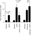
Average effects of MCPG, ACPD, and glutamate on spike adaptation in layer III neurons. MCPG (1 mm) alone increased the number of spikes per depolarizing pulse from a baseline value of 1.2 ± 0.1 to 5.3 ± 1 (n = 4; significant at p < 0.05 using pairedt test). ACPD (30 μm) increased the number of spikes per sweep from a baseline value of 1.2 ± 0.1 to 11.3 ± 2.8 (n = 4). Application of 1 mm MCPG significantly reduced the ACPD effect to 5.3 ± 2.7 spikes (p < 0.05, pairedt test). Glutamate (0.5 mm) increased the number of spikes per sweep from a baseline value of 1.5 ± 0.3 to 9.2 ± 1.1 (n = 7). Application of 1 mm MCPG did not inhibit the glutamate effect but rather tended to add to it (12.2 ± 3.1 spikes per sweep).

Similar articles

Cited by

References

    1. Abe T, Sugihara H, Nawa H, Shigemoto R, Mizuno N, Nakanishi S. Molecular characterization of a novel metabotropic glutamate receptor mGluR5 coupled to inositol phosphate/Ca2+ signal transduction. J Biol Chem. 1992;267:13361–13368. - PubMed
    1. Aizenman CD, Kirkwood A, Bear MF. Current source density analysis of evoked responses in visual cortex in vitro: implications for the regulation of long-term potentiation. Cereb Cortex. 1996;16:751–758. - PubMed
    1. Bashir ZI, Bortolotto ZA, Davies CH, Berretta N, Irving AJ, Seal AJ, Henley JM, Jane DS, Watkins JC, Collingridge GL. Induction of LTP in the hippocampus needs synaptic activation of glutamate metabotropic receptors. Nature. 1993;363:347–350. - PubMed
    1. Bashir ZI, Collingridge GL. An investigation of depotentiation of long-term potentiation in the CA1 region of the hippocampus. Exp Brain Res. 1994;100:437–443. - PubMed
    1. Baskys A, Malenka RC. Agonists at metabotropic glutamate receptors presynaptically inhibit EPSCs in neonatal rat hippocampus. J Physiol (Lond) 1991;444:687–701. - PMC - PubMed

MeSH terms