. 1998 Jan 1;18(1):36-47.
doi: 10.1523/JNEUROSCI.18-01-00036.1998.

Potassium channel distribution, clustering, and function in remyelinating rat axons

Affiliations

Potassium channel distribution, clustering, and function in remyelinating rat axons

M N Rasband et al. J Neurosci. .

Abstract

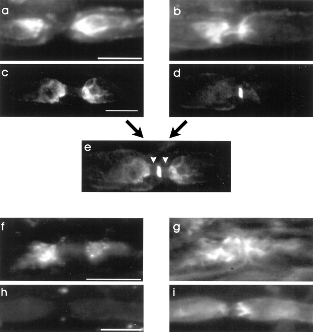
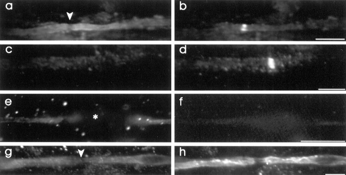
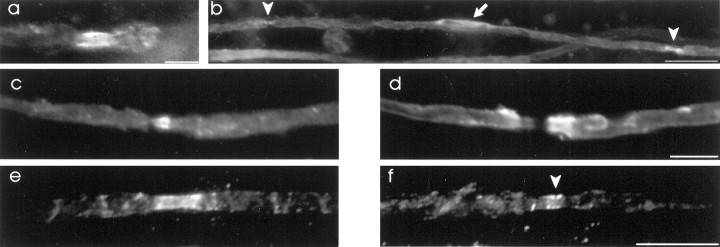
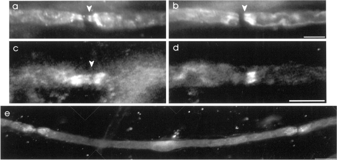
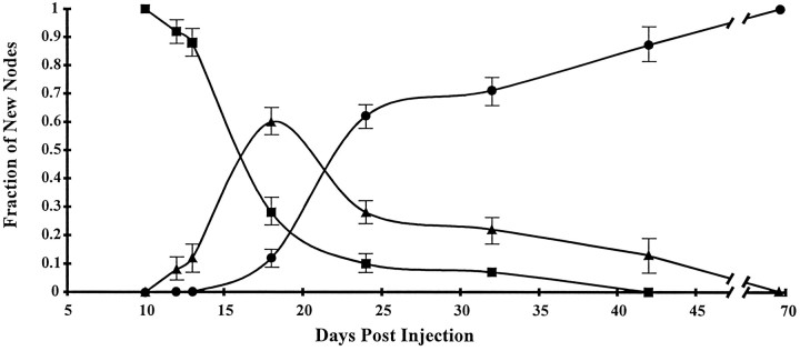
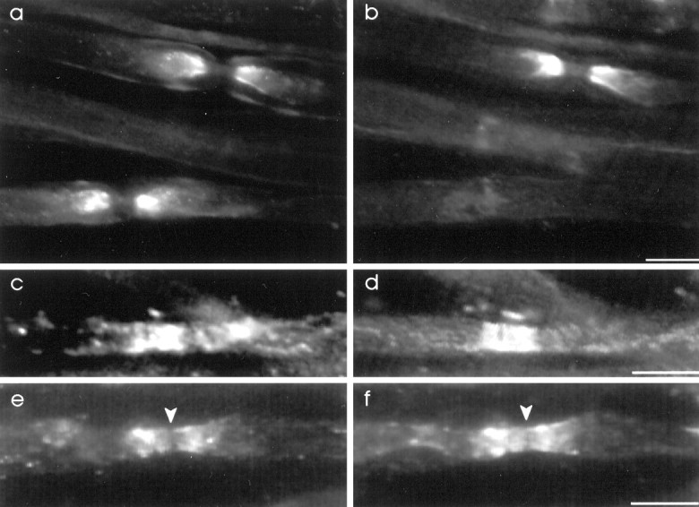
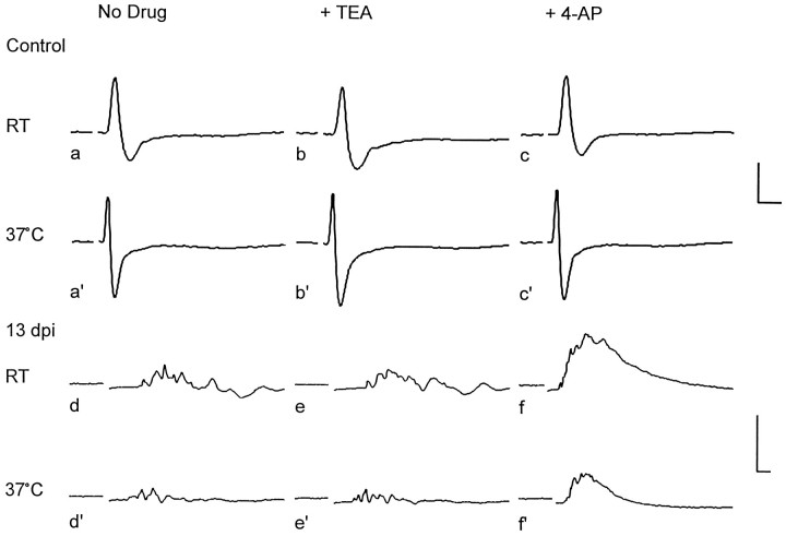
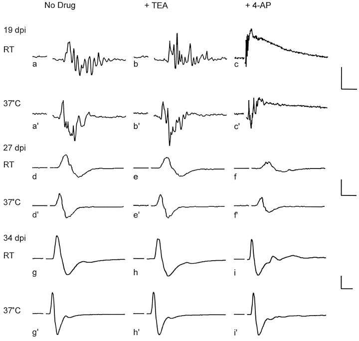
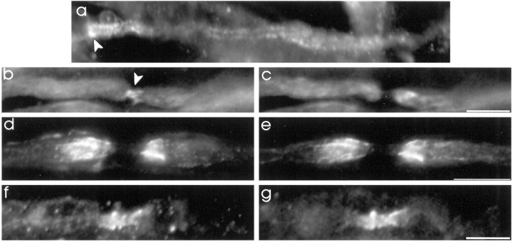
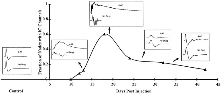
The K+ channel alpha-subunits Kv1.1 and Kv1.2 and the cytoplasmic beta-subunit Kvbeta2 were detected by immunofluorescence microscopy and found to be colocalized at juxtaparanodes in normal adult rat sciatic nerve. After demyelination by intraneural injection of lysolecithin, and during remyelination, the subcellular distributions of Kv1.1, Kv1.2, and Kvbeta2 were reorganized. At 6 d postinjection (dpi), axons were stripped of myelin, and K+ channels were found to be dispersed across zones that extended into both nodal and internodal regions; a few days later they were undetectable. By 10 dpi, remyelination was underway, but Kv1.1 immunoreactivity was absent at newly forming nodes of Ranvier. By 14 dpi, K+ channels were detected but were in the nodal gap between Schwann cells. By 19 dpi, most new nodes had Kv1.1, Kv1.2, and Kvbeta2, which precisely colocalized. However, this nodal distribution was transient. By 24 dpi, the majority of K+ channels was clustered within paranodal regions of remyelinated axons, leaving a gap that overlapped with Na+ channel immunoreactivity. Inhibition of Schwann cell proliferation delayed both remyelination and the development of the K+ channel distributions described. Conduction studies indicate that neither 4-aminopyridine (4-AP) nor tetraethylammonium alters normal nerve conduction. However, during remyelination, 4-AP profoundly increased both compound action potential amplitude and duration. The level of this effect matched closely the nodal presence of these voltage-dependent K+ channels. Our results suggest that K+ channels may have a significant effect on conduction during remyelination and that Schwann cells are important in K+ channel redistribution and clustering.


Figures

Fig. 1.
Fig. 1.
The distribution of Kv1.1 channels in normal adult rat sciatic nerve. a, b, A node double-labeled for Kv1.1 (a) and MAG (b). The paranodal localization of MAG indicates that Kv1.1 is mainly just outside of this zone. c,d, A node double-labeled for Kv1.1 (c) and Na+ channels (d). e, A merged image ofc and d illustrates the gaps (arrowheads) between Kv1.1 and Na+channel immunoreactivity. f, g, A cryosectioned node double-labeled for Kv1.1 (f) and MAG (g).h, i, A control node labeled with preabsorbed rabbit anti-Kv1.1 (h) and anti-MAG (i). Scale bars, 25 μm.
Fig. 2.
Fig. 2.
Kv1.1 distributions in demyelinated and early remyelinating axons. a, b, A node, at 6 dpi, double-labeled for Kv1.1 (a) and Na+ channels (b) has lost the restriction of Kv1.1 to the juxtaparanode. Instead, the Kv1.1 immunoreactivity has moved into nodal (arrowhead) and paranodal zones, whereas Na+ channels remain very focal. c, d, A node, at 6 dpi, double-labeled for Kv1.1 (c) and Na+ channels (d). Kv1.1 staining is undetectable in many demyelinated axons at this time.e, f, A demyelinated axon, at 7 dpi, double-labeled for Kv1.1 (e) and MAG (f). In e, weak Kv1.1 staining was observed adjacent to an associated cell (*). g,h, A new presumptive node, at 9 dpi, double-labeled for Kv1.1 (g) and MAG (h). Remyelinating Schwann cells had diffuse cytoplasmic Kv1.1 staining, but no immunoreactivity was detected at the presumptive node (arrowhead). Diffuse MAG immunoreactivity indicates early remyelination. Scale bars: a, b,g, h, 25 μm; c,d, e, f, 12 μm.
Fig. 3.
Fig. 3.
Kv1.1 is found at the node of Ranvier during remyelination. a, A node, at 14 dpi, labeled for Kv1.1.b (composite), A full internode, at 14 dpi, with Kv1.1 immunoreactivity at nodes (arrowheads) and in the perinuclear region of the Schwann cell (arrow).c, d, A node double-labeled for Kv1.1 (c) and MAG (d).e, f, Double-labeling for Kv1.1 (e) and Na+ channels (f) indicates that the two distributions overlap at the node (arrowhead) but that Kv1.1 staining extends beyond the Na+ channel immunoreactivity. Scale bars: a, c, d, 12 μm; b, e, f, 25 μm.
Fig. 4.
Fig. 4.
Kv1.1 channels are clustered at the paranodes by 24 d after lysolecithin injection. a,b, A node, at 24 dpi, double-labeled for Kv1.1 (a) and MAG (b) indicates that paranodal Kv1.1 and MAG overlap (arrowheads).c, d, A node, at 24 dpi, double-labeled for Kv1.1 (c) and Na+ channels (d). Kv1.1 is aggregated toward the paranodes, leaving a gap (arrowhead) that overlaps with Na+ channel immunoreactivity. e(composite), A full internode, at 32 dpi, has paranodal/juxtaparanodal Kv1.1 immunofluorescence. Further, the perinuclear region of the Schwann cell has Kv1.1 staining. Scale bars: a,b, 12 μm; c, d, 25 μm;e, 30 μm.
Fig. 5.
Fig. 5.
Kv1.1 channel distributions during remyelination plotted as the fraction of new nodes observed versus days postinjection of lysolecithin. ▪, No nodal, paranodal, or juxtaparanodal labeling; ▴, nodal immunoreactivity only; •, paranodal labeling (with a gap at the node). Error bars indicate ±SEM.
Fig. 6.
Fig. 6.
Kv1.1 and Kv1.2 are colocalized in most normal and remyelinated nerve fibers. a, b, Two normal nodes double-labeled for Kv1.1 (a) and Kv1.2 (b). Both nodes of Ranvier have juxtaparanodal Kv1.1, but only the top one has juxtaparanodal Kv1.2.c, d, At 19 dpi, double-labeling for Kv1.1 (c) and Kv1.2 (d) indicates that during remyelination both channel types may be present at newly forming nodes. e, f, A node, at 24 dpi, with an early gap (arrowheads) in both Kv1.1 (e) and Kv1.2 (f) immunoreactivity. Scale bars: a, b, 25 μm; cf, 12 μm.
Fig. 7.
Fig. 7.
Compound action potentials (CAPs) in control and remyelinating 13 dpi nerves. CAPs were measured both at room temperature (RT) and at 37°C, as indicated at the left of each row. At each temperature the traces are shown before the addition of drug (left) and in 10 mm tetraethylammonium (TEA;center) or 1 mm 4-aminopyridine (4-AP; right). Calibration: (Control), 0.25 mV, 1 msec; (13 dpi), 0.2 mV, 1 msec in traces d, d′,e, and e′ and 2 msec in fand f′.
Fig. 8.
Fig. 8.
Compound action potentials in remyelinating nerves at 19, 27, and 34 dpi. Temperature and drug conditions are as indicated and as in Figure 7. Calibration: (19 dpi) 0.05 mV, 1 msec in a, a′, b, andb′, 12 msec in c, and 3 msec inc′; (27 dpi), 0.5 mV, 1 msec; (34 dpi), 0.5 mV, 1 msec in g,g′, h, h′, andi′ and 2 msec in i.
Fig. 9.
Fig. 9.
Mitomycin-C delays remyelination and K+ channel clustering, and the K+channel subunit Kvβ2 is colocalized with Kv1.1 in normal and remyelinating nerve fibers. a, A demyelinated axon, at 24 dpi of mitomycin-C and lysolecithin, labeled for Kv1.1. A heminode in a (arrowhead) with the demyelinated zone to the right has diffuse labeling of Kv1.1 in the absence of Schwann cell interaction. b,c, A node, at 47 dpi with mitomycin-C and lysolecithin, illustrates Kv1.1 immunoreactivity within the nodal gap.d, e, A normal node double-labeled for Kv1.1 (d) and Kvβ2 (e).f, g, A node, at 19 dpi, double-labeled for Kv1.1 (f) and Kvβ2 (g), showing that Kvβ2 is colocalized with Kv1.1 at the node during remyelination. Scale bars: a,d, e, 25 μm; b,c, f, g, 12 μm.
Fig. 10.
Fig. 10.
Compound action potential amplitude and duration depend on the fraction of nodes with K+ channels (▴) and the consequent sensitivity to 4-aminopyridine (4-AP).

References

    1. Bekele-Arcuri Z, Matos MF, Manganas MM, Strassle BW, Monaghan MM, Rhodes KJ, Trimmer JS. Generation and characterization of subtype-specific monoclonal antibodies to K+ channel α- and β-subunit polypeptides. Neuropharmacology. 1996;35:851–865. - PubMed
    1. Bostock H, Sears TA. The internodal axon membrane: electrical excitability and continuous conduction in segmental demyelination. J Physiol (Lond) 1978;280:273–301. - PMC - PubMed
    1. Bostock H, Sears TA, Sherratt RM. The effects of 4-aminopyridine and tetraethylammonium ions on normal and demyelinated mammalian nerve fibres. J Physiol (Lond) 1981;313:301–315. - PMC - PubMed
    1. Castle NA, Fadous SR, Logothetis DE, Wang GK. 4-Aminopyridine binding and slow inactivation are mutually exclusive in rat Kv1.1 and Shaker potassium channels. Mol Pharmacol. 1994;46:1175–1181. - PubMed
    1. Chiu SY, Ritchie JM. Potassium channels in nodal and internodal axonal membrane of mammalian myelinated fibres. Nature. 1980;284:170–171. - PubMed

Publication types

MeSH terms

LinkOut - more resources