. 1998 Jan 1;18(1):237-50.
doi: 10.1523/JNEUROSCI.18-01-00237.1998.

Sox10, a novel transcriptional modulator in glial cells

Affiliations

Sox10, a novel transcriptional modulator in glial cells

K Kuhlbrodt et al. J Neurosci. .

Abstract

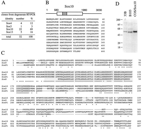
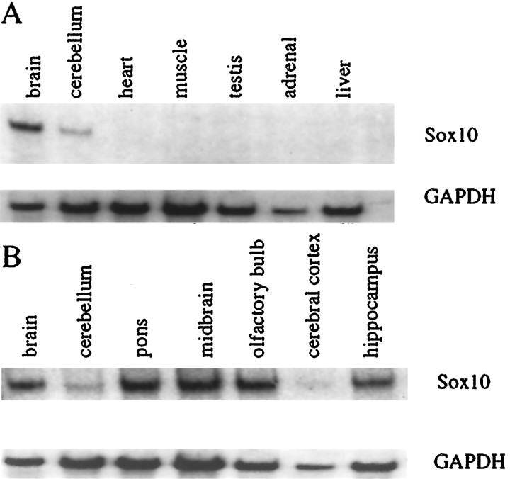
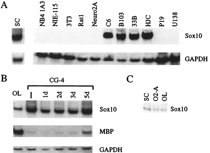
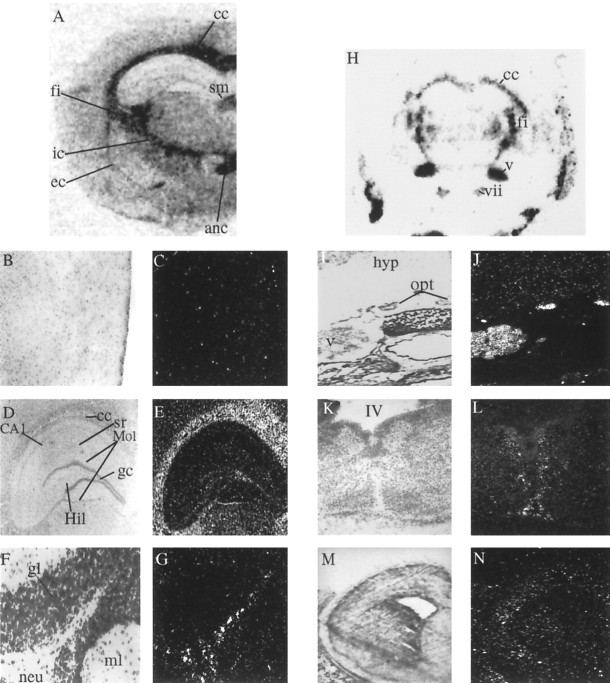
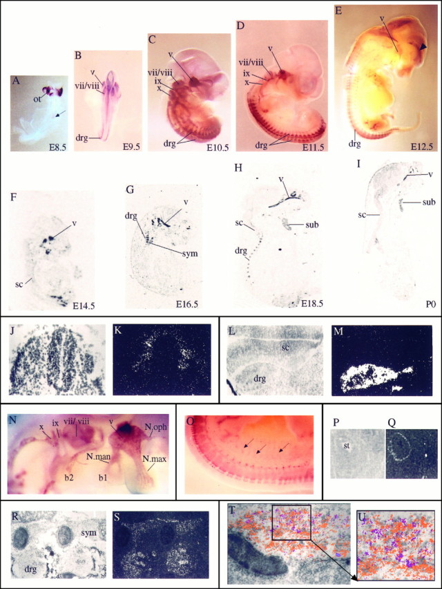
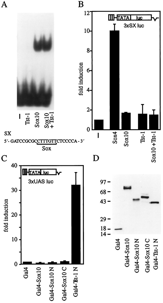
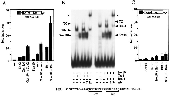
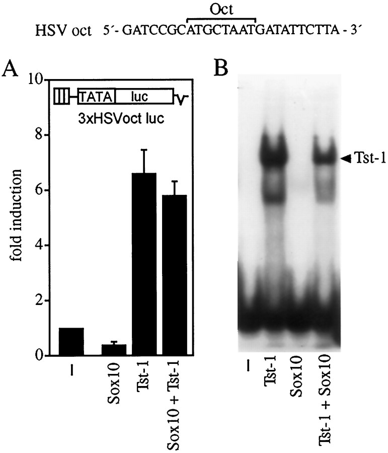
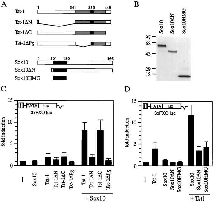
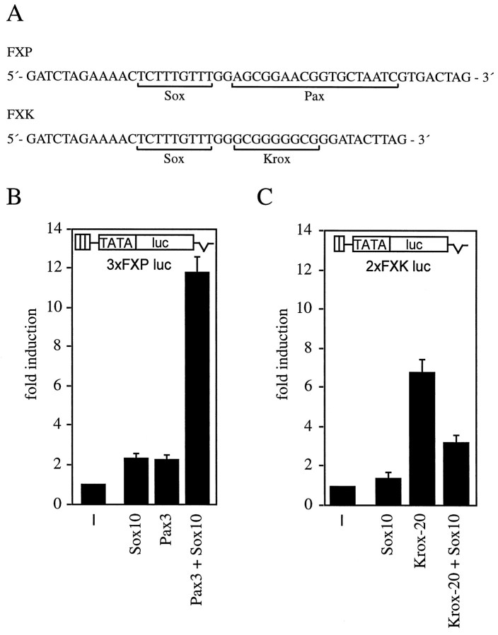
Sox proteins are characterized by possession of a DNA-binding domain with similarity to the high-mobility group domain of the sex determining factor SRY. Here, we report on Sox10, a novel protein with predominant expression in glial cells of the nervous system. During development Sox10 first appeared in the forming neural crest and continued to be expressed as these cells contributed to the forming PNS and finally differentiated into Schwann cells. In the CNS, Sox10 transcripts were originally confined to glial precursors and later detected in oligodendrocytes of the adult brain. Functional studies failed to reveal autonomous transcriptional activity for Sox10. Instead, Sox10 functioned synergistically with the POU domain protein Tst-1/Oct6/SCIP with which it is coexpressed during certain stages of Schwann cell development. Synergy depended on binding to adjacent sites in target promoters, was mediated by the N-terminal regions of both proteins, and could not be observed between Sox10 and several other POU domain proteins. Interestingly, Sox10 also modulated the function of Pax3 and Krox-20, two other transcription factors involved in Schwann cell development. We propose a role for Sox10 in conferring cell specificity to the function of other transcription factors in developing and mature glia.


Figures

Fig. 1.
Fig. 1.
Identification of Sox10. A, Sequence analysis of clones obtained from Schwann cell cDNA by PCR with Sox-specific degenerate primers. B, Structure of the Sox10 cDNA (EMBL/GenBank accession number AJ001029) and amino acid sequence deduced from the open reading frame between positions 583 and 1980. C, Comparison of the amino acid sequence of Sox10 with those of the related mouse Sox9 (Wright et al., 1995), mouse Sox8 (Wright et al., 1993), and rainbow trout SoxP1 (Ito et al., 1995). Exact matches between Sox10 and the aligned sequences are marked byasterisks. The amino acids corresponding to the HMG domain are boxed. D, Detection of endogenous Sox10 in nuclear extracts of 33B rat oligodendroglioma and B103 tumor cells by Western blot using a rabbit antiserum raised against Sox10 holoprotein. A nuclear extract from COS cells transfected with a Sox10 expression plasmid served as a positive control.Numbers on the left indicate sizes of molecular weight markers in kilodaltons.
Fig. 2.
Fig. 2.
Tissue distribution of Sox10 mRNA. Total cellular RNA from the indicated rat tissues (A) or regions of adult rat brain (B) was analyzed for the expression of Sox10 mRNA by Northern blot. Filters were hybridized consecutively with probes specific for Sox10 (top panel) and GAPDH (bottom panel). The region designated midbrain in Bcontained thalamus, hypothalamus, superior and inferior colliculi, as well as surrounding regions. Hippocampus included fimbria and part of the corpus callosum. Negative tissues not shown inA included spleen, lung, and kidney. Identical results were obtained with mouse tissues.
Fig. 3.
Fig. 3.
Expression of Sox10 in cultured cells.A, Total cellular RNA from Schwann cells (SC) and several cell lines was analyzed for the expression of Sox10 mRNA by Northern blot. Cell lines included NB4 1A3 mouse neuroblastoma, N1E-115 mouse neuroblastoma, 3T3 mouse fibroblasts, Rat1 fibroblasts, Neuro2A mouse neuroblastoma, C6 rat glioma, B103 rat tumor cells, 33B rat oligodendroglioma, HJC hamster glioma, P19 mouse embryonal carcinoma, and U138 human glioblastoma. Filters were hybridized consecutively with probes specific for Sox10 (top panel) and GAPDH (bottom panel). B, Total cellular RNA from mature oligodendrocytes (OL) and CG-4 cells was analyzed for the expression of Sox10 mRNA by Northern blot. CG-4 cells were either maintained in the undifferentiated state (−) or differentiated for various time intervals (1d–5d). Filters were hybridized consecutively with probes specific for Sox10 (top panel), MBP (middle panel), and GAPDH (bottom). C, Whole-cell extracts from purified rat Schwann cells (SC), primary oligodendrocyte progenitors (O2-A) and mature oligodendrocytes (OL) were analyzed for the presence of Sox10 protein by Western blot using a rabbit antiserum raised against Sox10 holoprotein.
Fig. 4.
Fig. 4.
Localization of Sox10 transcripts in adult and developing rodent brain. A, In situhybridization of a frontal section through an adult rat brain. Identical results were obtained on sections of adult mouse brain.B–G, High-power photomicrographs showing distribution of silver grains in the cerebral cortex (B, C), the hippocampal formation (D, E), and the cerebellar cortex (F, G). H, In situhybridization of a coronal section through a P7 mouse head. I, J, High-power photomicrographs exhibiting accumulation of silver grains over the optic nerves in a coronal section through a P7 mouse head. K, L, Distribution of Sox10 transcripts in a coronal section through the medulla oblongata at E14.5. M, N, Coronal section of the telencephalon at E16.5. Photomicrographs from overviews were taken from autoradiograms. High-resolution pictures are bright-field photomicrographs (left) and their corresponding dark-field photomicrographs (right). anc, Anterior commissure; CA1, pyramidal cells; cc, corpus callosum; ec, external capsule;fi, fimbria; gc, granular cells;gl, granular layer; Hil, hilus of the dentate gyrus; hyp, hypothalamus; ic, internal capsule; ml, molecular layer of the cerebellar cortex; Mol, molecular layer of the dentate gyrus;neu, neuropil; opt, optic nerve;sm, stria medullaris of the thalamus; sr, stratum radiatum; IV, fourth ventricle of the brain. All other Roman numerals mark cranial nerves and ganglia: v, trigeminal ganglion; vii, facial ganglion.
Fig. 5.
Fig. 5.
Localization of Sox10 transcripts in the developing PNS. A–I, In situhybridization of embryonic mice as whole mounts (A–E, from E8.5 to E12.5) or sagittal sections (F–I, from E14.5 to P0). A, B, Dorsal views; C–E, lateral views. The ventral surface in lateral views and sagittal sections is to the right; the dorsal surface is to theleft. Intense hybridization signals are detected over all ganglia and their corresponding nerve fibers. Thearrow in A marks the labeled cells in the already closed neural tube. The arrowhead inE points to a hybridization signal in the cortex, which was also seen with the sense probe but was never detected usingin situ hybridization on sections. Also note that no other hybridization signal was obtained with the sense probe. J, K, Corresponding bright- and dark-field photomicrographs of transverse section through E8.5 neural tube. L, M, Corresponding bright- and dark-field photomicrographs of cross-section through E11.5 spinal cord and the adjacent dorsal root ganglia.N, High magnification of the E11.5 embryo inD, showing hybridization over all facial–cranial ganglia and their fiber tracts. O, High magnification of the lower back region of the E12.5 embryo in E.Arrows point to some of the nerve fibers leaving the spinal cord area. P, Q, Corresponding bright- and dark-field photomicrographs of cross-section through the stomach of an E13.5 mouse. Transcripts were detected in the outer wall of the stomach. R, S, Corresponding bright- and dark-field photomicrographs of sagittal section through E16.5 sympathetic trunk and the dorsal root ganglia. T, Bright-field photomicrograph of E18.5 trigeminal ganglion with superimposed hybridization signals for Sox10 (orange) and SorLA (purple). Hybridization signals were obtained from dark-field images of adjacent sagittal sections and assigned false colors by computer imaging. U, Magnification of areaboxed in T. b1, Branchial arch 1; b2, branchial arch 2; drg, dorsal root ganglion; N.man, nervus mandibularis;N.max, nervus maxillaris; N.oph, nervus ophtalmicus; ot, otic vesicle; sc, spinal cord; st, stomach; sub, submandibulary gland; sym, sympathethic trunk. Cranial nerves and ganglia are in Roman numerals: v, trigeminal;vii, facial; viii, acoustic;ix, glossopharyngeal; x, vagus.
Fig. 6.
Fig. 6.
Functional characterization of Sox10.A, Purified Sox10–GST protein (Sox10) and nuclear extracts from COS cells transfected with Tst-1/Oct6/SCIP (Tst-1) were analyzed in electrophoretic mobility shift assays for their ability to bind to a radiolabeled oligonucleotide (SX, sequence as shown) that contained a consensus binding site for Sox proteins (van de Wetering et al., 1993).B, The Sox-responsive luciferase reporter plasmid 3xSX luc was transfected into U138 glioblastoma cells in combination with empty CMV expression plasmid (−), pCMV/Sox4 (2 μg/plate;Sox4), pCMV/Sox10 (2 μg/plate;Sox10), and pCMV/Tst-1 (2 μg/plate;Tst-1) as indicated. Luciferase activities were determined in three independent experiments, each performed in duplicate. Values from transfections with luciferase reporter and empty expression plasmid were arbitrarily set to 1. Data from all other transfections are presented as fold induction above this level.C, The Gal4-responsive luciferase reporter (3xUAS luc) was transfected into U138 glioblastoma cells together with expression plasmids for the Gal4 DNA-binding domain (Gal4) or for various Gal4 fusions (2 μg each). In addition to the Gal4 DNA-binding domain, fusions contained full-length Sox10 (Gal4–Sox10), amino acids 1–101 of Sox10 (Gal4–Sox10 N), amino acids 181–466 of Sox10 (Gal4–Sox10 C), or amino acids 1–240 of Tst-1/Oct6/SCIP (Gal4–Tst-1 N). Luciferase activities were determined in three independent experiments, each performed in duplicate. Data are presented for each Gal4 fusion as fold induction above the level of luciferase activity obtained in transfections with an expression plasmid for the Gal4 DNA-binding domain, which was given an arbitrary value of 1.D, Expression of Gal4 fusion proteins in transfected cells was confirmed by Western blot analyses of whole-cell extracts using a monoclonal antibody against the Gal4 DNA-binding domain.Numbers on the left indicate sizes of molecular weight markers in kilodaltons.
Fig. 7.
Fig. 7.
Synergistic action of Sox10 and POU domain proteins. A, The luciferase reporter plasmid 3xFXO luc was transfected into U138 glioblastoma cells in combination with empty CMV expression plasmid (−), pCMV/Sox2 (0.2 μg/plate;Sox2), pCMV/Oct-3/4 (0.2 μg/plate;Oct-3/4), pCMV/Sox10 (0.2 μg/plate;Sox10), and pCMV/Tst-1 (0.2 μg/plate;Tst-1; or 2 μg/plate,Tst-1) as indicated. Luciferase activities were determined in three independent experiments, each performed in duplicate. Values from transfections with luciferase reporter and empty expression plasmid were arbitrarily set to 1. Data for all other transfections are presented as fold induction above this level. B, Radiolabeled FXO oligonucleotide with adjacent Sox and POU domain binding sites (sequence as shown) was incubated in electrophoretic mobility shift assays with purified recombinant Sox10-GST (Sox10) and nuclear extracts from COS cells transfected with Tst-1/Oct6/SCIP (Tst-1) or Brn-1 (Brn-1). Antibodies (Ab) directed against Tst-1 (T), Brn-1 (B), or the GST portion of Sox10-GST (Sx) were added to the reactions as indicatedbelow the lanes. Specific complexes between a protein and an FXO are marked by the name of the respective protein, whereas the ternary complex of Sox10, POU domain protein, and DNA is labeled TC. The supershifted complexes are marked by asterisks. C, The luciferase reporter plasmid 3xFXO luc was transfected into U138 glioblastoma cells in combination with empty CMV expression plasmid (−), pCMV/Sox10 (0.2 μg/plate; Sox10), pCMV/Brn-1 (2 μg/plate;Brn-1), and pCMV/Brn-3.0 (2 μg/plate;Brn-3) as indicated. Luciferase activities were determined in three independent experiments and are presented as inA.
Fig. 8.
Fig. 8.
Binding site requirements for cooperativity between Sox10 and Tst-1/Oct6/SCIP. A, The POU-responsive luciferase reporter plasmid 3xHSVoct luc was transfected into U138 glioblastoma cells in combination with empty CMV expression plasmid (−), pCMV/Sox10 (0.2 μg/plate; Sox10), and pCMV/Tst-1 (0.2 μg/plate; Tst-1) as indicated. Luciferase activities were determined in three independent experiments, each performed in duplicate. Values from transfections with luciferase reporter and empty expression plasmid were arbitrarily set to 1. Data from all other transfections are presented as fold induction above this level. B, Purified Sox10-GST protein (Sox10) and nuclear extracts from COS cells transfected with Tst-1/Oct6/SCIP (Tst-1) were analyzed in electrophoretic mobility shift assays for their ability to bind to a radiolabeled HSVoct oligonucleotide, which contained the binding site for POU domain proteins from the HSV ICP0 promoter, as shown at thetop. The Tst-1/Oct6/SCIP-specific complex is marked by an arrowhead.
Fig. 9.
Fig. 9.
Protein domains involved in synergism between Sox10 and Tst-1/Oct6/SCIP. A, Summary of Tst-1/Oct6/SCIP and Sox10 mutants. Tst-1ΔN, Mutant Tst-1/Oct6/SCIP lacking amino acids 4–240;Tst-1ΔPS, mutant Tst-1/Oct6/SCIP lacking amino acids 241–319;Tst-1ΔC, mutant Tst-1/Oct6/SCIP lacking amino acids 396–448; Sox10ΔN, mutant Sox10 lacking amino acids 1–89; Sox10HMG, comprising amino acids 101–180 of Sox10. B, Comparison of expression levels between Sox10 and its mutants Sox10ΔN and Sox10HMG in nuclear extracts of transfected cells by Western blot using rabbit antiserum against Sox10. Numbers on theleft indicate sizes of molecular weight markers in kilodaltons. C, D, The luciferase reporter plasmid 3xFXO luc was transfected into U138 glioblastoma cells in combination with empty CMV expression plasmid (−), pCMV/Sox10 (Sox10), pCMV/Tst-1 (Tst-1), and various mutant versions of both plasmids (all 0.2 μg/plate) as indicated. Luciferase activities were determined in three independent experiments, each performed in duplicate. Values from transfections with luciferase reporter and empty expression plasmid were arbitrarily set to 1. Data for all other transfections are presented as fold induction above this level.
Fig. 10.
Fig. 10.
Synergistic interaction among Pax3, Krox-20, and Sox10. A, Arrangement of binding sites for Pax3, Krox-20, and Sox10 in the FXP and FXK oligonucleotides.B, The luciferase reporter plasmid 3xFXP luc, which contained adjacent binding sites for Pax3 and Sox10, was transfected into U138 glioblastoma cells in combination with empty CMV expression plasmid (−), pCMV/Sox10 (0.2 μg/plate; Sox10), and pCMV/Pax3 (50 ng/plate; Pax3) as indicated.C, The luciferase reporter plasmid 2xFXK luc, which contained adjacent binding sites for Krox-20 and Sox10, was transfected into U138 glioblastoma cells in combination with empty CMV expression plasmid (−), pCMV/Sox10 (0.2 μg/plate; Sox10), and pCMV/Krox-20 (50 ng/plate; Krox-20) as indicated. Luciferase activities were determined in three independent experiments, each performed in duplicate. Values from transfections with luciferase reporter and empty expression plasmid were arbitrarily set to 1. Data for all other transfections are presented as fold induction above this level.

References

    1. Bell DM, Leung KK, Wheatley SC, Ng LJ, Zhou S, Ling KW, Sham MH, Koopman P, Tam PP, Cheah KS. Sox9 directly regulates the type-II collagen gene. Nat Genet. 1997;16:174–178. - PubMed
    1. Bermingham JR, Scherer SS, O’Connell S, Arroyo E, Kalla KA, Powell FL, Rosenfeld MG. Tst-1/Oct-6/SCIP regulates a unique step in peripheral myelination and is required for normal respiration. Genes Dev. 1996;10:1751–1762. - PubMed
    1. Blanchard AD, Sinanan A, Parmantier E, Zwart R, Broos L, Meijer D, Meier C, Jessen KR, Mirsky R. Oct-6 (SCIP/Tst-1) is expressed in Schwann cell precursors, embryonic Schwann cells, and postnatal myelinating Schwann cells: comparison with Oct-1, Krox-20, and Pax-3. J Neurosci Res. 1996;46:630–640. - PubMed
    1. Brockes JP, Fields P, Raff MC. Studies on cultured rat Schwann cells. I. Establishment of purified populations from cultures of peripheral nerve. Brain Res. 1979;165:105–118. - PubMed
    1. Collarini EJ, Kuhn R, Marshall CJ, Monuki ES, Lemke G, Richardson WD. Down-regulation of the POU transcription factor SCIP is an early event in oligodendrocyte differentiation in vitro. Development. 1992;116:193–200. - PubMed

Publication types

MeSH terms

Associated data