

Masked presentations of emotional facial expressions modulate amygdala activity without explicit knowledge
- PMID: 9412517
- PMCID: PMC6793390
- DOI: 10.1523/JNEUROSCI.18-01-00411.1998
Masked presentations of emotional facial expressions modulate amygdala activity without explicit knowledge
Abstract
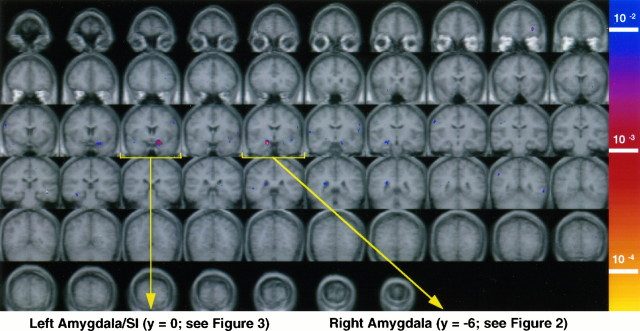
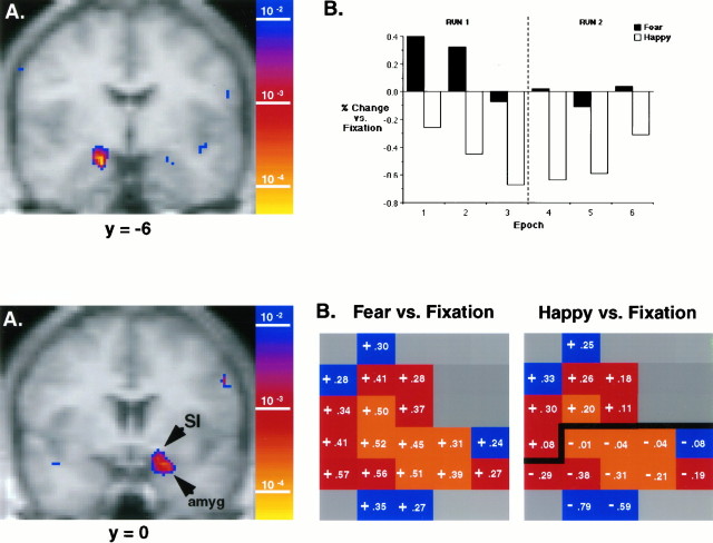
Functional magnetic resonance imaging (fMRI) of the human brain was used to study whether the amygdala is activated in response to emotional stimuli, even in the absence of explicit knowledge that such stimuli were presented. Pictures of human faces bearing fearful or happy expressions were presented to 10 normal, healthy subjects by using a backward masking procedure that resulted in 8 of 10 subjects reporting that they had not seen these facial expressions. The backward masking procedure consisted of 33 msec presentations of fearful or happy facial expressions, their offset coincident with the onset of 167 msec presentations of neutral facial expressions. Although subjects reported seeing only neutral faces, blood oxygen level-dependent (BOLD) fMRI signal in the amygdala was significantly higher during viewing of masked fearful faces than during the viewing of masked happy faces. This difference was composed of significant signal increases in the amygdala to masked fearful faces as well as significant signal decreases to masked happy faces, consistent with the notion that the level of amygdala activation is affected differentially by the emotional valence of external stimuli. In addition, these facial expressions activated the sublenticular substantia innominata (SI), where signal increases were observed to both fearful and happy faces--suggesting a spatial dissociation of territories that respond to emotional valence versus salience or arousal value. This study, using fMRI in conjunction with masked stimulus presentations, represents an initial step toward determining the role of the amygdala in nonconscious processing.
Figures
References
-
- Aggleton JP, editor. The amygdala: neurobiological aspects of emotion, memory, and mental dysfunction. Wiley; New York: 1992.
-
- Balaban MT. Affective influences on startle in five month old infants: reactions to facial expressions of emotion. Child Dev. 1995;66:28–36. - PubMed
-
- Berns GS, Cohen JD, Mintun MA. Brain regions responsive to novelty in the absence of awareness. Science. 1997;276:1272–1275. - PubMed
-
- Bordi F, LeDoux JE, Clugnet MC, Pavlides C. Single-unit activity in the lateral nucleus of the amygdala and overlying areas of striatum in freely behaving rats: rates, discharge patterns, and responses to acoustic stimuli. Behav Neurosci. 1993;107:757–769. - PubMed
Publication types
MeSH terms
Grants and funding
LinkOut - more resources
Full Text Sources
Other Literature Sources