. 1998 Jan 1;18(1):428-37.
doi: 10.1523/JNEUROSCI.18-01-00428.1998.

Dissociation Of working memory from decision making within the human prefrontal cortex

Affiliations

Dissociation Of working memory from decision making within the human prefrontal cortex

A Bechara et al. J Neurosci. .

Abstract

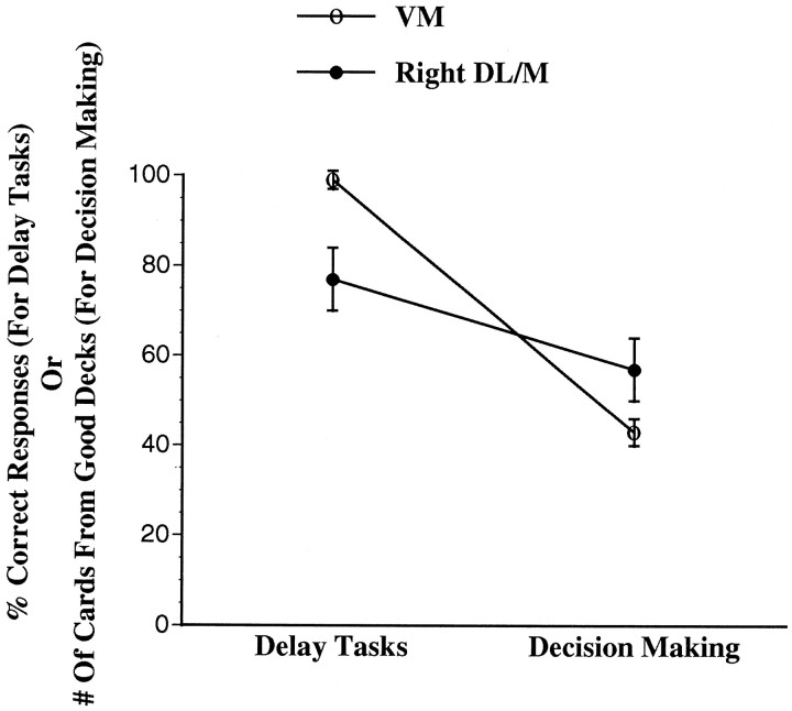
We tested the hypothesis that cognitive functions related to working memory (assessed with delay tasks) are distinct from those related to decision making (assessed with a gambling task), and that working memory and decision making depend in part on separate anatomical substrates. Normal controls (n = 21), subjects with lesions in the ventromedial (VM) (n = 9) or dorsolateral/high mesial (DL/M) prefrontal cortices (n = 10), performed on (1) modified delay tasks that assess working memory and (2) a gambling task designed to measure decision making. VM subjects with more anterior lesions (n = 4) performed defectively on the gambling but not the delay task. VM subjects with more posterior lesions (n = 5) were impaired on both tasks. Right DL/M subjects were impaired on the delay task but not the gambling task. Left DL/M subjects were not impaired on either task. The findings reveal a cognitive and anatomic double dissociation between deficits in decision making (anterior VM) and working memory (right DL/M). This presents the first direct evidence of such effects in humans using the lesion method and underscores the special importance of the VM prefrontal region in decision making, independent of a direct role in working memory.


Figures

Fig. 1.
Fig. 1.
Overlap of lesions in the three groups of brain-damaged subjects. A, Bilateral VM lesions.B, Right and left DL/M lesions. C, Color bar showing the color code corresponding to number of overlap of lesions.
Fig. 2.
Fig. 2.
Comparison of the performance of the three brain-damaged groups and the normals. Each graph represents mean ± SEM of the percent correct responses, or total number of cards selected from the good versus the bad decks, that were made by normal controls (n = 21), by subjects with bilateral orbital and VM frontal lobe lesions (n = 9), by subjects with lesions in the right DL/M sector of the prefrontal cortex (n = 4), or by subjects with lesions in the left DL/M sector of the prefrontal cortex (n = 6). We note that every participant in the delayed response and delayed nonmatching to sample tasks reached a 100% learning criterion at the 0 sec delay before the task could proceed to the 10, 30, or 60 sec delays.
Fig. 3.
Fig. 3.
Mean ± SEM of the percent correct responses, or total number of cards selected from the good versus the bad decks, that were made by normal controls (n = 21) and by subjects with bilateral orbital and VM frontal lobe lesions who were divided into two groups based on their performance on the delayed response and delayed nonmatching to sample tasks: group 1 (abnormal gambling and abnormal delay) (n = 5) and group 2 (abnormal gambling and normal delay) (n = 4).
Fig. 4.
Fig. 4.
Separate mapping of VM lesions for group 1 (A) and group 2 (B) subjects. The maximal overlap of subjects in A is seen spanning the whole extent of the mesial orbital surface of the frontal lobe. It reaches the most posterior sector (coronal slices3, 4) where basal forebrain structures are found. However, in B the maximal overlap is mostly anterior, extending only to slices 1 and2. Slices 3 and 4 do not show any lesion. Coronal sections are arranged according to radiological convention, i.e., right is left, and vice versa.
Fig. 5.
Fig. 5.
Mean ± SEM of the average of percent correct responses from the two delay tasks, or the total number of cards selected from the good decks, that were made by VM subjects with more anterior lesions (group 2), and by subjects with right DL/M lesions.
Fig. 6.
Fig. 6.
Examples of mapped individual cases from the VM and DL/M groups demonstrating that placement of the lesion, rather than the size of the lesion, is the crucial variable in producing the various combinations of deficits (see Discussion for detail).

References

    1. Anderson SW, Damasio H, Jones RD, Tranel D. Wisconsin card sorting test performance as a measure of frontal lobe damage. J Clin Exp Neuropsychol. 1991;3:909–922. - PubMed
    1. Anderson SW, Bechara A, Tranel D, Damasio H, Damasio AR. Characterization of the decision-making defect of subjects with ventromedial frontal lobe damage. Soc Neurosci Abstr. 1996;22:711.
    1. Baddeley A. Working memory. Science. 1992;255:556–559. - PubMed
    1. Bechara A, Damasio AR, Damasio H, Anderson SW. Insensitivity to future consequences following damage to human prefrontal cortex. Cognition. 1994;50:7–15. - PubMed
    1. Bechara A, Tranel D, Damasio H, Damasio AR. Failure to respond autonomically to anticipated future outcomes following damage to prefrontal cortex. Cereb Cortex. 1996;6:215–225. - PubMed

Publication types