

The promyelocytic leukemia protein PML has a pro-apoptotic activity mediated through its RING domain
- PMID: 9414089
- PMCID: PMC2398725
- DOI: 10.1016/s0014-5793(97)01344-6
The promyelocytic leukemia protein PML has a pro-apoptotic activity mediated through its RING domain
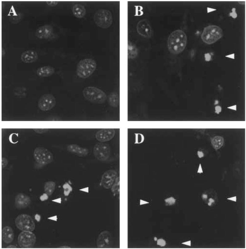
Abstract
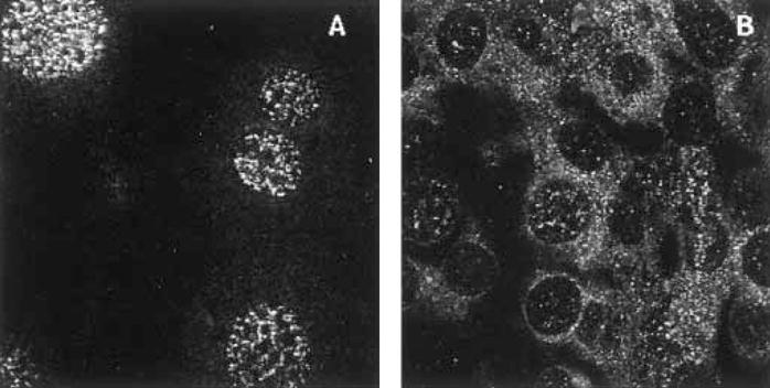
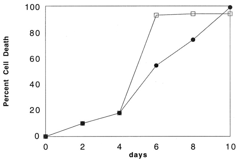
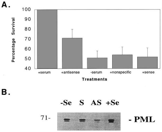
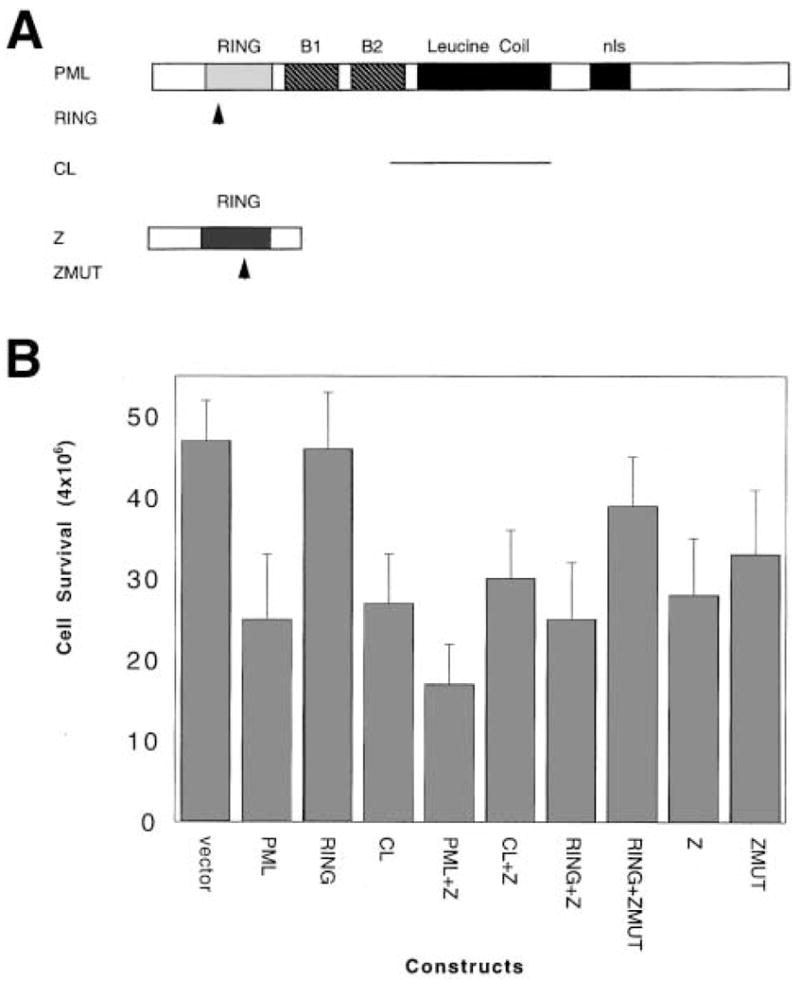
The promyelocytic leukemia protein PML is known to form nuclear multiprotein complexes which are compromised in several pathogenic conditions including acute promyelocytic leukemia. We show that in cells infected with a single stranded RNA virus, which relocates PML bodies to the cytoplasm, the infected cells are more resistant to serum starvation induced apoptosis than their uninfected counterparts. Antisense PML oligonucleotides increase cell survival under serum deprivation conditions indicating that PML is directly involved in the apoptotic activity. Transient transfection studies have indicated that this pro-apoptotic activity of PML is mediated through the zinc binding region known as the RING finger. Viral attack of PML nuclear bodies appears to allow the virus to deregulate host cell apoptotic machinery in order to establish chronic infection.
Figures
References
Publication types
MeSH terms
Substances
Grants and funding
LinkOut - more resources
Full Text Sources
Other Literature Sources
