

Antisecretory factor suppresses intestinal inflammation and hypersecretion
- PMID: 9414971
- PMCID: PMC1891580
- DOI: 10.1136/gut.41.5.642
Antisecretory factor suppresses intestinal inflammation and hypersecretion
Abstract
Background: Antisecretory factor (AF) is a recently identified regulatory protein which inhibits the intestinal fluid secretion induced by cholera toxin.
Aims: To test the effect of AF on: (a) inflammation and hypersecretion induced by toxin A from Clostridium difficile; and (b) morphological changes and hypersecretion induced by okadaic acid (the blue mussel toxin) in rat intestinal mucosa.
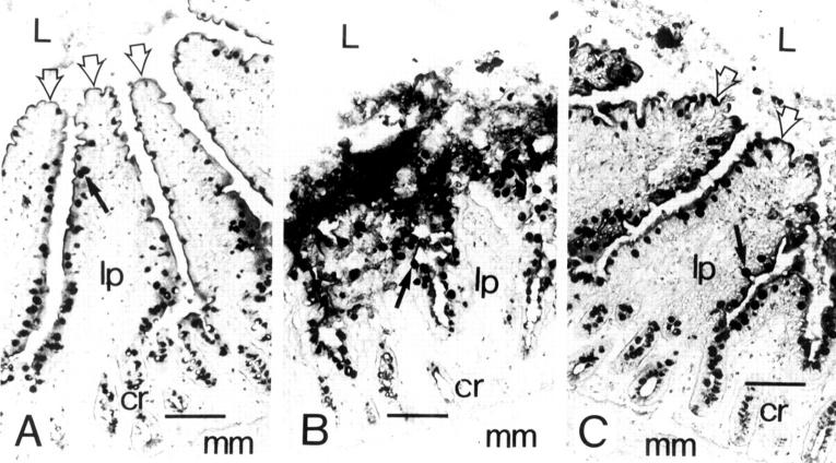
Methods: Morphological changes and fluid accumulation were observed in intestinal loops challenged with 1 microgram of toxin A or 3 micrograms of okadaic acid administered before or after injection of 0.1 microgram of recombinant AF (rAF).
Results: The cytotoxic and inflammatory reaction caused by toxin A was abolished after treatment with rAF given either intraveneously or intraluminally prior to the toxin or one hour after the toxin. The intestinal fluid response induced by toxin A and okadaic acid was reduced 55-80% by rAF. However, the characteristic increase in goblet cells at the tips of villi in the okadaic acid treated mucosa was not inhibited by rAF.
Conclusion: Results suggest that AF might be involved in protection against inflammation and in counteracting dehydration caused by enterotoxins. Both effects are probably mediated via the enteric nervous system.
Figures
Comment in
-
Treating diarrhoea: what might the pituitary offer?Gut. 1997 Nov;41(5):719-20. doi: 10.1136/gut.41.5.719. Gut. 1997. PMID: 9414990 Free PMC article. No abstract available.
References
MeSH terms
Substances
LinkOut - more resources
Full Text Sources
Medical
Research Materials
Miscellaneous