. 1998 Jan;111(1):127-38.
doi: 10.1085/jgp.111.1.127.

Protease modulation of the activity of the epithelial sodium channel expressed in Xenopus oocytes

Affiliations

Protease modulation of the activity of the epithelial sodium channel expressed in Xenopus oocytes

A Chraïbi et al. J Gen Physiol. 1998 Jan.

Abstract

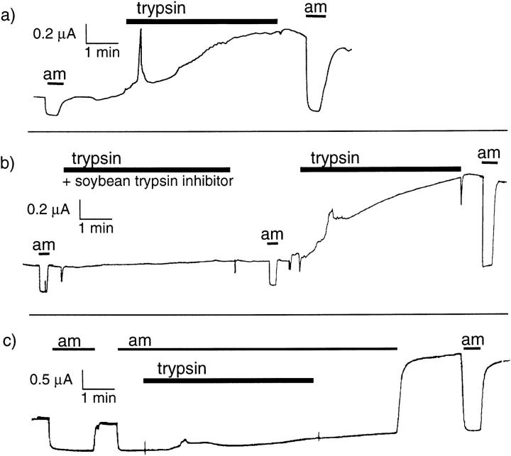
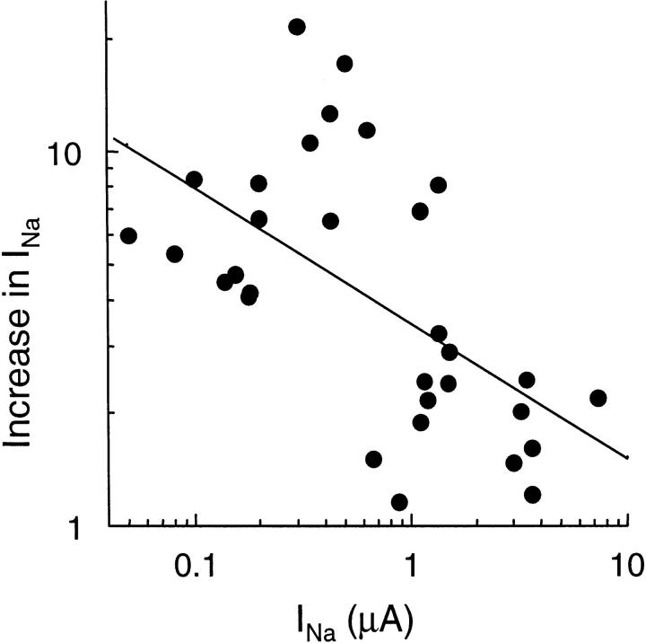
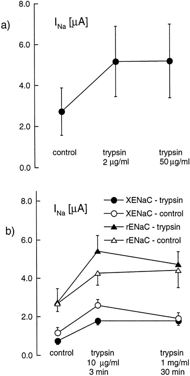
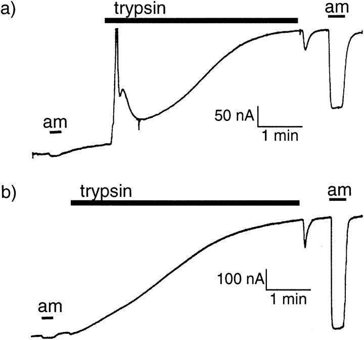
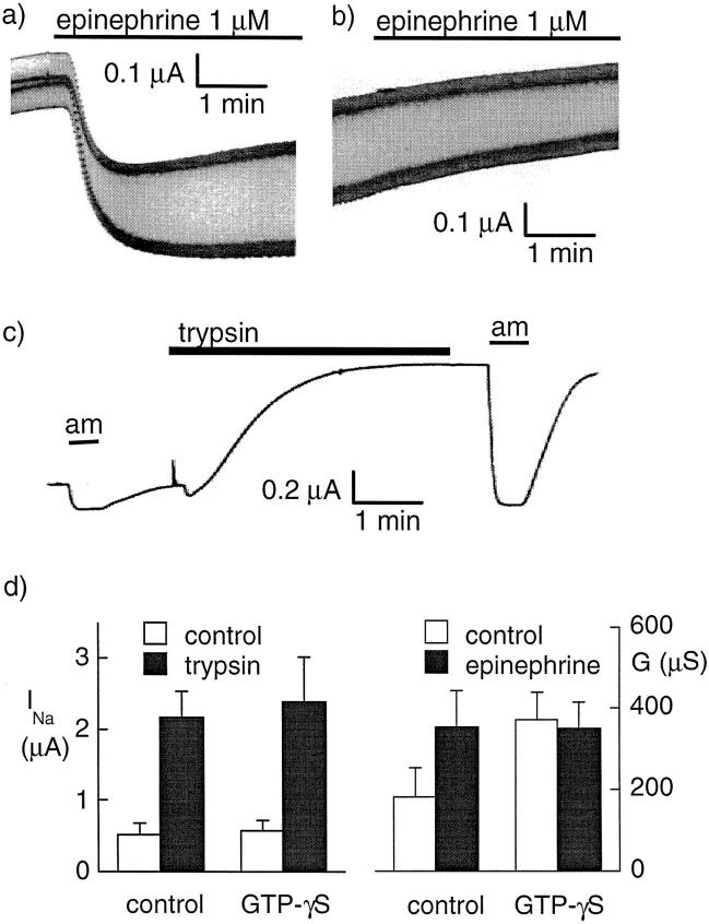
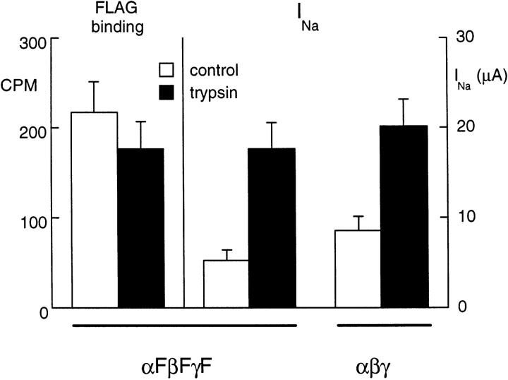
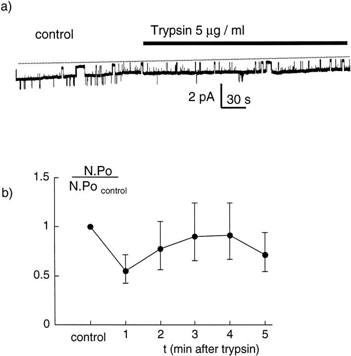
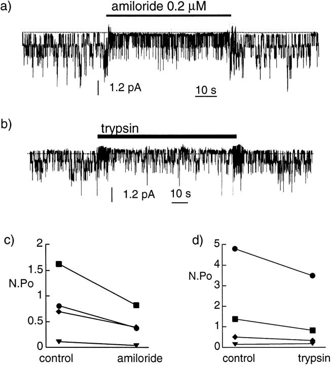
We have investigated the effect of extracellular proteases on the amiloride-sensitive Na+ current (INa) in Xenopus oocytes expressing the three subunits alpha, beta, and gamma of the rat or Xenopus epithelial Na+ channel (ENaC). Low concentrations of trypsin (2 microg/ml) induced a large increase of INa within a few minutes, an effect that was fully prevented by soybean trypsin inhibitor, but not by amiloride. A similar effect was observed with chymotrypsin, but not with kallikrein. The trypsin-induced increase of INa was observed with Xenopus and rat ENaC, and was very large (approximately 20-fold) with the channel obtained by coexpression of the alpha subunit of Xenopus ENaC with the beta and gamma subunits of rat ENaC. The effect of trypsin was selective for ENaC, as shown by the absence of effect on the current due to expression of the K+ channel ROMK2. The effect of trypsin was not prevented by intracellular injection of EGTA nor by pretreatment with GTP-gammaS, suggesting that this effect was not mediated by G proteins. Measurement of the channel protein expression at the oocyte surface by antibody binding to a FLAG epitope showed that the effect of trypsin was not accompanied by an increase in the channel protein density, indicating that proteolysis modified the activity of the channel present at the oocyte surface rather than the cell surface expression. At the single channel level, in the cell-attached mode, more active channels were observed in the patch when trypsin was present in the pipette, while no change in channel activity could be detected when trypsin was added to the bath solution around the patch pipette. We conclude that extracellular proteases are able to increase the open probability of the epithelial sodium channel by an effect that does not occur through activation of a G protein-coupled receptor, but rather through proteolysis of a protein that is either a constitutive part of the channel itself or closely associated with it.


Figures

Figure 1
Figure 1
Effect of trypsin on the amiloride-sensitive current in oocytes expressing αβγENaC. (a) Original current recording in an oocyte expressing αβγrENaC. The current sensitive to 10 μM amiloride was measured at −100 mV before and after a 4-min exposure to 2 μg/ml trypsin. A transient spike of current, attributed to Ca-activated Cl channels occurs in the 10–20 s after the beginning of trypsin treatment, and there is an approximately threefold increase in the amiloride-sensitive current. (b) Original current recording in an oocyte expressing αβγXENaC. The current sensitive to 10 μM amiloride was measured at −100 mV before and after 4-min exposures to 2 μg/ml trypsin, first in the presence of 10 μg/ml soybean trypsin inhibitor, and then without inhibitor. Trypsin has no detectable effect on either INa or a chloride current in the presence of the inhibitor. (c) Original current recording in an oocyte expressing αβγrENaC. The current sensitive to 10 μM amiloride was measured at −100 mV before and after a 4-min exposure to 2 μg/ml trypsin in the presence of 10 μM amiloride. A transient increase in current, corresponding to the Ca-activated Cl current can be observed ∼1 min after the beginning of exposure to trypsin. A more than twofold increase is seen when amiloride is removed after trypsin treatment.
Figure 2
Figure 2
Trypsin-induced increase in amiloride-sensitive Na current (INa): relationship to the initial INa value. The increase in INa after a 3–5-min exposure to 2 μg/ml trypsin is reported as a function of the initial value of INa in 31 oocytes expressing αβγ Xenopus ENaC. Note that both scales are logarithmic. The straight line is the regression line of log(increase in INa) versus log(INa). The corresponding r = 0.5794 (n = 31), indicating a statistically significant inverse relationship (P < 0.001) between these two variables.
Figure 3
Figure 3
Response to high concentration of trypsin. The amiloride-sensitive current (INa) was measured before exposure to trypsin, first after a 3-min exposure to a 2-μg/ml concentration of trypsin, and then after a 3-min exposure to 50 μg/ml trypsin. Although the low concentration induced a significant increase of INa (P < 005, n = 8, paired student's t test), there was no significant further increase nor decrease of INa after the high concentration of trypsin. The amiloride-sensitive current (INa) was measured before exposure to trypsin, first after a 3-min exposure to a 2 μg/ml concentration of trypsin, and then after a 30-min incubation in a 1 mg/ml-containing bath solution (experimental group, filled symbols) or in the same solution without trypsin (control group, open symbols). Results are shown for oocytes expressing Xenopus ENaC (circles; experimental, n = 18; control, n = 8) and rat ENaC (open squares; experimental, n = 14; control, n = 12). In all groups, the first treatment with trypsin induced a significant increase of INa (P < 0.01 in each case). However, the long exposure to the high concentration of trypsin did not induce any significant further increase or decrease of INa. In the XENaC control group only, there was a small decrease (P < 0.02) of INa after the 30-min incubation in the control medium.
Figure 4
Figure 4
Effect of intracellular EGTA injection. Original current recordings obtained in oocytes expressing αXENaC and βγrENaC. The current sensitive to 10 μM amiloride was measured at −100 mV before and after a 4-min exposure to 2 μg/ml trypsin. In these oocytes, the amiloride-sensitive sodium current (INa) is hardly detectable before trypsin treatment. (a) The recording obtained in a control oocyte injected with 50 nl of water before the electrophysiological measurement; a large transient current is observed almost immediately after trypsin treatment and a >20-fold increase in INa was induced. (b) The recording obtained with an oocyte injected with 50 nl of 200 mM EGTA; no transient Ca-activated Cl current could be observed, but the activation INa was similar to that observed in the absence of intracellular EGTA.
Figure 5
Figure 5
Effect of G protein stimulation with GTP-γS. (a) Original current recording showing the effect of 1 μM epinephrine on the conductance of an oocyte expressing the β-adrenergic receptor and CFTR. The holding potential was alternating between −40 and −60 mV with a 1 Hz frequency. (b) The same protocol was applied to an oocyte injected 13 min before with 50 nl of a 1.8-mM GTP-γS solution. (c) Effect of trypsin (2 μg/ml for 3.5 min) on the amiloride-sensitive Na+ current (INa) at −100 mV after intracellular injection with GTP-γS (50 nl, 1.8 mM). (d) The effect of a 3-min trypsin treatment (left) on INa (before trypsin, white bars; after trypsin, black bars) in oocytes expressing αβγXENaC. Nine oocytes were injected with GTP-γS, and seven control oocytes were noninjected. The effect of trypsin was not affected by previous GTP-γS intracellular injection. (right) The whole oocyte conductance in oocytes expressing CFTR and the β2-adrenergic receptor before (open bars) and after (filled bars) stimulation with 1 μM epinephrine. In control oocytes (n = 11) (i.e., without previous GTP-γS injection), epinephrine induced an increase of the whole oocyte conductance (P < 0.005, paired t test). Intracellular GTP-γS injection (n = 13) increased the oocyte conductance (P < 0.05, unpaired t test) and completely prevented the effect of epinephrine.
Figure 6
Figure 6
Effect of trypsin on channel density. The channel density estimated by anti–FLAG antibody in oocytes injected with αF, βF, and γF ENaC cRNA (F indicates insertion of a FLAG epitope) was not modified by a 10-min exposure to trypsin (control group, n = 43; trypsin group, n = 44), indicating no effect of trypsin on the surface channel density, while the amiloride-sensitive Na current (INa) increased more than threefold in a parallel group of oocytes (P < 0.01, n = 24). For comparison, the effect of trypsin was tested in a group of oocytes injected with wild-type (without FLAG epitope) αβγ rat ENaC in which there was no specific anti– FLAG antibody binding. Trypsin had a similar effect on INa in oocytes expressing the channel with FLAG epitope (middle columns) and in the wild-type channel (right columns).
Figure 7
Figure 7
Effect of oocyte superfusion with trypsin on the Na+ channel activity. (a) Original channel recording showing the activity of Na+ channels in a cell-attached patch. At the time indicated by the bar, a solution containing 5 μg/ml trypsin was perfused around the oocyte. Trypsin did not induce any evident change in the channel activity. The pipette was filled with a solution containing 100 mM LiCl and the pipette potential was +100 mV. A downward deflection indicates current flowing from the pipette into the cell. The closed-channel level is indicated by the dotted line. (b) Mean Na+ channel activity expressed as n.Po in the absence and presence of the trypsin in the bath. Because of the large range and the highly asymmetrical distribution of the n.Po values, the mean n.Po was calculated as exp(mean(log(n.Po))). The control value is the mean n.Po over a 3-min period before trypsin and during each minute after superfusion with trypsin. The number of measurements is 16 for the control and 1-min values, and between 15 and 11 for the 2–5-min values.
Figure 8
Figure 8
Effect of extracellular trypsin on Na+ channel activity in outside-out excised patches. (a) In this original recording, amiloride (200 nM) added to the bath (extracellular side) solution, reduced the activity of a single Na+ channel, an effect that was quickly reversible. (b) With the same patch, trypsin (5 μg/ml) added to the external solution did not produce any obvious increase either in the single channel conductance or in the channel activity. The pipette was filled with a K+ gluconate solution and the bath solution contained 100 mM LiCl solution. The pipette potential was −100 mV. A downward deflection indicates a current flowing into the pipette and the closed-channel level is indicated by the dotted line. (c and d) The n.Po values in four patches in which recordings were obtained before, during, and after exposure to amiloride (c) and before and during exposure to trypsin (d) are shown.

Similar articles

Cited by

References

    1. Awayda MS, Ismailov II, Berdiev BK, Fuller CM, Benos DJ. Protein kinase regulation of a cloned epithelial Na+ channel. J Gen Physiol. 1996;108:49–65. - PMC - PubMed
    1. Canessa CM, Horisberger J-D, Rossier BC. Functional cloning of the epithelial sodium channel: relation with genes involved in neurodegeneration. Nature. 1993;361:467–470. - PubMed
    1. Canessa CM, Schild L, Buell G, Thorens B, Gautshi Y, Horisberger J-D, Rossier BC. The amiloride-sensitive epithelial sodium channel is made of three homologous subunits. Nature. 1994;367:463–467. - PubMed
    1. Durieux ME, Salafranca MN, Lynch KR. Trypsin induces Ca2+-activated Cl− currents in X. laevisoocytes. FEBS Lett. 1994;337:235–238. - PubMed
    1. Firsov D, Schild L, Gautschi I, Merillat AM, Schneeberger E, Rossier BC. Cell surface expression of the epithelial Na channel and a mutant causing Liddle syndrome—a quantitative approach. Proc Natl Acad Sci USA. 1996;93:15370–15375. - PMC - PubMed

Publication types

MeSH terms