. 1998 Jan;18(1):130-40.
doi: 10.1128/MCB.18.1.130.

Myotonic dystrophy kinase-related Cdc42-binding kinase acts as a Cdc42 effector in promoting cytoskeletal reorganization

Affiliations

Myotonic dystrophy kinase-related Cdc42-binding kinase acts as a Cdc42 effector in promoting cytoskeletal reorganization

T Leung et al. Mol Cell Biol. 1998 Jan.

Abstract

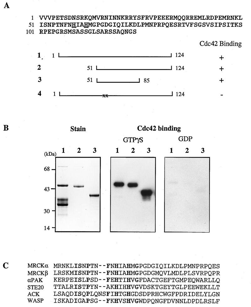
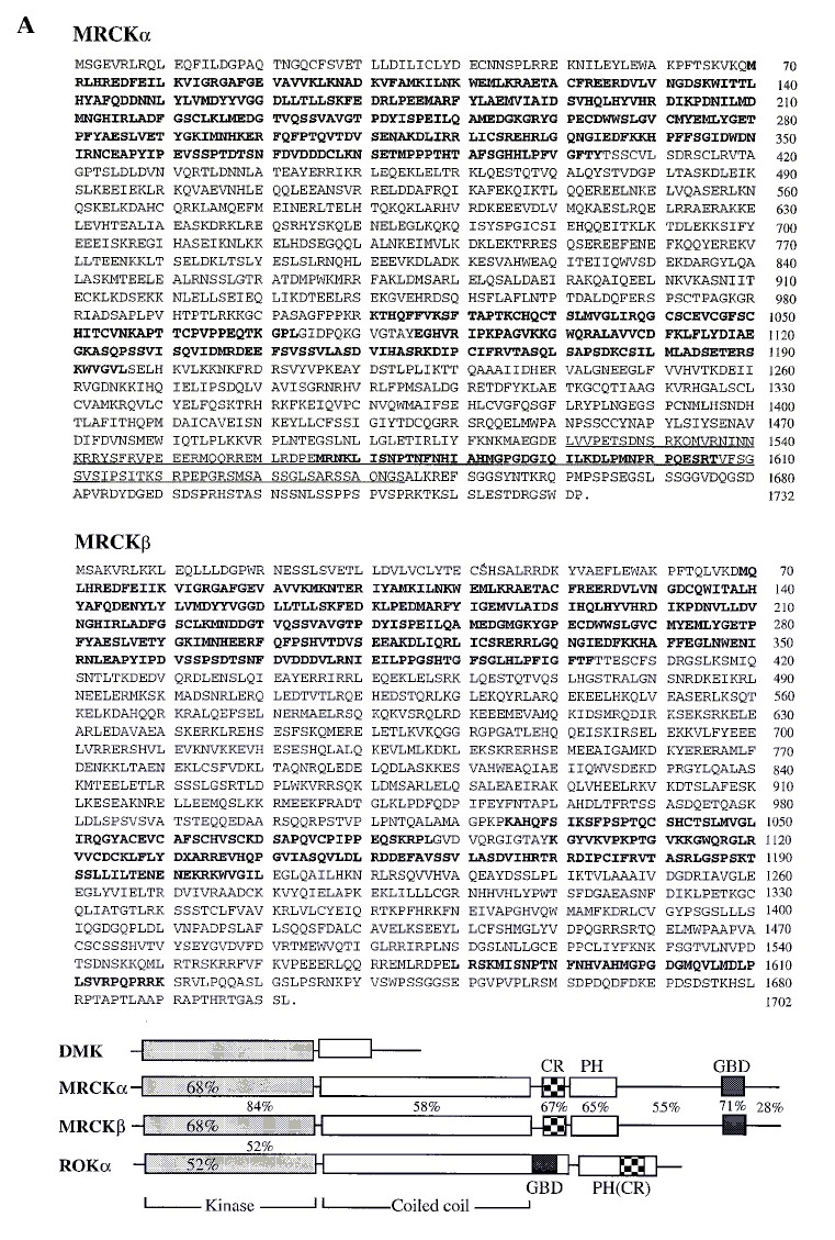
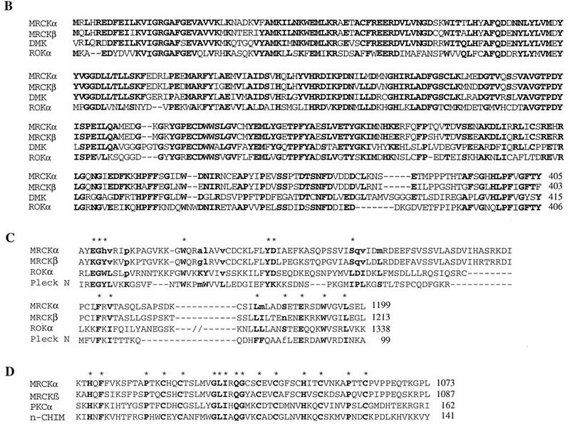
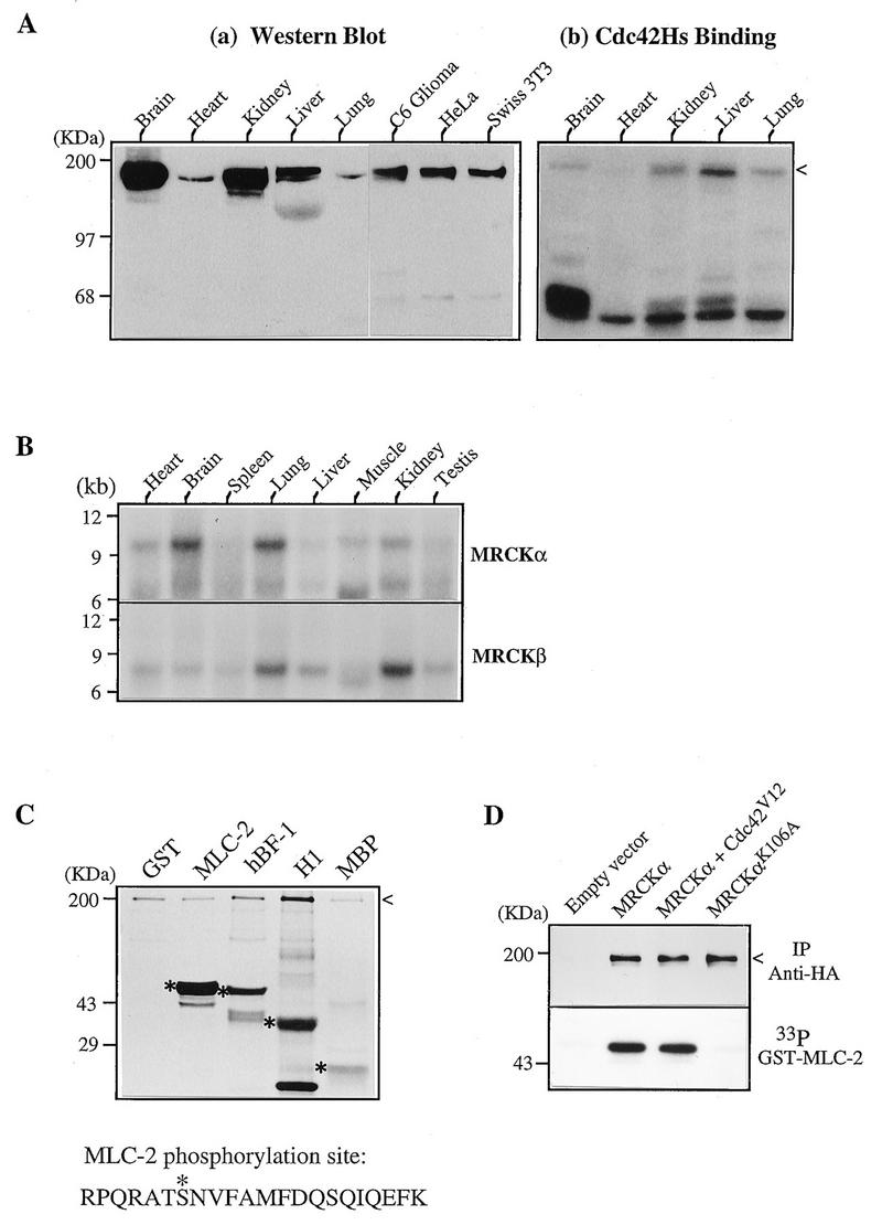
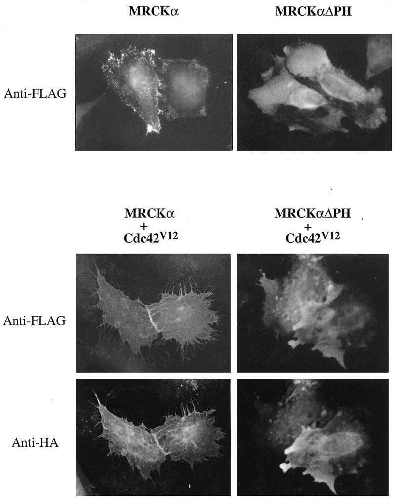
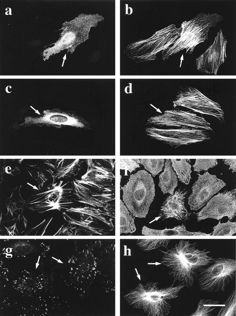
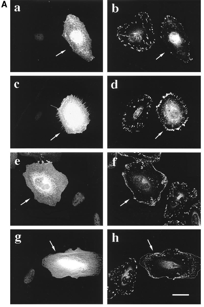
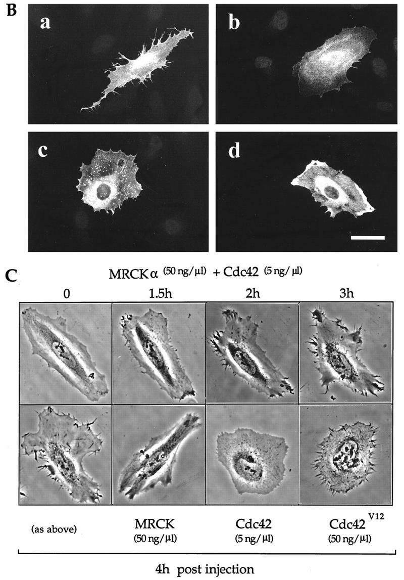
The Rho GTPases play distinctive roles in cytoskeletal reorganization associated with growth and differentiation. The Cdc42/Rac-binding p21-activated kinase (PAK) and Rho-binding kinase (ROK) act as morphological effectors for these GTPases. We have isolated two related novel brain kinases whose p21-binding domains resemble that of PAK whereas the kinase domains resemble that of myotonic dystrophy kinase-related ROK. These approximately 190-kDa myotonic dystrophy kinase-related Cdc42-binding kinases (MRCKs) preferentially phosphorylate nonmuscle myosin light chain at serine 19, which is known to be crucial for activating actin-myosin contractility. The p21-binding domain binds GTP-Cdc42 but not GDP-Cdc42. The multidomain structure includes a cysteine-rich motif resembling those of protein kinase C and n-chimaerin and a putative pleckstrin homology domain. MRCK alpha and Cdc42V12 colocalize, particularly at the cell periphery in transfected HeLa cells. Microinjection of plasmid encoding MRCK alpha resulted in actin and myosin reorganization. Expression of kinase-dead MRCK alpha blocked Cdc42V12-dependent formation of focal complexes and peripheral microspikes. This was not due to possible sequestration of the p21, as a kinase-dead MRCK alpha mutant defective in Cdc42 binding was an equally effective blocker. Coinjection of MRCK alpha plasmid with Cdc42 plasmid, at concentrations where Cdc42 plasmid by itself elicited no effect, led to the formation of the peripheral structures associated with a Cdc42-induced morphological phenotype. These Cdc42-type effects were not promoted upon coinjection with plasmids of kinase-dead or Cdc42-binding-deficient MRCK alpha mutants. These results suggest that MRCK alpha may act as a downstream effector of Cdc42 in cytoskeletal reorganization.


Figures

FIG. 1
FIG. 1
Identification of a family of Cdc42Hs-binding proteins. (A) Deduced amino acid sequence of a human brain partial cDNA clone isolated by expression screening with [γ-32P]GTP-Cdc42. GST fusion proteins were made with wild-type (construct 1), deleted (constructs 2 and 3), or mutated (construct 4) variants. In construct 4, two histidines (underlined) were mutated to alanine. Binding of [γ-32P]GTP-Cdc42 was performed as described previously (35). (B) Nucleotide-dependent binding by two related rat Cdc42-binding proteins. Nitrocellulose filters with 50 ng of GST fusion protein containing the binding domain of human MRCKα (residues 1 to 124; lane 1), rat MRCKβ (residues 1569 to 1690; lane 2), and αPAK (residues 67 to 150; lane 3) were assayed for binding with a 32P-phosphorylated Cdc42Hs (from pGEX-2TK) exchanged with either GTPγS or GDP (31). (C) Consensus sequence of Cdc42-binding motifs of different proteins (9).
FIG. 2
FIG. 2
Sequence of a family of Ser/Thr kinases containing a Cdc42-binding domain and other functional domains. (A) Deduced amino acid sequences of rat MRCKα and MRCKβ. Regions in boldface represent, in order, kinase, cysteine-rich (CR), PH, and p21 GTPase-binding (GBD) domains. In MRCKα, the region underlined is identical, apart from an initial L→V, to the human sequence shown in Fig. 1A. Domain organization of MRCKs, myotonic dystrophy kinase (DMK), and ROKα, along with percent identities of related domains, is also shown. (B) Kinase domains of MRCKα, MRCKβ, myotonic dystrophy kinase (DMK), and ROKα. (C) PH domains in MRCKα, MRCKβ, ROKα, and pleckstrin N terminus (Pleck N). Amino acids identical to the most commonly occurring consensus sequence in PH domains (in boldface and uppercase) are marked with asterisks. (D) Cysteine-rich domains of MRCKs, PKCα, and n-chimaerin (n-CHIM). Conserved residues (17) are indicated by asterisks.
FIG. 2
FIG. 2
Sequence of a family of Ser/Thr kinases containing a Cdc42-binding domain and other functional domains. (A) Deduced amino acid sequences of rat MRCKα and MRCKβ. Regions in boldface represent, in order, kinase, cysteine-rich (CR), PH, and p21 GTPase-binding (GBD) domains. In MRCKα, the region underlined is identical, apart from an initial L→V, to the human sequence shown in Fig. 1A. Domain organization of MRCKs, myotonic dystrophy kinase (DMK), and ROKα, along with percent identities of related domains, is also shown. (B) Kinase domains of MRCKα, MRCKβ, myotonic dystrophy kinase (DMK), and ROKα. (C) PH domains in MRCKα, MRCKβ, ROKα, and pleckstrin N terminus (Pleck N). Amino acids identical to the most commonly occurring consensus sequence in PH domains (in boldface and uppercase) are marked with asterisks. (D) Cysteine-rich domains of MRCKs, PKCα, and n-chimaerin (n-CHIM). Conserved residues (17) are indicated by asterisks.
FIG. 3
FIG. 3
Expression and biochemical characterization of MRCKs. (A) Expression of MRCK in tissues and cultured cells. (a) Soluble protein extracts from various rat tissues and cells were separated by polyacrylamide gel electrophoresis and transferred to nitrocellulose filters for Western analysis using antibodies against the Cdc42-binding domain of human MRCKα (C-terminal 124 residues). (b) A similar blot showing Cdc42 binding. The arrowhead indicates the positions of the immunoreactive and Cdc42-binding regions. (B) Northern (mRNA) blot from rat tissues (Clontech) hybridized to the 32P-labeled MRCKα and MRCKβ cDNA probes. (C) Kinase activity toward different substrates. GST–MLC-2, GST-MRCKα p21-binding domain hBF-1 (residues 1 to 124; Fig. 1), histone H1, and myelin basic protein (MBP) were used as substrates in a kinase assay with purified MRCKα expressed in baculovirus as a GST fusion protein. The 33P-labeled bands corresponding to the Coomassie blue-stained substrate proteins are marked with asterisks, and the autophosphorylated GST-MRCKα band is indicated by an arrowhead. The sequence at the bottom shows the Lys-C peptide with the serine 19 (asterisk) phosphorylation site in MLC-2. (D) Kinase activities in transfected cells. COS-7 cells were transfected with vector pXJ40HA or with a vector containing MRCKα alone, MRCKα in combination with Cdc42V12, or kinase-inactive MRCKαK106A. Tagged proteins were immunoprecipitated (IP) with anti-HA antibody, and kinase activity (lower panel) was assayed with [γ-33P]ATP as described elsewhere (36).
FIG. 4
FIG. 4
Cellular localization of MRCKα and the effects of Cdc42V12. HeLa cells grown in minimal essential medium with 10% fetal bovine serum were transfected with pXJ40-FLAG plasmids encoding either MRCKα alone or MRCKαΔPH (the latter with the PH domain deleted). Cells were fixed with 4% paraformaldehyde and stained with anti-FLAG antibody after 16 h. For cotransfection experiments, plasmid encoding FLAG-tagged MRCKα or MRCKαΔPH was cotransfected with plasmid encoding HA-tagged Cdc42V12. Cells were fixed and doubly stained with antibodies against FLAG for MRCKα and HA for Cdc42V12.
FIG. 5
FIG. 5
MRCKα affects the organization of cellular structures. HeLa cells grown on coverslips were microinjected with a plasmid encoding HA-tagged wild-type MRCKα (a and b), kinase-dead MRCKαK106A (c and d), or kinase domain alone (e to h). Two hours after incubation, cells were fixed and stained with anti-HA antibody (a and c) or doubly stained with phalloidin (b, d, and e) or antibodies against myosin light chain (f), vinculin (g), or tubulin (h). Arrows indicate the injected cells located by HA staining (not shown in panels e to h). Bar = 10 μm.
FIG. 6
FIG. 6
MRCKα potentiates the effects of Cdc42 on microspike formation. (A) Kinase-dead MRCKα blocks Cdc42-mediated effects on focal complexes and morphology. Serum-starved HeLa cells were injected with plasmid encoding FLAG-tagged kinase-dead/p21-binding-deficient mutant MRCKαK106A,H1579A,H1581A (50 ng/μl); 3 h later, these preinjected cells (a, b, e, and f) and uninjected control cells (c, d, g, and h) were injected with plasmid pXJ40-HA (50 ng/μl) encoding Cdc42V12 (a to d) or Rac1V12 (e to h). Cells were fixed and stained with antibodies against FLAG (a and e), HA (c and g), or paxillin (b, d, f, and h) after incubating for 2 h. Essentially similar results were obtained with kinase-dead MRCKαK106A. (B) Morphological effect of expression of MRCKα and limiting amounts of Cdc42. HeLa cells grown in serum-containing medium were injected with plasmids encoding FLAG-tagged Cdc42 (5 ng/μl) together with plasmid encoding either HA-tagged wild-type MRCKα (a), MRCKαK106A (b), p21-binding-defective MRCKαH1579A,H1582A (c), or ROKα (d) at 50 ng/μl. Cells incubated for 2 h were fixed and stained with anti-HA antibody. Bar = 10 μm. (C) Time-lapse phase-contrast microscopy of HeLa cells coinjected with plasmids encoding MRCKα and Cdc42 as in panel B. Morphological changes in a typical coinjected cell are shown up to 4 h after the coinjection. Cells 4 h after injection with plasmids encoding MRCKα (50 ng/μl) alone, Cdc42 (5 ng/μl) alone, or Cdc42V12 (50 ng/μl) are included for comparison.
FIG. 6
FIG. 6
MRCKα potentiates the effects of Cdc42 on microspike formation. (A) Kinase-dead MRCKα blocks Cdc42-mediated effects on focal complexes and morphology. Serum-starved HeLa cells were injected with plasmid encoding FLAG-tagged kinase-dead/p21-binding-deficient mutant MRCKαK106A,H1579A,H1581A (50 ng/μl); 3 h later, these preinjected cells (a, b, e, and f) and uninjected control cells (c, d, g, and h) were injected with plasmid pXJ40-HA (50 ng/μl) encoding Cdc42V12 (a to d) or Rac1V12 (e to h). Cells were fixed and stained with antibodies against FLAG (a and e), HA (c and g), or paxillin (b, d, f, and h) after incubating for 2 h. Essentially similar results were obtained with kinase-dead MRCKαK106A. (B) Morphological effect of expression of MRCKα and limiting amounts of Cdc42. HeLa cells grown in serum-containing medium were injected with plasmids encoding FLAG-tagged Cdc42 (5 ng/μl) together with plasmid encoding either HA-tagged wild-type MRCKα (a), MRCKαK106A (b), p21-binding-defective MRCKαH1579A,H1582A (c), or ROKα (d) at 50 ng/μl. Cells incubated for 2 h were fixed and stained with anti-HA antibody. Bar = 10 μm. (C) Time-lapse phase-contrast microscopy of HeLa cells coinjected with plasmids encoding MRCKα and Cdc42 as in panel B. Morphological changes in a typical coinjected cell are shown up to 4 h after the coinjection. Cells 4 h after injection with plasmids encoding MRCKα (50 ng/μl) alone, Cdc42 (5 ng/μl) alone, or Cdc42V12 (50 ng/μl) are included for comparison.

References

    1. Amano M, Chihara K, Kimura K, Fukata Y, Nakamura N, Matsuura N, Kaibuchi K. Formation of actin stress fibers and focal adhesions enhanced by Rho-kinase. Science. 1997;275:1308–1311. - PubMed
    1. Amano M, Ito M, Kimura K, Fukata Y, Chihara K, Nakano T, Matsuura Y, Kaibuchi K. Phosphorylation and activation of myosin by Rho-associated kinase (Rho-kinase) J Biol Chem. 1996;271:20246–20249. - PubMed
    1. Amano M, Mukai H, Ono Y, Chihara K, Matsui T, Hamajima Y, Okawa K, Iwamatsu A, Kaibuchi K. Identification of a putative target for Rho as the serine-threonine kinase protein kinase N. Science. 1996;271:648–650. - PubMed
    1. Aspenstrom P, Lindberg U, Hall A. Two GTPases, Cdc42 and Rac, bind directly to a protein implicated in the immunodeficiency disorder Wiskott-Aldrich syndrome. Curr Biol. 1995;6:70–75. - PubMed
    1. Bragodia S, Derijard B, Davis R J, Cerione R A. Cdc42 and PAK-mediated signalling leads to JNK and p38 mitogen-activated protein kinase activation. J Biol Chem. 1995;270:27995–27998. - PubMed

Publication types

Associated data