. 1998 Jan;18(1):141-51.
doi: 10.1128/MCB.18.1.141.

Mutations of the Drosophila dDP, dE2F, and cyclin E genes reveal distinct roles for the E2F-DP transcription factor and cyclin E during the G1-S transition

Affiliations

Mutations of the Drosophila dDP, dE2F, and cyclin E genes reveal distinct roles for the E2F-DP transcription factor and cyclin E during the G1-S transition

R J Duronio et al. Mol Cell Biol. 1998 Jan.

Erratum in

  • Mol Cell Biol 1998 May;18(5):3101

Abstract

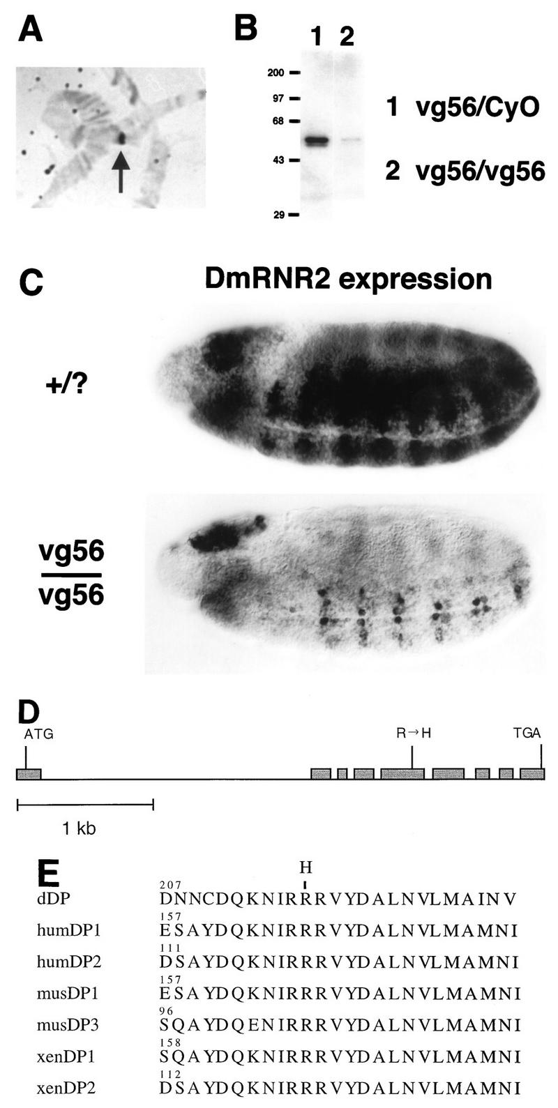
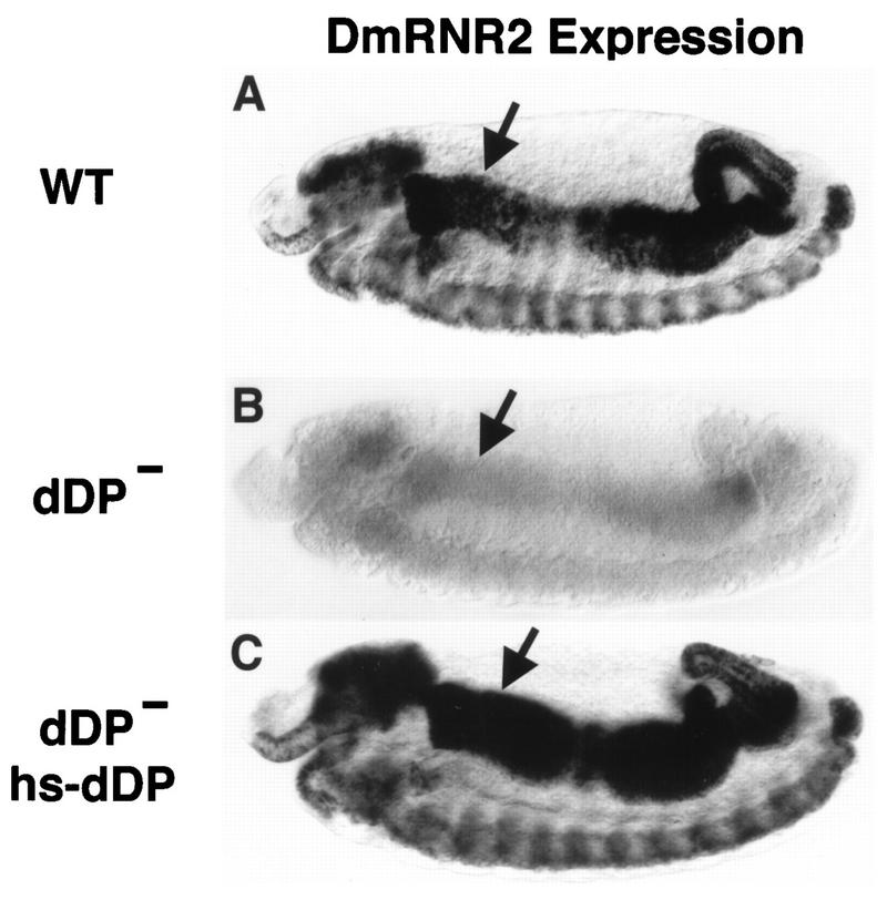
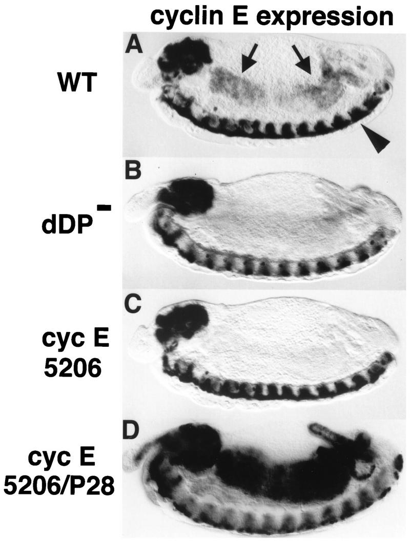
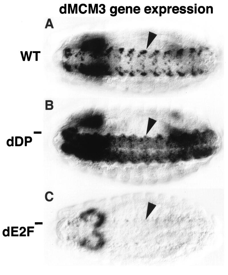
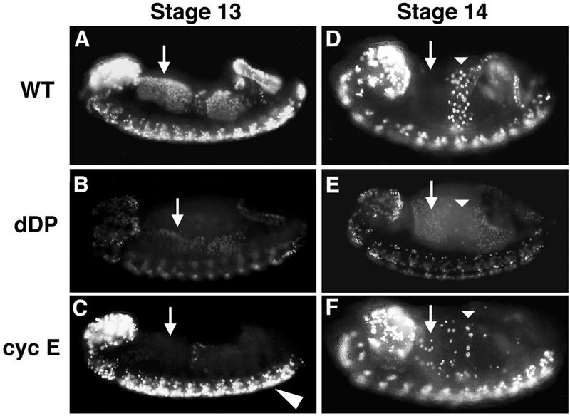
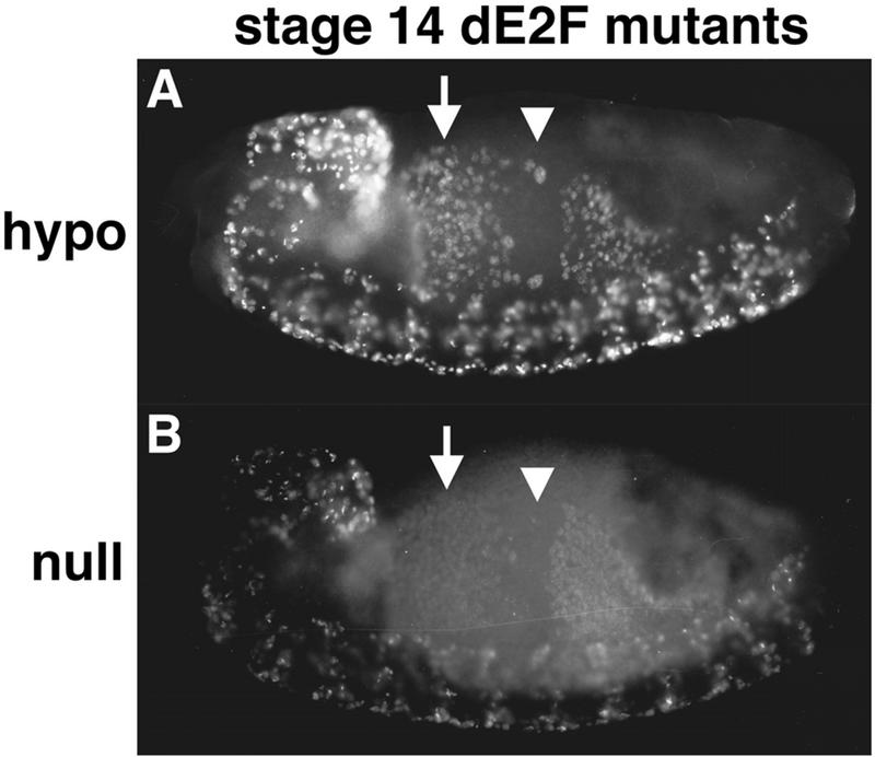
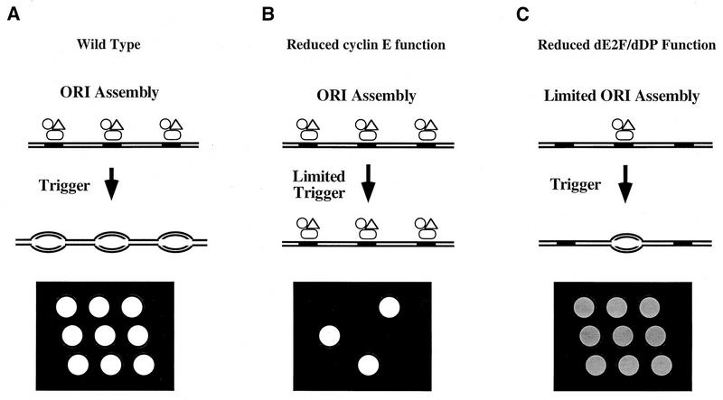
Activation of heterodimeric E2F-DP transcription factors can drive the G1-S transition. Mutation of the Drosophila melanogaster dE2F gene eliminates transcriptional activation of several replication factors at the G1-S transition and compromises DNA replication. Here we describe a mutation in the Drosophila dDP gene. As expected for a defect in the dE2F partner, this mutation blocks G1-S transcription of DmRNR2 and cyclin E as previously described for mutations of dE2F. Mutation of dDP also causes an incomplete block of DNA replication. When S phase is compromised by reducing the activity of dE2F-dDP by either a dE2F or dDP mutation, the first phenotype detected is a reduction in the intensity of BrdU incorporation and a prolongation of the labeling. Notably, in many cells, there was no detected delay in entry into this compromised S phase. In contrast, when cyclin E function was reduced by a hypomorphic allele combination, BrdU incorporation was robust but the timing of S-phase entry was delayed. We suggest that dE2F-dDP contributes to the expression of two classes of gene products: replication factors, whose abundance has a graded effect on replication, and cyclin E, which triggers an all-or-nothing transition from G1 to S phase.


Figures

FIG. 1
FIG. 1
Identification of dDP mutations. (A) Hybridization of a Df(2R)vg56/+ salivary gland polytene chromosome with a dDP cDNA probe. The hybridization signal (arrow) is lost in the Df(2R)vg56 deletion and consequently appears over half the width of the heterozygous polytene chromosome. (B) Western blot of extract of germ band-retracted embryos collected from the Df(2R)vg56/CyO stock probed with anti-dDP monoclonal antibody. Since Df(2R)vg56 deletes the dDP gene, any dDP protein present in Df(2R)vg56/Df(2R)vg56 embryos must be of maternal origin. (C) Germ band-extended control (+/?) and Df(2R)vg56/Df(2R)vg56 mutant stage 11 embryos subjected to in situ hybridization with a digoxigenin-labeled probe derived from the DmRNR2 gene. DmRNR2 expression is failing in the developing ventral (toward the bottom) nerve cord of the mutant embryo. (D) Intron and exon structure of the transcribed region of the dDP gene. Exons are represented by shaded boxes, and introns are represented by a straight line. ATG and TGA indicate the beginning and end of the dDP open reading frame, respectively. The Arg-to-His missense mutation in exon 5 of the dDPvr10 allele is indicated. (E) Alignment of a highly conserved region of the heterodimerization domain of DPs from Drosophila (this paper), human (hum) (30, 70), mouse (mus) (30, 49), and Xenopus (xen) (24). The numbers indicate the position in each primary amino acid sequence. The position of the Arg-to-His change in the dDPvr10 allele is indicated at the top.
FIG. 2
FIG. 2
dDP is required for expression of DmRNR2. Each panel shows a stage 13 embryo (anterior at the left and dorsal at the top) subjected to in situ hybridization with a digoxigenin-labeled probe derived from the DmRNR2 gene. (A) Wild type (WT). The pattern of DmRNR2 expression is coincident with the pattern of DNA replication (Fig. 5A). This includes internal endoreduplicating cells (e.g., midgut cells) and proliferating cells in the CNS. (B) dDPvr10/dDPvr10 (dDP). DmRNR2 expression is not activated in the dDP mutant. (C) A dDPvr10/dDPvr10; hsp70-dDP/hsp70-dDP (dDP hs-dDP) embryo subjected to a 30-min 37°C heat shock followed by a 70-min recovery at room temperature. Expression of dDP cDNA in the mutant restores the normal pattern of DmRNR2 expression. Each of the images has an internal focal plane in order to observe the anterior midgut (arrows).
FIG. 3
FIG. 3
dDP is required for cyclin E expression at the G1-S transition in endoreduplicating cells. Each panel shows a stage 13 embryo (anterior at the left and dorsal at the top) subjected to in situ hybridization with a digoxigenin-labeled probe derived from cyclin E cDNA. (A) Wild type (WT). The arrows indicate the midgut. The arrowhead indicates the ventral nerve cord. (B) dDPvr10/Df(2R)vg56 (dDP). The cells of the midgut normally express cyclin E at this stage but fail to do so in the dDP mutant. CNS expression of cyclin E is normal in the dDP mutant. (C) cyclin E5206/cyclin E5206 mutant embryo. This allele contains a P-element insertion into the upstream regulatory region of the cyclin E gene (33). cyclin E is not expressed in the midgut or other cells that normally endoreduplicate but is expressed in the CNS. (D) cyclin E5206/cyclin EP28 transheterozygote. Expression of cyclin E, presumably from the cyclin EP28 homolog, initiates at the correct developmental time in the midgut. However, transcription is not down regulated as usual, leading to persistently high levels of cyclin E mRNA.
FIG. 4
FIG. 4
Mutation of dDP and dE2F causes distinct phenotypes. Each panel shows a stage 14 embryo (anterior at the left) subjected to in situ hybridization with a digoxigenin-labeled probe derived from dMCM3 cDNA. The perspective is ventral to view the nerve cord (VNC) of the CNS (arrowheads). (A) Wild type (WT). Expression occurs coincident with replication, generating the observed pattern. (B) dDPvr10/dDPvr10 mutant. dMCM3 expression continues in the VNC, but occurs in an expanded domain of cells. This suggests that dDP functions to prevent dMCM3 from becoming derepressed in this tissue. (C) dE2F91/dE2F91 null mutant. Very little if any dMCM3 expression can be detected in the VNC, indicating that dE2F is required for normal dMCM3 expression. No ectopic dMCM3 expression is observed, perhaps because dE2F does not perform an essential repression function. Residual dMCM3 expression in the brain accounts for the out-of-focus staining at the left.
FIG. 5
FIG. 5
Different cell cycle roles for cyclin E and dDP. Embryos were collected and aged until stage 13 (A to C) or stage 14 (D to F), pulse labeled for 15 min with BrdU, and then immediately fixed. BrdU that incorporated into replicating nuclei during the pulse was subsequently detected by indirect immunofluorescence. The orientation of each embryo is the same as in Fig. 2. (A and D) Wild type (WT). (B and E) dDPvr10/dDPvr10 (dDP). (C and F) cyclin EP28/cyclin E05206 (cyc E). The arrows indicate the cells of the anterior midgut (AMG). These cells begin S phase 17 during stage 12, continue replication into stage 13 (A), and exit S phase by stage 14 (D). In the dDP mutant, all or most of the AMG cells enter S phase at the correct time but incorporate much less BrdU (B). By stage 14 these cells are still replicating (E), indicating that S phase is longer than it is in the wild type. In the hypomorphic cyclin E mutant, the AMG cells do not begin S phase 17 on schedule (C) but eventually enter S phase in a random fashion beginning in stage 14 (F). Thus, G1 of cycle 17 is longer than usual in these cells. The actual length of G1 depends upon when the cells reach the critical level of cyclin E activity that triggers S phase. Also note that replication in the CNS appears normal, consistent with wild-type cyclin E expression in the CNS of cyclin E05206 mutants.
FIG. 6
FIG. 6
Prolonged S phase in dE2F mutants. Stage 14 dE2F164/Def(3R)eD7 (hypo) (A) and dE2F7172/dE2F7172 (null) (B) embryos pulse labeled for 15 min with BrdU. S phase 17 continues in the anterior midgut (arrows) when in the wild type it has already finished (Fig. 5D). This is an indication that S phase is prolonged in the mutants. In the central portion of the midgut (arrowheads) S phase 18 has not started on schedule for most of the cells (Fig. 5D). This indicates that dE2F also contributes to correct timing of the G1-S transition.
FIG. 7
FIG. 7
Model of dE2F-dDP and cyclin E function at the G1-S transition and schematic representation of the G1-S transition and BrdU incorporation in the embryonic midgut. (A) Wild type. Components of the prereplicative complex (open symbols) assemble on chromosomes (parallel lines) at origins of DNA replication (solid rectangles). Since E2F-DP is required for the transcription of genes encoding known components of prereplicative complexes like dMCM3 in Drosophila and HsOrc1 in humans (46), we propose that dE2F-dDP contributes to prereplicative complex assembly. Cells remain in G1 until they are induced to progress through the cell cycle by a developmental signal. DNA synthesis is then triggered at the beginning of S phase via cyclin E action, and bidirectional replication forks appear in the chromosome (ovals). After BrdU pulse labeling and in situ immunofluorescent detection of incorporated BrdU, the nuclei in a field of cells that synchronously entered S phase stain brightly and uniformly (nine open circles on the black background). (B) Reduced cyclin E function. Prereplicative complexes form normally at a frequency identical to that of the wild type, because dE2F-dDP is activated independently of cyclin E (16). If cyclin E function is eliminated, replication is not triggered and cells remain in G1. If the cyclin E function is reduced but not eliminated, S phase will still be triggered provided cyclin E function can eventually achieve the critical threshold. In the latter situation, G1 is prolonged and DNA replication is normal once S phase begins. In a field of cells of this type, the length of G1 might vary stochastically as cells approach the S phase threshold. The pattern of BrdU-labeled nuclei would appear random, and each nucleus would have the wild-type intensity of staining (three open circles instead of nine on the black background). (C) Reduced dE2F-dDP function. Decreased provision of components causes limited assembly of prereplicative complexes. If those that assemble are triggered at the usual time in development, the length of G1 does not change. However, fewer prereplicative complexes initiate fewer bidirectional replication forks. Consequently, S phase is prolonged and BrdU incorporation during pulse labeling is significantly reduced. A field of cells of this type appears uniformly and weakly labeled (nine shaded circles on the black background). (Note that the symbols designating components of the prereplicative complex are not shown on the DNA after bidirectional DNA synthesis has begun for the purpose of clarity. Some components of this complex [e.g., ORC] are thought to remain bound to the DNA throughout the entire cell cycle [11].)

References

    1. Andrews B J, Herskowitz I. Regulation of cell cycle-dependent gene expression in yeast. J Biol Chem. 1990;265:14057–14060. - PubMed
    1. Ashburner M, Thompson P, Roote J, Lasko P F, Grau Y, El Messal M, Roth S, Simpson P. The genetics of a small autosomal region of Drosophila melanogaster containing the structural gene for alcohol dehydrogenase. VII. Characterization of the region around the snail and cactus loci. Genetics. 1990;126:679–694. - PMC - PubMed
    1. Bandara L R, Buck V M, Zamanian M, Johnston L H, La T N. Functional synergy between DP-1 and E2F-1 in the cell cycle-regulating transcription factor DRTF1/E2F. EMBO J. 1993;12:4317–4324. - PMC - PubMed
    1. Botz J, Zerfass-Thome K, Spitkovsky D, Delius H, Vogt B, Eilers M, Hatzigeorgiou A, Jansen-Durr P. Cell cycle regulation of the murine cyclin E gene depends on an E2F binding site in the promoter. Mol Cell Biol. 1996;16:3401–3409. - PMC - PubMed
    1. Breeden L. Start-specific transcription in yeast. Curr Top Microbiol Immunol. 1996;208:95–127. - PubMed

Publication types

MeSH terms

Associated data