. 1998 Jan;18(1):368-77.
doi: 10.1128/MCB.18.1.368.

The large subunit of basal transcription factor SNAPc is a Myb domain protein that interacts with Oct-1

Affiliations

The large subunit of basal transcription factor SNAPc is a Myb domain protein that interacts with Oct-1

M W Wong et al. Mol Cell Biol. 1998 Jan.

Abstract

The human RNA polymerase II and III snRNA promoters have similar enhancers, the distal sequence elements (DSEs), and similar basal promoter elements, the proximal sequence elements (PSEs). The DSE, which contains an octamer motif, binds broadly expressed activator Oct-1. The PSE binds a multiprotein complex referred to as SNAPc or PTF. On DNAs containing both an octamer site and a PSE, Oct-1 and SNAPc bind cooperatively. SNAPc consists of at least four stably associated subunits, SNAP43, SNAP45, SNAP50, and SNAP190. None of the three small subunits, which have all been cloned, can bind to the PSE on their own. Here we report the isolation of cDNAs corresponding to the largest subunit of SNAPc, SNAP190. SNAP190 contains an unusual Myb DNA binding domain consisting of four complete repeats (Ra to Rd) and a half repeat (Rh). A truncated protein consisting of the last two SNAP190 Myb repeats, Rc and Rd, can bind to the PSE, suggesting that the SNAP190 Myb domain contributes to recognition of the PSE by the SNAP complex. SNAP190 is required for snRNA gene transcription by both RNA polymerases II and III and interacts with SNAP45. In addition, SNAP190 interacts with Oct-1. Together, these results suggest that the largest subunit of the SNAP complex is involved in direct recognition of the PSE and is a target for the Oct-1 activator. They also provide an example of a basal transcription factor containing a Myb DNA binding domain.


Figures
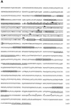
FIG. 1
FIG. 1
Amino acid sequence and schematic structure of SNAP190. (A) Amino acid sequence of SNAP190. The shaded regions correspond to peptide sequences obtained from the purified protein. The dashed arrow corresponds to the Myb half repeat, Rh, and the other arrows indicate Myb repeats Ra, Rb, Rc, and Rd, with the conserved tryptophan (or phenylalanine in the Ra repeat and tyrosines in Rb and Rc repeats) indicated in boldface. The arginine-rich and serine-rich regions are boxed. The leucines spaced as in a leucine zipper are indicated by asterisks. The synthetic peptides used to generate antibodies in rabbits are underlined: peptide 190-1 (aa 1452 to 1469) gave antibodies 398 and 399a; peptide 190-2 (aa 1309 to 1328) gave antibodies 400 and 401; peptide 190-3 (aa 843 to 865) gave antibodies 402 and 403; and peptide 190-4 (aa 712 to 735) gave antibodies 439 and 440. (B) Schematic structure of p190. The location of the Myb domain with the half repeat (Rh) and the four complete repeats (Ra, Rb, Rc, and Rd), as well as the locations of the arginine-rich, serine-rich, and leucine zipper-like regions, are indicated. The region of the protein that interacts with Oct-1 in a yeast one-hybrid assay is indicated by a bracket. (C) Alignment of the p190 half Myb repeat, Rh, and repeats Ra, Rb, Rc, and Rd and Myb repeats R1, R2, and R3 from the human A-Myb, B-Myb (19), and c-Myb (12, 28) proteins. The conserved tryptophans (replaced in some cases by tyrosines or phenylalanines) are indicated in boldface. In this alignment, the p190 Ra repeat is 17, 25, and 22% identical to the Myb R1, R2, and R3 repeats, respectively; the p190 Rb repeat is 14, 23, and 23% identical to the Myb R1, R2, and R3 repeats, respectively; the p190 Rc repeat is 27, 38, and 23% identical to the Myb R1, R2, and R3 repeats, respectively; and the p190 Rd repeat is 21, 38, and 30% identical to the Myb R1, R2, and R3 repeats, respectively.
FIG. 1
FIG. 1
Amino acid sequence and schematic structure of SNAP190. (A) Amino acid sequence of SNAP190. The shaded regions correspond to peptide sequences obtained from the purified protein. The dashed arrow corresponds to the Myb half repeat, Rh, and the other arrows indicate Myb repeats Ra, Rb, Rc, and Rd, with the conserved tryptophan (or phenylalanine in the Ra repeat and tyrosines in Rb and Rc repeats) indicated in boldface. The arginine-rich and serine-rich regions are boxed. The leucines spaced as in a leucine zipper are indicated by asterisks. The synthetic peptides used to generate antibodies in rabbits are underlined: peptide 190-1 (aa 1452 to 1469) gave antibodies 398 and 399a; peptide 190-2 (aa 1309 to 1328) gave antibodies 400 and 401; peptide 190-3 (aa 843 to 865) gave antibodies 402 and 403; and peptide 190-4 (aa 712 to 735) gave antibodies 439 and 440. (B) Schematic structure of p190. The location of the Myb domain with the half repeat (Rh) and the four complete repeats (Ra, Rb, Rc, and Rd), as well as the locations of the arginine-rich, serine-rich, and leucine zipper-like regions, are indicated. The region of the protein that interacts with Oct-1 in a yeast one-hybrid assay is indicated by a bracket. (C) Alignment of the p190 half Myb repeat, Rh, and repeats Ra, Rb, Rc, and Rd and Myb repeats R1, R2, and R3 from the human A-Myb, B-Myb (19), and c-Myb (12, 28) proteins. The conserved tryptophans (replaced in some cases by tyrosines or phenylalanines) are indicated in boldface. In this alignment, the p190 Ra repeat is 17, 25, and 22% identical to the Myb R1, R2, and R3 repeats, respectively; the p190 Rb repeat is 14, 23, and 23% identical to the Myb R1, R2, and R3 repeats, respectively; the p190 Rc repeat is 27, 38, and 23% identical to the Myb R1, R2, and R3 repeats, respectively; and the p190 Rd repeat is 21, 38, and 30% identical to the Myb R1, R2, and R3 repeats, respectively.
FIG. 2
FIG. 2
Recombinant p190 comigrates with SNAP190. Three (lane 1), 10 (lane 2), and 30 (lane 3) μl of a SNAPc-containing P11C fraction (7) and 5 (lane 4), 10 (lane 5), and 30 (lane 6) μl of reticulocyte lysate programmed with the p190 cDNA or a mixture of 10 μl of the P11C fraction and 10 μl of p190 cDNA-programmed reticulocyte lysate (lane 7) were fractionated on an SDS–5% polyacrylamide gel. The proteins were transferred to nitrocellulose, and the filter was immunoblotted with the anti-p190 402 antibody. Unprogrammed reticulocyte lysate gave no signal (data not shown). The positions of molecular weight markers and of SNAP190 in the P11C fraction are indicated.
FIG. 3
FIG. 3
p190 corresponds to the SNAP190 subunit of SNAPc. (A) EMSA performed with a probe containing the wild-type mouse U6 PSE (lanes 1 and 3 to 8) or a mutated mouse U6 PSE (ABC mutation [25];lane 2), a fraction highly enriched in SNAPc (mono-Q fraction) (7), and either no antibodies (lanes 1 and 2), 1 μl of preimmune antibodies (lane 3), or 1 μl of anti-p190 (402) antibodies (lanes 4 to 8). Lanes 5 and 6 also contained 1 and 3 μg, respectively, of specific peptide (peptide 190-3), and lanes 7 and 8 contained 1 and 3 μg, respectively, of nonspecific peptide (peptide CSH375). The locations of the free probe and the complexes containing SNAPc or SNAPc and anti-p190 antibodies (SNAPc + Ab) bound to the PSE are indicated. (B) Beads coated with either preimmune (lanes 3 to 6) or anti-p190 (402) (lanes 7 to 10) antibodies were incubated with the SNAPc-enriched mono-Q fraction. The bound material was then eluted either without (lanes 3, 4, 7, and 8) or with (lanes 5, 6, 9, and 10) a specific peptide (peptide 190-3) and used in an EMSA with a probe containing the wild-type mouse U6 PSE. For lanes 1 and 2, the EMSA was performed with a probe containing the wild-type (lane 1) or mutated (lane 2) mouse U6 PSE and the SNAPc-enriched mono-Q fraction.
FIG. 4
FIG. 4
SNAP190 is required for RNA polymerase II and III transcription from snRNA promoters. (A) Schematic representation of the four promoters used for in vitro transcription. (B) SNAP190 is required for human snRNA gene transcription. For the AdML, VAI, and U6 snRNA gene transcription experiments, 100 μl of HeLa whole-cell extract was depleted with either 100 μl of rabbit preimmune antibody beads (lane 4), 67 μl of preimmune antibody beads plus 33 μl of anti-SNAP190 antibody (antibody 402) beads (lane 5), 33 μl of preimmune antibody beads plus 67 μl of anti-SNAP190 antibody beads (lane 6), or 100 μl of anti-SNAP190 antibody beads (lanes 7 to 12). In lanes 8 and 9, the anti-SNAP190 antibody beads were first incubated with an excess of a nonspecific (CSH483) and a specific (peptide 190-3) peptide, respectively. In lanes 10 to 12, 2, 4, and 8 μl, respectively, of the SNAPc-enriched mono-Q fraction were added to transcription reaction mixtures. Lanes 1 to 3 show transcription performed with undiluted extract (lane 1), extract diluted 1:2 with buffer D (lane 2), and extract diluted 1:4 with buffer D (lane 3). Lane 2 is directly comparable to lanes 4 to 12. The U1 snRNA gene transcription was sensitive to the effects of dilution (lanes 2 and 3); therefore, 40 μl of whole-cell extract was depleted with 20 μl of antibody beads by using the same ratio of preimmune to anti-SNAP190 antibody beads as that described above. The bands corresponding to correctly initiated RNA are labeled AdML for the Ad2 major-late promoter, U1 5′ for the U1 snRNA promoter, U6 5′ for the U6 snRNA promoter, and VAI for the Ad2 VAI promoter. The band labeled RT corresponds to transcripts derived from cryptic promoters located within vector sequences (25).
FIG. 5
FIG. 5
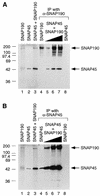
SNAP190 interacts with SNAP45. (A) A cDNA encoding SNAP190 (lanes 1 and 4), SNAP45 (lanes 2 and 8), or a mixture of cDNAs encoding SNAP190 and SNAP45 (lanes 3 and 5 to 7) were expressed by coupled in vitro transcription and translation in reticulocyte lysates. In lanes 1 to 3, 1 μl of the products was loaded directly on the SDS-polyacrylamide gel. In lanes 4 to 8, 10, 3, 10, 30, and 30 μl, respectively, of the products were first incubated with beads coated with the anti-SNAP190 (402) antibody. The beads were washed, and the bound proteins were eluted by boiling the beads in Laemmli buffer and were fractionated on the SDS-polyacrylamide gel. (B) The same experiment as in panel A was performed, except that in lanes 4 to 8, the products were first incubated with beads coated with an anti-SNAP45 antibody (α-CSH467; rabbit 234).
FIG. 6
FIG. 6
DNA binding of the SNAP190 Myb repeats. (A) An EMSA was performed with a probe containing a wild-type (lanes 1, 3 to 5, and 9 to 13) or mutated (lanes 2, 6 to 8, and 14 to 18) mouse U6 PSE and either 2 μl of a fraction enriched in SNAPc (mono-Q fraction; 0.3 mg of protein/ml [7]) (lanes 1 and 2) or 0.1 (lanes 3, 6, 10, and 15), 0.3 (lanes 4, 7, 11, and 16), 1 (lanes 5, 8, 12, and 17) or 3 (lanes 13 and 18) μl of the bacterially expressed proteins indicated above the lanes. In lanes 9 and 14, no proteins were added to the probes. The locations of free probes, complexes containing SNAPc, GST-190RcRd, and 190RcRd are indicated. (B) The sequences of the PSEs present in the wild-type and mutant probes are shown. Uppercase letters correspond to sequences derived from the mouse U6 promoter, with underlined characters corresponding to mutations. Flanking sequences are in lowercase characters. The c-Myb consensus binding site (c-Myb BS) is also indicated.
FIG. 6
FIG. 6
DNA binding of the SNAP190 Myb repeats. (A) An EMSA was performed with a probe containing a wild-type (lanes 1, 3 to 5, and 9 to 13) or mutated (lanes 2, 6 to 8, and 14 to 18) mouse U6 PSE and either 2 μl of a fraction enriched in SNAPc (mono-Q fraction; 0.3 mg of protein/ml [7]) (lanes 1 and 2) or 0.1 (lanes 3, 6, 10, and 15), 0.3 (lanes 4, 7, 11, and 16), 1 (lanes 5, 8, 12, and 17) or 3 (lanes 13 and 18) μl of the bacterially expressed proteins indicated above the lanes. In lanes 9 and 14, no proteins were added to the probes. The locations of free probes, complexes containing SNAPc, GST-190RcRd, and 190RcRd are indicated. (B) The sequences of the PSEs present in the wild-type and mutant probes are shown. Uppercase letters correspond to sequences derived from the mouse U6 promoter, with underlined characters corresponding to mutations. Flanking sequences are in lowercase characters. The c-Myb consensus binding site (c-Myb BS) is also indicated.
FIG. 7
FIG. 7
The carboxy-terminal half of SNAP190 interacts with DNA-bound Oct-1 both in vivo and in vitro. (A) The strategy for isolating proteins interacting with DNA-bound Oct-1 (29) is depicted. An interaction between Oct-1, which is transcriptionally inactive in yeast, and a fusion protein containing the VP16 activation domain results in induction of HIS3 transcription, which in turn allows cells to grow on AT-containing medium. The Oct-1 protein and the VP16 cDNA library were expressed from single-copy plasmids marked with the URA3 and TRP1 genes, respectively. Expression of the VP16 hybrid proteins was placed under control of the galactose-inducible GAL1, 10 regulatory sequences (UASG). (B) Yeast cells expressing Oct-1, SNAP190C, and OBF-1 as indicated above the lanes were plated on medium without AT or containing 40 mM AT. Only cells expressing induced levels of the HIS3 gene product can grow in the presence of AT. (C) EMSA performed with a probe containing an octamer motif and the in vitro-translated proteins indicated above the lanes. The total amount of reticulocyte lysate was kept constant in all lanes. Lane 1 contains unprogrammed reticulocyte lysate only, lanes 2 to 5 contain 1 μl of Oct-1 POU, lane 3 contains in addition 1 μl of OBF-1, and lanes 4 and 5 contain 1 and 2 μl, respectively, of SNAP190C.

References

    1. Aasland R, Stewart A F, Gibson T. The SANT domain: a putative DNA-binding domain in the SWI-SNF and ADA complexes, the transcriptional co-repressor N-CoR and TFIIIB. Trends Biochem Sci. 1996;21:87–88. - PubMed
    1. Bai L, Wang Z, Yoon J-B, Roeder R G. Cloning and characterization of the β subunit of human proximal sequence element-binding transcription factor and its involvement in transcription of small nuclear RNA genes by RNA polymerases II and III. Mol Cell Biol. 1996;16:5419–5426. - PMC - PubMed
    1. Ford E, Hernandez N. Characterization of a trimeric complex containing Oct-1, SNAPc, and DNA. J Biol Chem. 1997;272:16048–16055. - PubMed
    1. Gstaiger M, Knoepfel L, Georgiev O, Schaffner W, Hovens C M. A B-cell coactivator of octamer-binding transcription factors. Nature. 1995;373:360–362. - PubMed
    1. Harlow E, Lane D. Antibodies: a laboratory manual. Cold Spring Harbor, N.Y: Cold Spring Harbor Laboratory Press; 1988.

Publication types

Associated data