. 1998 Jan;18(1):409-19.
doi: 10.1128/MCB.18.1.409.

Opposite transcriptional effects of cyclic AMP-responsive elements in confluent or p27KIP-overexpressing cells versus serum-starved or growing cells

Affiliations

Opposite transcriptional effects of cyclic AMP-responsive elements in confluent or p27KIP-overexpressing cells versus serum-starved or growing cells

L Deleu et al. Mol Cell Biol. 1998 Jan.

Abstract

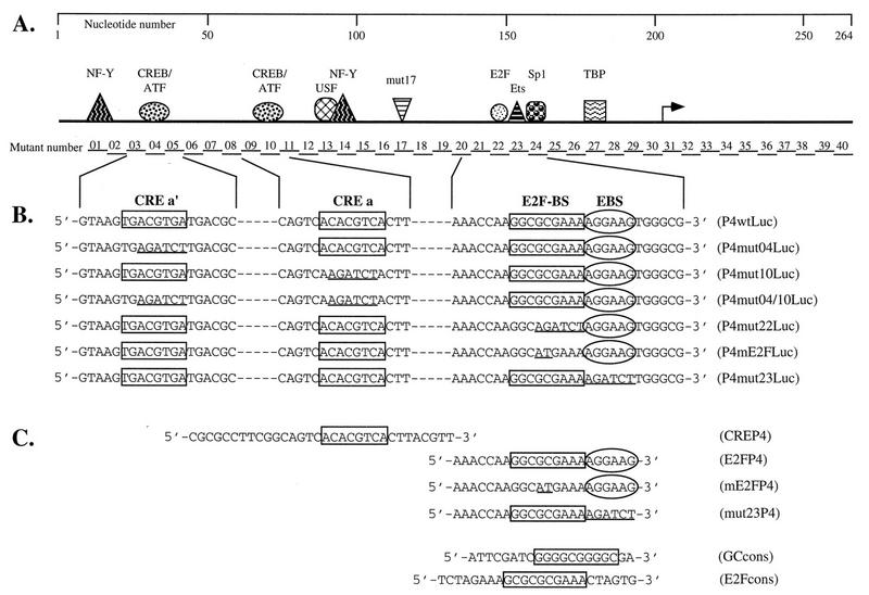
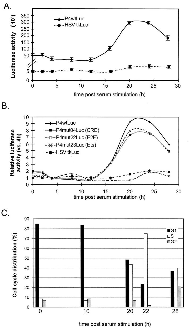
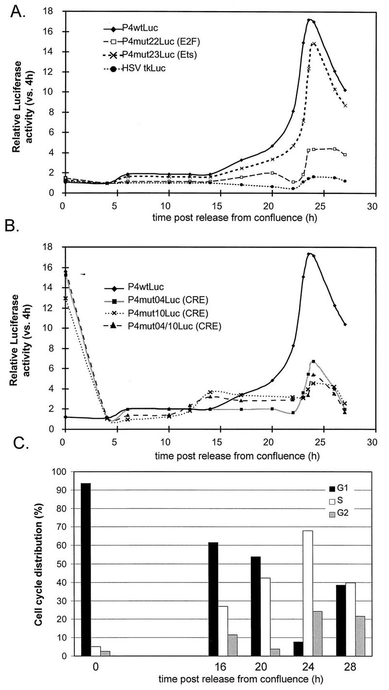
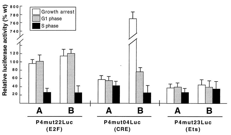
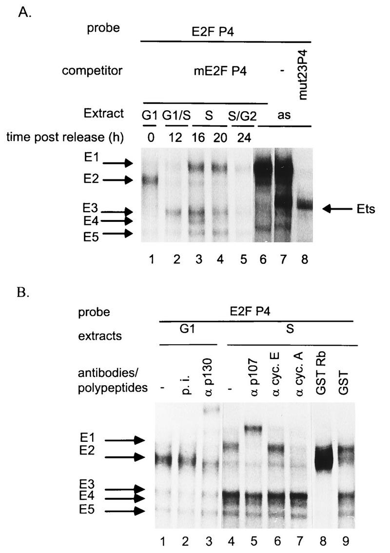
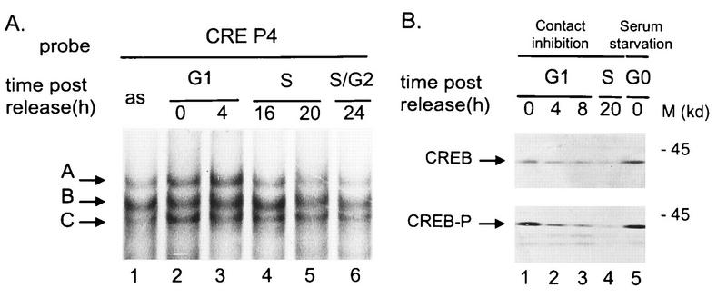
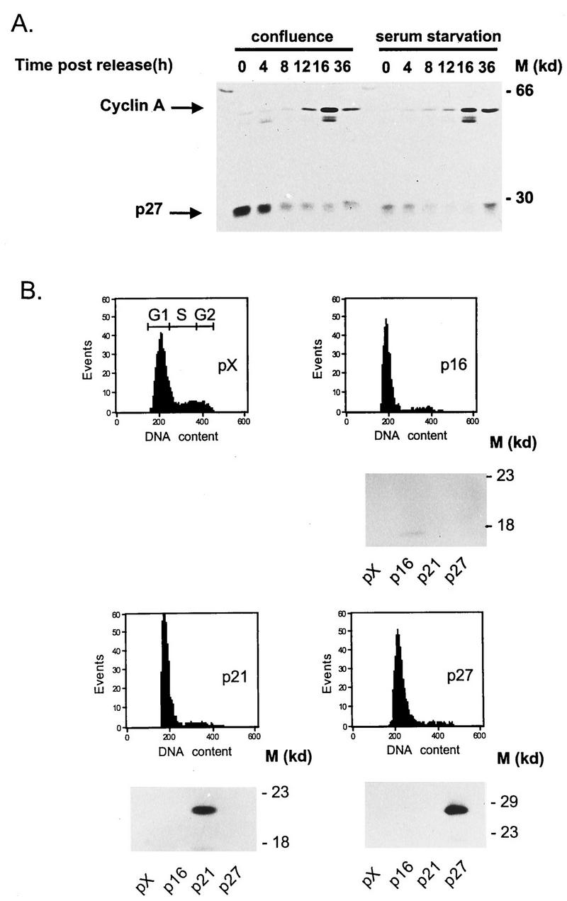
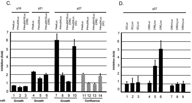
The minute virus of mice, an autonomous parvovirus, requires entry of host cells into the S phase of the cell cycle for its DNA to be amplified and its genes expressed. This work focuses on the P4 promoter of this parvovirus, which directs expression of the transcription unit encoding the parvoviral nonstructural polypeptides. These notably include protein NS1, necessary for the S-phase-dependent burst of parvoviral DNA amplification and gene expression. The activity of the P4 promoter is shown to be regulated in a cell cycle-dependent manner. At the G1/S-phase transition, the promoter is activated via a cis-acting DNA element which interacts with phase-specific complexes containing the cellular transcription factor E2F. It is inhibited, on the other hand, in cells arrested in G1 due to contact inhibition. This inhibitory effect is not observed in serum-starved cells. It is mediated in cis by cyclic AMP response elements (CREs). Unlike serum-starved cells, confluent cells accumulate the cyclin-dependent kinase inhibitor p27, suggesting that the switch from CRE-mediated activation to CRE-mediated repression involves the p27 protein. Accordingly, plasmid-driven overexpression of p27 causes down-modulation of promoter P4 in growing cells, depending on the presence of at least two functional CREs. No such effect is observed with two other cyclin-dependent kinase inhibitors, p16 and p21. Given the importance of P4-driven synthesis of protein NS1 in parvoviral DNA amplification and gene expression, the stringent S-phase dependency of promoter P4 is likely a major determinant of the absolute requirement of the minute virus of mice for host cell proliferation.


Figures

FIG. 1
FIG. 1
Schematic representation of the MVMp P4 promoter, of the mutants derived from it, and of the oligonucleotides used in this study. (A) The upper panel depicts the P4 promoter from nt 1 to the translation initiation site at nt 261 (numbering according to Astell et al. [2]). The arrow indicates the transcription initiation site. Symbols represent transcription factors shown to interact with specific P4 promoter sequence elements: TBP, TATA-box-binding protein (1, 16); Sp1, GC-box-binding proteins (1, 17); Ets, Ets family of transcription factors (17); E2F, E2F-binding-site-specific protein complexes (reference and this study); mut17, as yet unidentified proteins binding to and activating promoter P4 via the DNA element mutated in P4mut17 (15); NF-Y, Y-box-binding protein (18); USF, E-box-binding protein (19); CREB/ATF, CRE-binding proteins (36). The 40 contiguous BglII substitutions introduced in mutants P4mut01 to P4mut40, respectively, are located along the promoter. (B and C) The sequence elements analyzed in this study are framed and aligned beneath the line diagram of the promoter. Underlined sequences indicate mutations introduced in the various elements. (B) P4 promoter constructs driving expression of the luciferase reporter gene. (C) Oligonucleotides used as probes or competitors in electrophoretic mobility assays.
FIG. 2
FIG. 2
Cell cycle-dependent activity of promoter P4 in cells synchronized by serum starvation. Pools of FR3T3 cells stably transfected with reporter construct P4wtLuc, P4mutLuc, or HSVtkLuc were synchronized by serum starvation. After release from the block, luciferase activities were measured at 4-h intervals. (A) Absolute luciferase activities achieved by P4wtLuc and HSVtkLuc are given in arbitrary light units, after correction for the number of cells and the background of the luminometer. Averages of six independent experiments are shown with standard deviation bars. (B) Relative luciferase activities achieved by P4wtLuc, derived mutants (P4mut04/22/23-Luc), or HSVtkLuc. Each activity level is expressed as the ratio of the value measured for the construct concerned at the indicated time to the value measured 4 h postrelease. (C) Cell cycle distribution of stably transfected FR3T3 cultures at different times (0, 10, 20, 22, and 28 h) after release from serum starvation, as determined by FACS analysis.
FIG. 3
FIG. 3
Cell cycle-dependent activity of promoter P4 in cells synchronized by contact inhibition. Pools of FR3T3 cells stably transfected with a reporter construct (P4wtLuc, a derived mutant [P4mutxLuc], or HSVtkLuc) were synchronized by contact inhibition. After release from the block, luciferase activities were measured at 4-h or shorter intervals. (A and B) Relative luciferase activities for a given construct are expressed as ratios of the values measured at the indicated time versus 4 h postrelease. (C) Cell cycle distribution of stably transfected FR3T3 cultures at different times (0, 16, 20, 24, and 28 h) after release from confluence, as determined by FACS analysis.
FIG. 4
FIG. 4
Comparison of wild-type and mutant forms of promoter P4: activities during different phases of the cell cycle. FR3T3 cultures were growth arrested by serum starvation (A) or contact inhibition (B), transfected with equal amounts of wild-type or mutant (mut04, mut22, or mut23) P4 promoter-driven luciferase gene constructs, and released into the cell cycle. Transient expression assays were carried out by measuring luciferase activities prior to release from the block (growth arrest) and at different intervals thereafter (corresponding to the G1 and S phases [Fig. 2C and 3C]). Levels of mutant-P4-driven luciferase gene expression are shown as percentages of the values determined for the wild-type promoter at the same time points.
FIG. 5
FIG. 5
Association of FR3T3 cell proteins with the P4 promoter E2F-binding site. Whole FR3T3 cell extracts were incubated with 32P-end-labeled oligonucleotide E2FP4 containing the E2F-like motif of promoter P4 (Fig. 1). Specific DNA-protein complexes (arrows) were separated by electrophoresis and revealed by autoradiography. The free probe ran out of the gel. (A) Extracts were prepared from FR3T3 cultures at various intervals after release from confluence (corresponding to the indicated phases of the cell cycle) and supplemented with a 100-fold molar excess of unlabeled mE2FP4 competitor oligonucleotide. Asynchronous cell extracts (as) supplemented (or not) with a 100-fold molar excess of unlabeled mE2FP4 or mut23P4 competitor oligonucleotides were included for comparison. The mE2FP4 competitor prevents formation of the Ets-specific complex, whereas mut23P4 competes for formation of E2F-specific complexes. (B) Supershift assays were conducted, in the presence of a 100-fold molar excess of unlabeled mE2FP4 oligonucleotide, using either antibodies (α) directed against proteins known to interact with E2F (p130, p107, and cyclins [cyc.] A and E) or recombinant proteins purified from E. coli (GST and GST-Rb fusion polypeptide). p.i., preimmune serum.
FIG. 6
FIG. 6
FR3T3 cell proteins interacting with the P4 promoter CRE motif. (A) Extracts from asynchronous FR3T3 cultures (as) or from FR3T3 cells harvested at different time points after release from contact inhibition were incubated with 32P-end-labeled oligonucleotide CREP4 (Fig. 1). Specific DNA-protein complexes (arrows; see reference 36) were fractionated by electrophoretic mobility shift assays and revealed by autoradiography. The free probe ran out of the gel. (B) Extracts from FR3T3 cells released from contact inhibition (lanes 1 to 4) or blocked by serum starvation (lanes 5) were analyzed by Western blotting for their content of CREB1. Upper panel, total CREB1; lower panel, CREB1 phosphorylated on serine residue 133. The antibody specifically recognizing phosphorylated CREB1 did not react with CREBs when the extracts were pretreated with phage lambda phosphatase (data not shown). M, apparent molecular mass of proteins standards.
FIG. 7
FIG. 7
Influence of p27 overexpression on whole and minimal P4 promoter construct activities. (A) Whole protein extracts were prepared from FR3T3 cells at increasing times after release from confluence or serum starvation (see Fig. 2C and 3C for cell cycle progression). Proteins were fractionated by sodium dodecyl sulfate-polyacrylamide gel electrophoresis and transferred to a nitrocellulose membrane. The upper and lower parts of the blot were incubated with antibodies directed against cyclin A and CKI p27, respectively. Immunocomplexes were revealed with the ECL detection system. M, apparent molecular masses of protein standards. (B) FACS analysis of FR3T3 cultures cotransfected with either of the CKI-expressing plasmid (p16, p21, or p27) or with the corresponding empty vector (pX). CKI overexpression was verified by Western blot analysis shown below the FACS profiles. (C) Growing or confluent FR3T3 cell cultures were cotransfected with each of the indicated P4Luc reporter plasmids (Fig. 1B) and either p16, p21, or p27 CKI-expressing vector. Transient expression assays were carried out at 48 h posttransfection and are presented as ratios of luciferase activities achieved in the presence of empty versus CKI-expressing vector (i.e., as factors of CKI-induced promoter repression). (D) Growing FR3T3 cell cultures were cotransfected with each of the indicated minimal promoter constructs (see Materials and Methods) and p27-expressing vector. Transient expression assays were carried out at 48 h posttransfection and are presented as ratios of luciferase activities achieved in the presence of empty versus p27-expressing vector (i.e., as factors of p27-induced promoter repression).
FIG. 7
FIG. 7
Influence of p27 overexpression on whole and minimal P4 promoter construct activities. (A) Whole protein extracts were prepared from FR3T3 cells at increasing times after release from confluence or serum starvation (see Fig. 2C and 3C for cell cycle progression). Proteins were fractionated by sodium dodecyl sulfate-polyacrylamide gel electrophoresis and transferred to a nitrocellulose membrane. The upper and lower parts of the blot were incubated with antibodies directed against cyclin A and CKI p27, respectively. Immunocomplexes were revealed with the ECL detection system. M, apparent molecular masses of protein standards. (B) FACS analysis of FR3T3 cultures cotransfected with either of the CKI-expressing plasmid (p16, p21, or p27) or with the corresponding empty vector (pX). CKI overexpression was verified by Western blot analysis shown below the FACS profiles. (C) Growing or confluent FR3T3 cell cultures were cotransfected with each of the indicated P4Luc reporter plasmids (Fig. 1B) and either p16, p21, or p27 CKI-expressing vector. Transient expression assays were carried out at 48 h posttransfection and are presented as ratios of luciferase activities achieved in the presence of empty versus CKI-expressing vector (i.e., as factors of CKI-induced promoter repression). (D) Growing FR3T3 cell cultures were cotransfected with each of the indicated minimal promoter constructs (see Materials and Methods) and p27-expressing vector. Transient expression assays were carried out at 48 h posttransfection and are presented as ratios of luciferase activities achieved in the presence of empty versus p27-expressing vector (i.e., as factors of p27-induced promoter repression).

Similar articles

Cited by

References

    1. Ahn J K, Gavin B J, Kumar G, Ward D C. Transcriptional analysis of minute virus of mice P4 promoter mutants. J Virol. 1989;63:5425–5439. - PMC - PubMed
    1. Astell C R, Thomson M, Merchlinsky M, Ward D C. The complete DNA sequence of minute virus of mice, an autonomous parvovirus. Nucleic Acids Res. 1983;11:999–1018. - PMC - PubMed
    1. Ausubel I, Frederick M. Introduction of DNA into mammalian cells. In: Ausubel F M, Brent R, Kingston R E, Moore D D, Seidman J G, Smith J A, Struhl K, editors. Current protocols in molecular biology. Vol. 1. New York, N.Y: John Wiley & Sons, Inc.; 1987. pp. 9.7.12–9.7.18.
    1. Beijersbergen R L, Bernards R. Cell cycle regulation by the retinoblastoma family of growth inhibitory proteins. Biochim Biophys Acta. 1996;1287:103–120. - PubMed
    1. Boeuf H, Reimund B, Jansen Durr P, Kedinger C. Differential activation of the E2F transcription factor by the adenovirus E1a and EIV products in F9 cells. Proc Natl Acad Sci USA. 1990;87:1782–1786. - PMC - PubMed

Publication types

Substances

LinkOut - more resources