. 1998 Jan;18(1):468-76.
doi: 10.1128/MCB.18.1.468.

A subunit of the anaphase-promoting complex is a centromere-associated protein in mammalian cells

Affiliations

A subunit of the anaphase-promoting complex is a centromere-associated protein in mammalian cells

P M Jörgensen et al. Mol Cell Biol. 1998 Jan.

Abstract

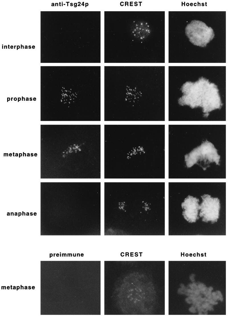
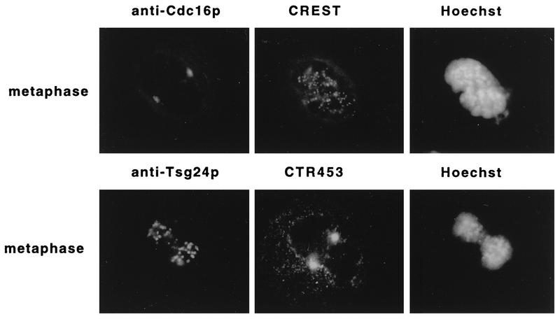
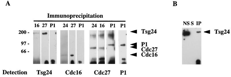
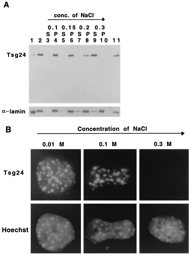
Sister chromatids in early mitotic cells are held together mainly by interactions between centromeres. The separation of sister chromatids at the transition between the metaphase and the anaphase stages of mitosis depends on the anaphase-promoting complex (APC), a 20S ubiquitin-ligase complex that targets proteins for destruction. A subunit of the APC, called APC-alpha in Xenopus (and whose homologs are APC-1, Cut4, BIME, and Tsg24), has recently been identified and shown to be required for entry into anaphase. We now show that the mammalian APC-alpha homolog, Tsg24, is a centromere-associated protein. While this protein is detected only during the prophase to the anaphase stages of mitosis in Chinese hamster cells, it is constitutively associated with the centromeres in murine cells. We show that there are two forms of this protein in mammalian cells, a soluble form associated with other components of the APC and a centromere-bound form. We also show that both the Tsg24 protein and the Cdc27 protein, another APC component, are bound to isolated mitotic chromosomes. These results therefore support a model in which the APC by ubiquitination of a centromere protein regulates the sister chromatid separation process.


Figures

FIG. 1
FIG. 1
The Tsg24 protein transiently accumulates in the centromeric regions of mitotic chromosomes. Interphase or mitotic CHO cells were fixed with methanol-acetone (50:50 [vol/vol]) and triple stained with the anti-Tsg24 antibody (1:20) or preimmune serum (1:20), a CREST antiserum (1:500), and Hoechst 33258 in an indirect immunofluorescence microscopy experiment. The secondary antibodies were a fluorescein isothiocyanate-conjugated swine anti-rabbit IgG and a rhodamine-conjugated goat anti-human IgG. The cells were analyzed by indirect immunofluorescence microscopy.
FIG. 2
FIG. 2
The Tsg24 protein binds to murine centromeres in a cell cycle-independent manner. Mitotic or interphase murine Swiss-3T3 cells were fixed with methanol-acetone (50:50 [vol/vol]), triple stained with different combinations of antibodies, and analyzed by indirect immunofluorescence microscopy. Rows 1 and 2 show mitotic cells labelled with the anti-Tsg24 antibody (1:20), a CREST antiserum (1:500), and Hoechst 33258. Rows 3 to 5 show interphase cells and late-S-phase cells triple stained with the anti-Tsg24 antibody (1:20) or a preimmune serum, a monoclonal anti-PCNA antibody (1:500), and Hoechst 33258. The anti-PCNA antibody labels early- (e) and late-S-phase (l) cells. Late-S-phase cells were also analyzed at a higher magnification (rows 4 and 5). The secondary antibodies used were the same as in Fig. 1, except that a rhodamine-conjugated goat anti-mouse IgG was used to label the anti-PCNA antibody. Fixation of cells with paraformaldehyde gave identical results but weaker signals. Staining of murine L cells gave identical results.
FIG. 3
FIG. 3
The Tsg24 protein is uniformly expressed throughout the cell cycle. Protein extracts were prepared from a serum-deprived synchronized population of CHO cells, following the addition of serum to the cells. Equal numbers of cells were sampled at the indicated time points (0 to 24 h). The extracts were analyzed by immunoblotting with the anti-Tsg24 serum. To ensure that equal amounts of proteins had been loaded onto the gel at all time points, the same immunoblot was labelled in parallel with an anti-α-lamin antibody. m, mitotic extract.
FIG. 4
FIG. 4
The APC subunits Tsg24 and Cdc16 are differently located in mitotic cells. Mitotic Swiss-3T3 cells were fixed with methanol-acetone (50:50 [vol/vol]), triple stained with different combinations of antibodies, and analyzed by indirect immunofluorescence microscopy. Row 1 shows a metaphase cell labelled with the anti-Cdc16 antibody (1:150), a CREST antiserum (1:500), and Hoechst 33258. Row 2 shows a metaphase cell labelled with the anti-Tsg24 antibody (1:20), an antibody against centrosomal protein CTR453 (1:1), and Hoechst 33258. The secondary antibodies used were the same as for Fig. 1 and 2.
FIG. 5
FIG. 5
The APC subunits Tsg24, Cdc16, and Cdc27 are coimmunoprecipitated from mammalian cells. (A) Protein extracts were prepared from Swiss-3T3 cells and immunoprecipitated with anti-Tsg24, anti-Cdc16, anti-Cdc27, or anti-P1 antibodies. The immunoprecipitates were detected by Western blot analysis with the same antibodies. The material precipitated with the anti-Cdc16 antibodies included Cdc16, the anti-Tsg24 antibody precipitated Tsg24, the anti-Cdc27 precipitated Cdc27, and the anti-P1 antibody immunoprecipitated the P1 protein (only the anti-P1–P1 results are shown). The arrowheads indicate the protein bands precipitated. The filters were probed sequentially with different antibodies, and some of the anti-P1 antibody signal remained after washing, explaining the P1 band seen on the filters probed with the anti-Cdc27 antibody. The anti-P1 antibody detects a replication protein in murine cells and was used here as a negative control (45). Molecular masses (indicated at the left) are in kilodaltons. (B) In order to test the solubility of the Tsg24 protein in these experiments, protein extracts were solubilized either with RIPA buffer or with mild lysis buffer (data not shown). Three fractions were tested in parallel by Western blotting: proteins not solubilized by the extraction buffer (NS), proteins solubilized and immunoprecipitated with the anti-Tsg24 antibody (IP), and proteins solubilized but not immunoprecipitated with the anti-Tsg24 antibody (5).
FIG. 6
FIG. 6
The binding of Tsg24 to the centromere is salt dependent. (A) Nuclei were prepared from asynchronously growing Swiss-3T3 cells and extracted with hypotonic buffers containing increasing NaCl concentrations (conc.). The proteins which had been eluted into a supernatant (S) were concentrated and subjected to Western blot analysis. The extracted nuclei, the pellet (P), were also analyzed by Western blotting. The Western blots were labelled with anti-Tsg24 and anti-α-lamin antibodies. Total cellular protein extract (lane 11), total cytoplasm (lane 1), and total nuclear proteins prior to extraction (lane 2) are also shown. (B) Nuclei extracted with different salt concentrations were centrifuged onto glass slides, fixed, and stained with the anti-Tsg24 antibody and Hoechst 33258.
FIG. 7
FIG. 7
The APC components Tsg24 and Cdc27 are bound to mitotic chromosomes. Protein extracts were prepared from mitotic CHO cells (m) and from isolated chromosomes prepared from mitotic CHO cells (pmc). The protein extracts were analyzed by immunoblotting with the anti-Tsg24, anti-P1, antipericentrin, anti-α-tubulin, and anti-Cdc27 antibodies. It is likely that the band with a unique molecular weight detected by the anti-Cdc27 antibody in extracts prepared from isolated mitotic cells is due to the artificial removal of posttranslational modifications (see the text) during the purification procedure, as this protein has been shown to be phosphorylated in mitotic cells (39).

References

    1. Ault J G, Rieder C L. Centrosome and kinetochore movement during mitosis. Curr Opin Cell Biol. 1994;6:41–49. - PubMed
    1. Bailly E, Doree E, Nurse P, Bornens M. p34-cdc2 is located in both nucleus and cytoplasm: part is centrosomally associated and enters vesicles at anaphase. EMBO J. 1989;8:3985–3995. - PMC - PubMed
    1. Bhat M A, Philp A V, Glover D M, Bellen H J. Chromatid segregation at anaphase requires the barren product, a novel chromosome-associated protein that interacts with topoisomerase II. Cell. 1996;87:1103–1114. - PubMed
    1. Bickel S E, Orr-Weaver T L. Holding chromatids together to ensure they go their separate ways. Bioessays. 1996;18:293–300. - PubMed
    1. Bravo R, Macdonald-Bravo H. Existence of two populations of cyclin/proliferating cell nuclear antigen during the cell cycle: association with DNA replication sites. J Cell Biol. 1987;105:1549–1554. - PMC - PubMed

Publication types

LinkOut - more resources