. 1998 Jan;18(1):576-89.
doi: 10.1128/MCB.18.1.576.

Role of UEV-1, an inactive variant of the E2 ubiquitin-conjugating enzymes, in in vitro differentiation and cell cycle behavior of HT-29-M6 intestinal mucosecretory cells

Affiliations

Role of UEV-1, an inactive variant of the E2 ubiquitin-conjugating enzymes, in in vitro differentiation and cell cycle behavior of HT-29-M6 intestinal mucosecretory cells

E Sancho et al. Mol Cell Biol. 1998 Jan.

Abstract

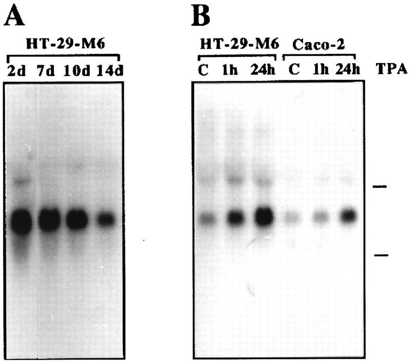
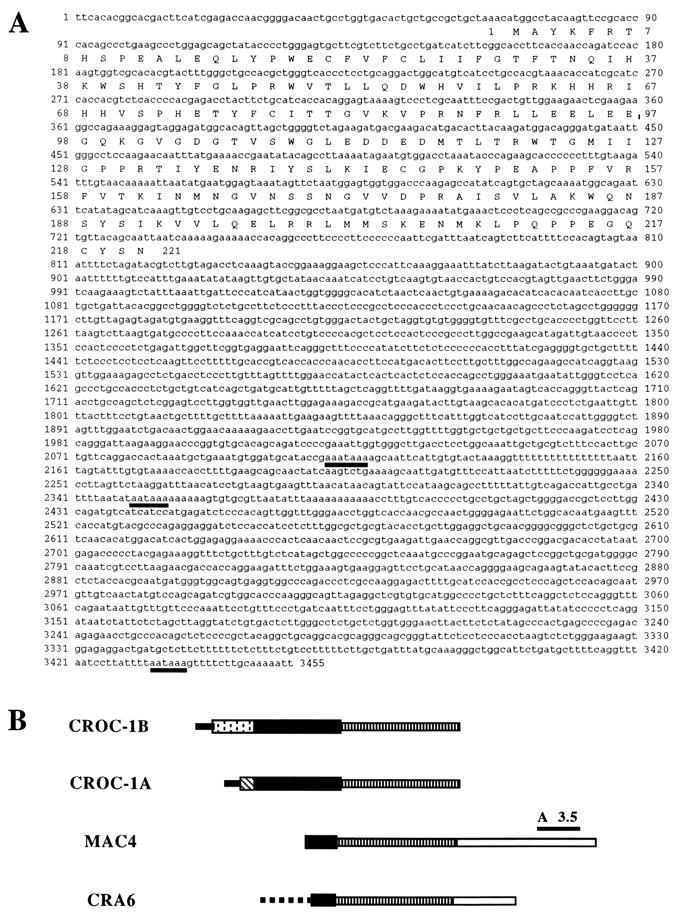
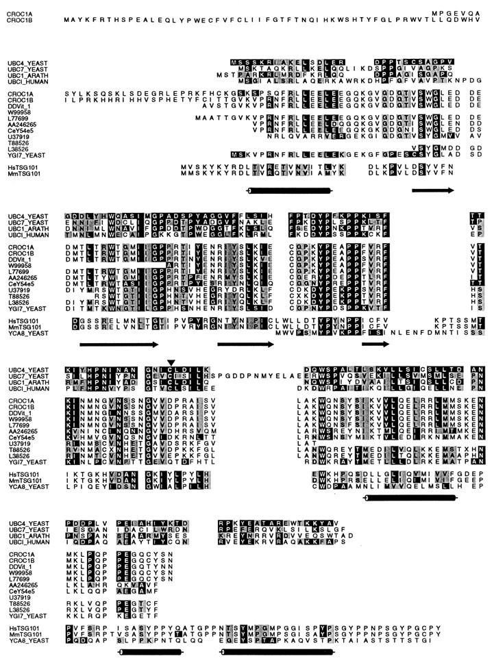
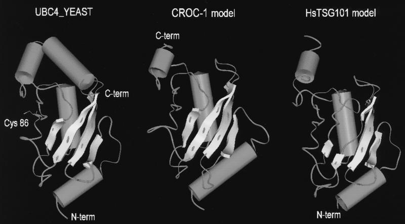
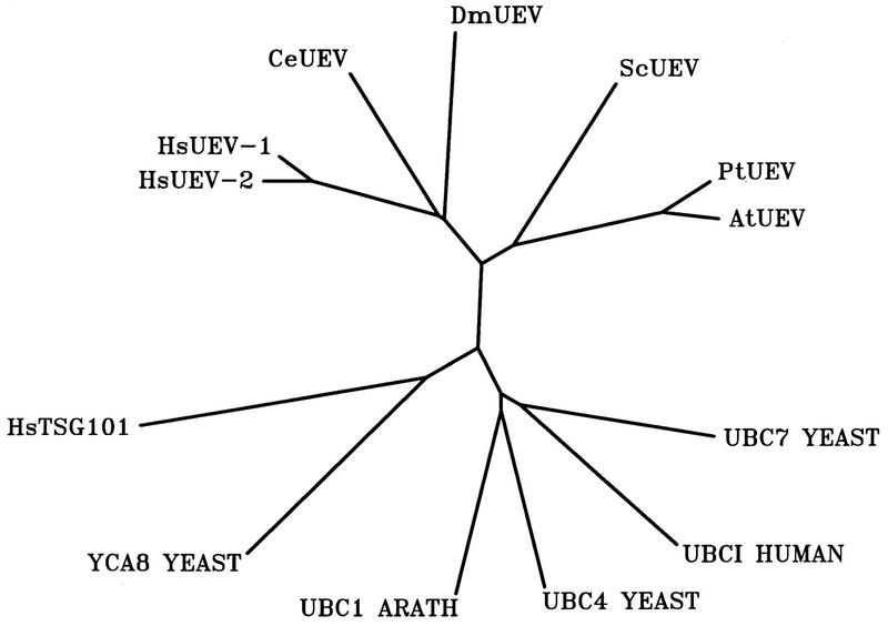
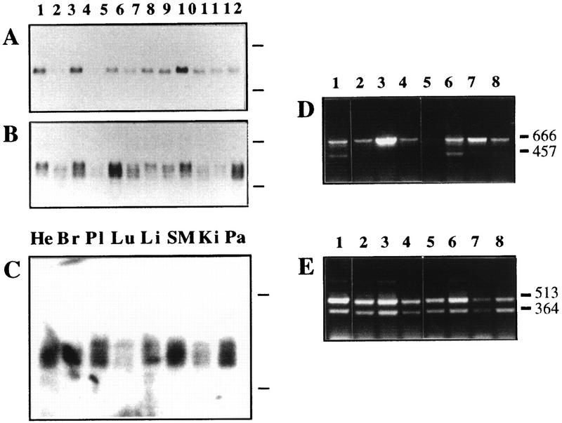
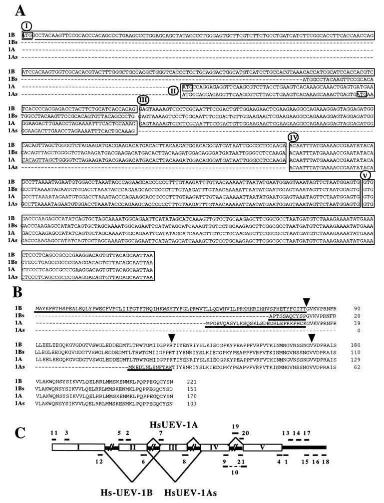
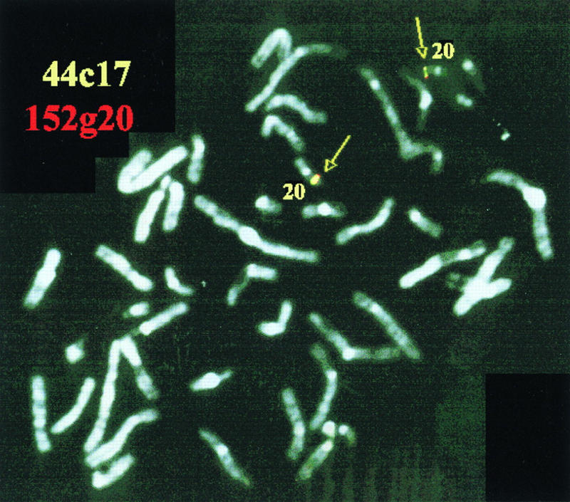
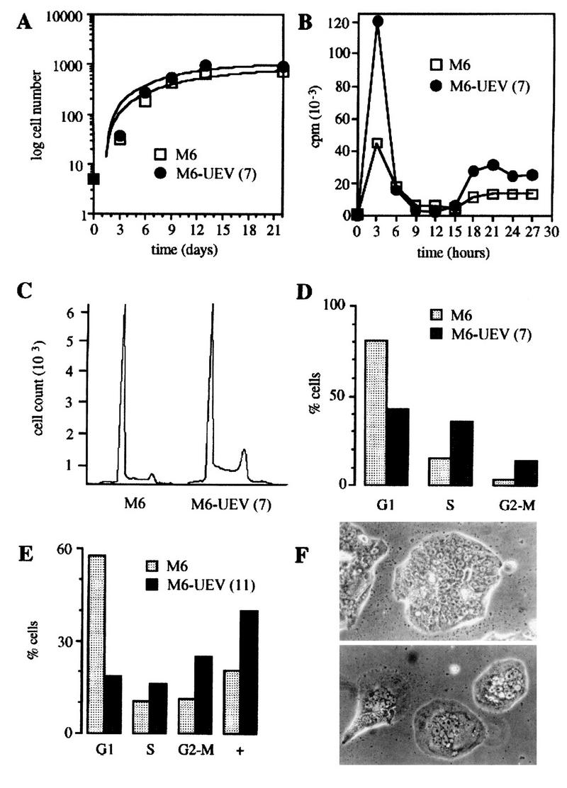
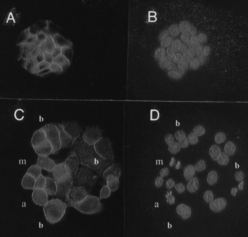
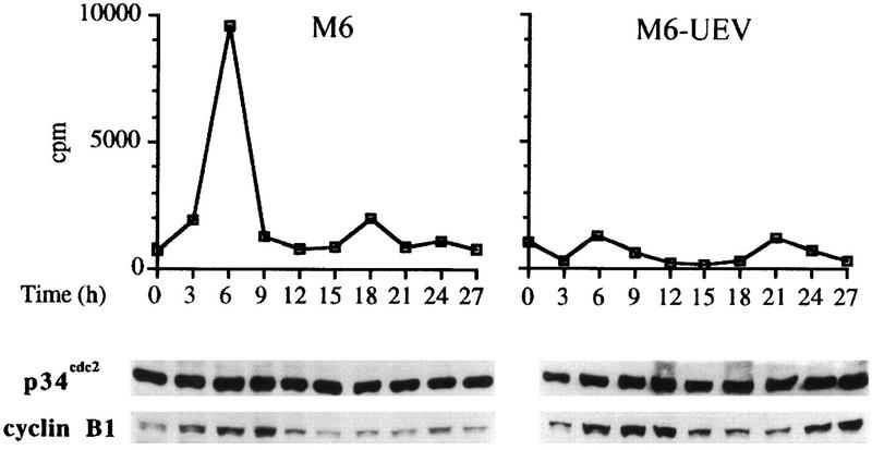
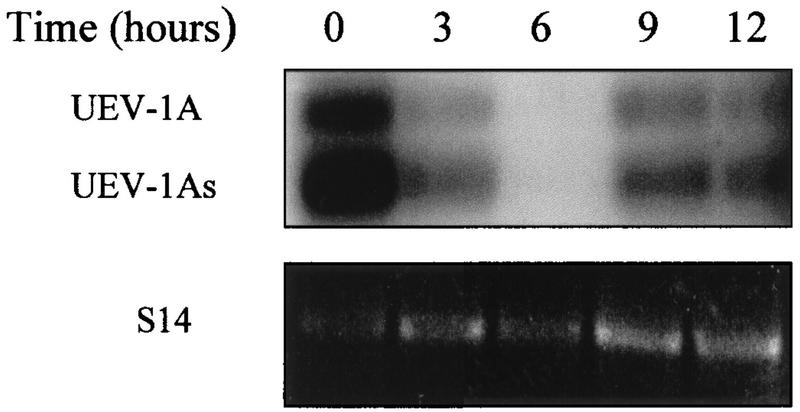
By means of differential RNA display, we have isolated a cDNA corresponding to transcripts that are down-regulated upon differentiation of the goblet cell-like HT-29-M6 human colon carcinoma cell line. These transcripts encode proteins originally identified as CROC-1 on the basis of their capacity to activate transcription of c-fos. We show that these proteins are similar in sequence, and in predicted secondary and tertiary structure, to the ubiquitin-conjugating enzymes, also known as E2. Despite the similarities, these proteins lack a critical cysteine residue essential for the catalytic activity of E2 enzymes and, in vitro, they do not conjugate or transfer ubiquitin to protein substrates. These proteins constitute a distinct subfamily within the E2 protein family and are highly conserved in phylogeny from yeasts to mammals. Therefore, we have designated them UEV (ubiquitin-conjugating E2 enzyme variant) proteins, defined as proteins similar in sequence and structure to the E2 ubiquitin-conjugating enzymes but lacking their enzymatic activity (HW/GDB-approved gene symbol, UBE2V). At least two human genes code for UEV proteins, and one of them, located on chromosome 20q13.2, is expressed as at least four isoforms, generated by alternative splicing. All human cell types analyzed expressed at least one of these isoforms. Constitutive expression of exogenous human UEV in HT-29-M6 cells inhibited their capacity to differentiate upon confluence and caused both the entry of a larger proportion of cells in the division cycle and an accumulation in G2-M. This was accompanied with a profound inhibition of the mitotic kinase, cdk1. These results suggest that UEV proteins are involved in the control of differentiation and could exert their effects by altering cell cycle distribution.


Figures

FIG. 1
FIG. 1
Probe A3.5 recognizes a transcript down-regulated during differentiation of HT-29-M6 cells. (A) HT-29-M6 cells were preconfluent or cultured to confluence for the indicated periods of time (days [d]); (B) 17-day postconfluent HT-29-M6 cells and 5-day postconfluent Caco-2 cells were treated with 200 nM TPA for the indicated times. Total RNA (12 μg/lane) was isolated and processed and filters hybridized with 32P-labeled band A3.5 as described in Materials and Methods. Marks to the right of the autoradiograms indicate migration of 28S and 18S rRNAs. C, untreated control cells.
FIG. 2
FIG. 2
Phage clones isolated by screening with probe A3.5 are identical at their 5′ ends to CROC-1A and CROC-1B. (A) Composite nucleotide and deduced amino acid sequences of CROC-1B (positions 1 to 2136), MAC4 (positions 504 to 3455), and CRA6 (positions 588 to 2388). (B) Diagram depicting the relationships between CROC-1A, CROC-1B, MAC4, and CRA6. Solid lines, 5′ untranslated sequences; dotted line, intervening sequence (intron); stippled and striped boxes, coding regions unique to either CROC-1A or CROC-1B; solid box, coding region common to all forms; striped thin box, common 3′ untranslated sequence; open thin box, sequences unique to MAC4 and CRA6 (not present in CROC-1 cDNAs) at the 3′ untranslated region.
FIG. 3
FIG. 3
Alignment of CROC-1A, CROC-1B, and related proteins with four E2 enzymes for which structure has been determined, S. cerevisiae UBC4 and UBC7, A. thaliana UBC1, and human UBCI, and the product of the human tumor suppressor gene TSG101 and its mouse and yeast homologs. The long carboxy termini of the products of human and mouse TSG101, unrelated to E2 enzymes, and CROC-1 proteins, are not represented in the alignment. Accession numbers and organisms with represented sequence: S. cerevisiae UBC4, P15732; S. cerevisiae UBC7, Q02159; A. thaliana UBC1, P25865; Homo sapiens UBCI, P50550; CROC-1B, U39361; CROC-1A, U39360; DDVit 1, X98091; W99958, Mus musculus EST; L77699, Gallus gallus cDNA; AA246265, D. melanogaster EST; CeY54E5, C. elegans genomic cosmid; U37919, Oriza sativa EST; T88528, A. thaliana EST; L38756, Pisolinum tintorium EST; YGI7_YEAST, P53152; HsTSG101, U82130; MmTSG101, U52945; YCA_8, P25604. The C. elegans gene and protein were deduced from unannotated genomic sequence both manually and with the aid of the gene prediction algorithm GeneWise (see Materials and Methods). Secondary structure predictions are shown below the sequences as cylinders (alpha helices) and arrows (beta pleated sheets). The E2 catalytic Cys is marked with an arrowhead.
FIG. 4
FIG. 4
Three-dimensional molecular models of CROC-1 (middle) and TSG101 (right), built on the basis of the experimental three-dimensional structure of S. cerevisiae UBC4 (12) (left). The critical Cys, essential for the catalytic activity of ubiquitin-conjugating enzymes, is highlighted in the latter. Beta pleated sheets are represented as flat arrows, and alpha helices are represented as barrels. The models were built on sequences that could be aligned with UBC4, and thus the amino terminus (N-term) of CROC-1 and the carboxy terminus (C-term) of TSG101 are not represented.
FIG. 5
FIG. 5
Unrooted tree of phylogenetic relationships between the Ubc domain-like regions of UEV/CROC-1 and TSG101-related proteins and the Ubc domains of selected ubiquitin-conjugating enzymes. Accession numbers for the corresponding sequences are as in Fig. 3.
FIG. 6
FIG. 6
HsUEV-1 does not promote the conjugation of ubiquitin to protein substrates in vitro. Purified E1 only (lane 1), E1 with fraction IIA (containing E3 enzymes) (lane 2), E1 plus fraction IIA plus E2-F1 (lane 3), and E1 plus fraction IIA and increasing concentrations of recombinant GST–UEV-1 (lanes 4 to 8) were used in in vitro ubiquitination assays for the conjugation of 125I-ubiquitin. High-molecular-weight conjugates are formed only in the presence of E2-F1 (lane 2). Bands in lane 2 are a result of endogenous E2 contaminating fraction IIA and are taken as background signal for this assay.
FIG. 7
FIG. 7
Expression range of HsUEV-1. Northern blotting for the expression of HsUEV-1 in human cultured cell lines (A and B) and tissues (C). Probe A3.5 (A) or MAC4 (B) were used to hybridize a filter with RNAs from the following cell lines: 1, SK-CO-15 (colon carcinoma); 2, Caco-2 (colon carcinoma); 3, SK-PC-1 (pancreas carcinoma); 4, SK-PC-3 (pancreas carcinoma); 5, HepG2 (liver carcinoma); 6, T24 (bladder carcinoma); 7, HeLa (cervix carcinoma); 8, EW-1 (Ewing’s sarcoma); 9, RD-ES (Ewing’s sarcoma); 10, SK-MEL-28 (melanoma); 11, HEL (erythroleukemia); and 12, K562 (erythroleukemia). (C) Probe MAC4 was used to hybridize a filter with poly(A)-enriched RNA from the following human tissues: He, heart; Br, breast; Pl, placenta; Lu, lung; Li, liver; SM, skeletal muscle; Ki, kidney; Pa, pancreas. Marks to the right of the autoradiograms represent the migration of 28S and 18S rRNAs. (D and E) RT-PCR analysis with primers specific for HsUEV-1B (D) and HsUEV-1A (E), using as templates cDNAs from the following cell lines: 1, SK-MEL-28; 2, EW-1; 3, HepG2; 4, HT-29-M6 (colon carcinoma, mucosecretory); 5, H-29 (colon carcinoma, undifferentiated); 6, K562; 7, HEL; and 8, HDF (diploid fibroblasts). The sizes of the major amplified bands are indicated in nucleotides on the right.
FIG. 8
FIG. 8
Alternative splice variants and genomic arrangement of exons of HsUEV-1. (A) Alignment of sequences of the major RT-PCR products as shown in panels C and D. Sequence blocks I through IV were assigned on the basis of presence or absence in the different forms. Block V is present in clone CRA6, a partially processed transcript (Fig. 2). Boxes show the initial codons for open reading frames in each form. (B) Deduced amino acid sequences for the four forms of HsUEV-1. The amino-terminal residues unique to each form are underlined. Arrowheads correspond to the junctions of the blocks in panel A. (C) Diagram for the arrangement of HsUEV-1 exons in genomic DNA and PAC clone 44c17. PCR analyses were performed with the primers indicated on the diagram and described in Table 1.
FIG. 9
FIG. 9
Chromosomal assignment of HsUEV-1 by FISH analysis. Human metaphase chromosomes were probed with two biotinylated independent PAC clones (yellow, 44c17; red, 152g20). Both probes yielded signals that colocalized on chromosome 20q13.2 (arrows and paired green spots).
FIG. 10
FIG. 10
Effect of the constitutive expression of UEV-1 on the contact-induced expression of MUC5AC in HT-29-M6 cells. Control (lane pair 1) and independent clones of UEV-1-transfected HT-29-M6 cells (lane pairs 2 to 5) were harvested 2 days after seeding (lanes a) or 10 days after reaching confluence (lanes b). Transfected clones expressed different levels of the exogenous UEV-1, as determined by RT-PCR, the lowest levels corresponding to clone 2. Total RNA was analyzed by Northern blotting with a probe for the apomucin gene MUC5AC.
FIG. 11
FIG. 11
Effects of the constitutive expression of UEV-1 on the growth and cell cycle patterns of HT-29-M6 cells. (A) Growth curves of control (empty squares) and UEV-1-transfected (filled circles) HT-29-M6 cells. Equal numbers of cells were seeded in triplicate plates and counted at 3-day intervals as attached and trypsinized cells. (B) Rates of DNA synthesis of synchronized control (empty squares) and UEV-1-transfected (filled circles) HT-29-M6 cells. Cells were seeded at equal numbers in triplicate wells, arrested at G1-S by double thymidine block, and allowed to enter S by removal of excess thymidine. [3H]thymidine was added 30 min before harvesting, which was done at 3-h intervals (see Materials and Methods). (C) Flow cytometry analysis for the DNA content of asynchronous cultures of control (left) and UEV-1-transfected (right) HT-29-M6 cells. Clone 7 of UEV-1-transfected cells was analyzed for DNA content 2 days after seeding. (D) Integrative multicycle analysis of single-fluorescence histograms corresponding to panel C. (E) Multicycle analysis of histograms from flow cytometry of clone 11 of UEV-1-transfected HT-29-M6 cells, analyzed 5 days after seeding. (F) Phase-contrast micrographs of control (top) and UEV-1-transfected clone 11 (bottom) HT-29-M6 cells, 7 days after seeding (magnification, ×400).
FIG. 12
FIG. 12
Effects of the constitutive expression of UEV-1 on the number and appearance of nuclei in HT-29-M6 cells. Control (A and B) and UEV-transfected (C and D) cells were decorated with anti-E-cadherin antibodies (A and C) to delimit the cell periphery. The same preparations were stained with Hoechst 33258 (B and D) for the visualization of nuclei. In UEV-1 transfected, but not in control, cells, binucleated cells (b), nuclei with apoptotic morphology (a), and frequent mitotic figures (m) are observed. Magnification, ×400.
FIG. 13
FIG. 13
Effect of the constitutive expression of UEV-1 on the activity of the mitotic kinase cdk1 in HT-29-M6 cells. Synchronized control (left) and UEV-1-transfected (right) HT-29-M6 cells were analyzed for p13suc1-associated (cdk1) activity at 3-h intervals after release from the block at G1-S (top panel). Extracts from parallel cultures were analyzed by Western blotting for the subunit components of cdk1, p34cdc2 and cyclin B1, as indicated.
FIG. 14
FIG. 14
Cell cycle-regulated expression of endogenous UEV-1A. HT-29-M6 cells were synchronized by double thymidine block, allowed to enter the S phase, and analyzed for expression of UEV-1 at the indicated times by RT-PCR followed by hybridization with a specific labeled oligonucleotide (top). For normalization, the same samples were subjected to parallel RT-PCRs with primers for the ribosomal protein S14 (bottom). Entry of the cells in S was monitored in parallel experiments by incorporation of [3H]thymidine, which produced a curve equivalent to that shown in Fig. 11B, with a peak at 3 h. The experiment shown is representative of three independent experiments.

Similar articles

Cited by

References

    1. Arion D, Meijer L, Brizuela L, Beach D. cdc2 is a component of the M phase-specific histone H1 kinase: evidence for identity with MPF. Cell. 1988;55:371–378. - PubMed
    1. Bailly V, Lamb J, Sung P, Prakash S, Prakash L. Specific complex formation between yeast RAD6 and RAD18 proteins: a potential mechanism for targeting RAD6 ubiquitin-conjugating activity to DNA damage sites. Genes Dev. 1994;8:811–820. - PubMed
    1. Banerjee A, Deshaies R J, Chau V. Characterization of a dominant negative mutant of the cell cycle ubiquitin-conjugating enzyme Cdc34. J Biol Chem. 1995;270:26209–26215. - PubMed
    1. Barila D, Murgia C, Nobili F, Gaetani S, Perozzi G. Subtractive hybridization cloning of novel genes differentially expressed during intestinal development. Eur J Biochem. 1994;223:701–709. - PubMed
    1. Bartz S R, Rogel M E, Emerman M. Human immunodeficiency virus type 1 cell cycle control: Vpr is cytostatic and mediates G2 accumulation by a mechanism which differs from DNA damage checkpoint control. J Virol. 1996;70:2324–2331. - PMC - PubMed

Publication types

MeSH terms