Case Reports
. 1998 Jan 5;187(1):71-7.
doi: 10.1084/jem.187.1.71.

Mutations in the human lambda5/14.1 gene result in B cell deficiency and agammaglobulinemia

Affiliations
Case Reports

Mutations in the human lambda5/14.1 gene result in B cell deficiency and agammaglobulinemia

Y Minegishi et al. J Exp Med. .

Abstract

B cell precursors transiently express a pre-B cell receptor complex consisting of a rearranged mu heavy chain, a surrogate light chain composed of lambda5/14.1 and VpreB, and the immunoglobulin (Ig)-associated signal transducing chains, Igalpha and Igbeta. Mutations in the mu heavy chain are associated with a complete failure of B cell development in both humans and mice, whereas mutations in murine lambda5 result in a leaky phenotype with detectable humoral responses. In evaluating patients with agammaglobulinemia and markedly reduced numbers of B cells, we identified a boy with mutations on both alleles of the gene for lambda5/14.1. The maternal allele carried a premature stop codon in the first exon of lambda5/14.1 and the paternal allele demonstrated three basepair substitutions in a 33-basepair sequence in exon 3. The three substitutions correspond to the sequence in the lambda5/14. 1 pseudogene 16.1 and result in an amino acid substitution at an invariant proline. When expressed in COS cells, the allele carrying the pseudogene sequence resulted in defective folding and secretion of mutant lambda5/14.1. These findings indicate that expression of the functional lambda5/14.1 is critical for B cell development in the human.


Figures

Figure 1
Figure 1
Comparison of the normal and mutant λ5/14.1 genes. (A) Partial DNA sequence of the first (left) and third (right) exon of λ5/14.1 in the patient and in a normal control. The basepair substitutions are indicated by an asterisk. (B) Sequence alignment of the third exon of the functional λ5/14.1 gene, the patient's λ5/14.1 gene, and the 16.1 pseudogene. Only the basepairs at which λ5/14.1 and the pseudogene 16.1 differ are indicated. The basepairs at which the patient's λ5/14.1 gene correspond to the 16.1 gene are shown in bold type.
Figure 2
Figure 2
Southern blot analysis of the lambda and lambda-like genes. Genomic DNA from the patient (lane 1) and four controls (lanes 2–5) was digested with EcoRI (A) or HindIII (B) and analyzed with a probe from the third exon of λ5/14.1. The molecular size standards are shown at the right side of the figure. In the EcoRI digest, the genes for λ5/14.1 and the pseudogene 16.1 are indicated at the left side of the figure. The bands at 8, 18, and 23 kb in the EcoRI digest represent previously reported polymorphic variants in the number of lambda constant region genes (49).
Figure 3
Figure 3
Analysis of λ5/14.1 cDNA from the patient's bone marrow. RT-PCR was used to amplify the 3′ portion of the λ5/14.1 transcripts from an age-matched control (lane 1), the patient (lane 2), or a negative control (lane 3). Molecular size standards (lane M), the undigested PCR products (A), and the MspI-digested products (B) were separated by electrophoresis. All of the 266-basepair product from the control, but none from the patient, was digested to yield 143- and 123-basepair fragments.
Figure 4
Figure 4
Flow cytometric analysis of bone marrow B cell precursors from a normal age-matched control (left) and the patient with mutations in λ5/14.1 (right). (A) Isometric contour plots of lymphoid-gated cells stained with antibodies to CD19 and surface immunoglobulin (kappa + lambda light chains). In the control, 46% of cells were CD19+, of which 29% were surface Ig (sIg)+. In the patient, 6% of cells were CD19+, with only 0.7% of which were sIg+. (B) Three-color immunofluorescence was used to stain cells for CD19, CD34, and sIg and CD19+ cells were analyzed. In the control sample, 11% of CD19+ cells were CD34+ and 60% were CD34, sIg. In the patient, 87% of CD19+ cells were CD34+. (C) Permeabilized cells were stained for TdT and surface or cytoplasmic IgM. In the control, 5% of cells were TdT+, IgM, 1% were TdT+, IgM+, and 40% were TdT, IgM+. In the patient, 5% of cells were TdT+, IgM, 0.2% were TdT+, IgM+, and 0.4% were TdT, IgM+.
Figure 5
Figure 5
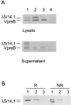
Transfection of COS7 cells with expression vectors for normal or mutant λ5/ 14.1 and normal VpreB. (A) Cells were transfected with VpreB and normal λ5/14.1 (lane 1), VpreB and mutant λ5/14.1 (lane 2), VpreB alone (lane 3), or an empty vector (lane 4). After metabolic labeling, both lysates and supernatants were immunoprecipitated with antibody to VpreB. (B) COS7 cells were transfected with normal λ5/14.1 (lane 1), mutant λ5/14.1 (lane 2), or an empty vector (lane 3), immunoprecipitated using a polyclonal antilambda antibody, and electrophoresed under reducing (R) or nonreducing (NR) conditions.

Similar articles

Cited by

References

    1. Spanopoulou E, Roman CA, Corcoran LM, Schlissel MS, Silver DP, Nemazee D, Nussenzweig MC, Shinton SA, Hardy RR, Baltimore D. Functional immunoglobulin transgenes guide ordered B-cell differentiation in Rag-1–deficient mice. Genes Dev. 1994;8:1030–1042. - PubMed
    1. Young F, Ardman B, Shinkai Y, Lansford R, Blackwell TK, Mendelsohn M, Rolink A, Melchers F, Alt FW. Influence of immunoglobulin heavy- and light-chain expression on B-cell differentiation. Genes Dev. 1994;8:1043–1057. - PubMed
    1. Rajewsky K. Clonal selection and learning in the antibody system. Nature. 1996;381:751–758. - PubMed
    1. Burrows PD, Cooper MD. B cell development and differentiation. Curr Opin Immunol. 1997;9:239–244. - PubMed
    1. Yel L, Minegishi Y, Coustan-Smith E, Buckley RH, Trubel H, Pachman LM, Kitchingman GR, Campana D, Rohrer J, Conley MD. Mutations in the mu heavy chain gene in patients with agammaglobulinemia. N Engl J Med. 1996;335:1486–1493. - PubMed

Publication types