. 1998 Jan 6;95(1):156-61.
doi: 10.1073/pnas.95.1.156.

Shortage of mitogen-activated protein kinase is responsible for resistance to AP-1 transactivation and transformation in mouse JB6 cells

Affiliations

Shortage of mitogen-activated protein kinase is responsible for resistance to AP-1 transactivation and transformation in mouse JB6 cells

C Huang et al. Proc Natl Acad Sci U S A. .

Abstract

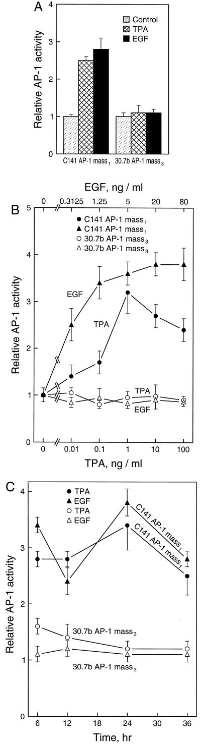
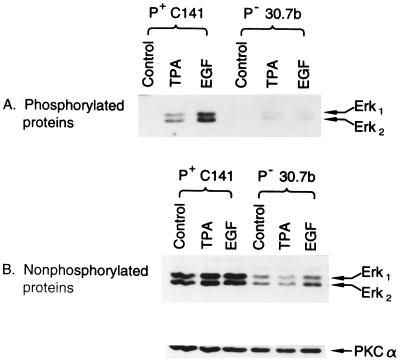
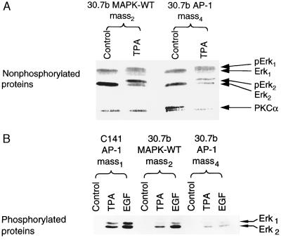
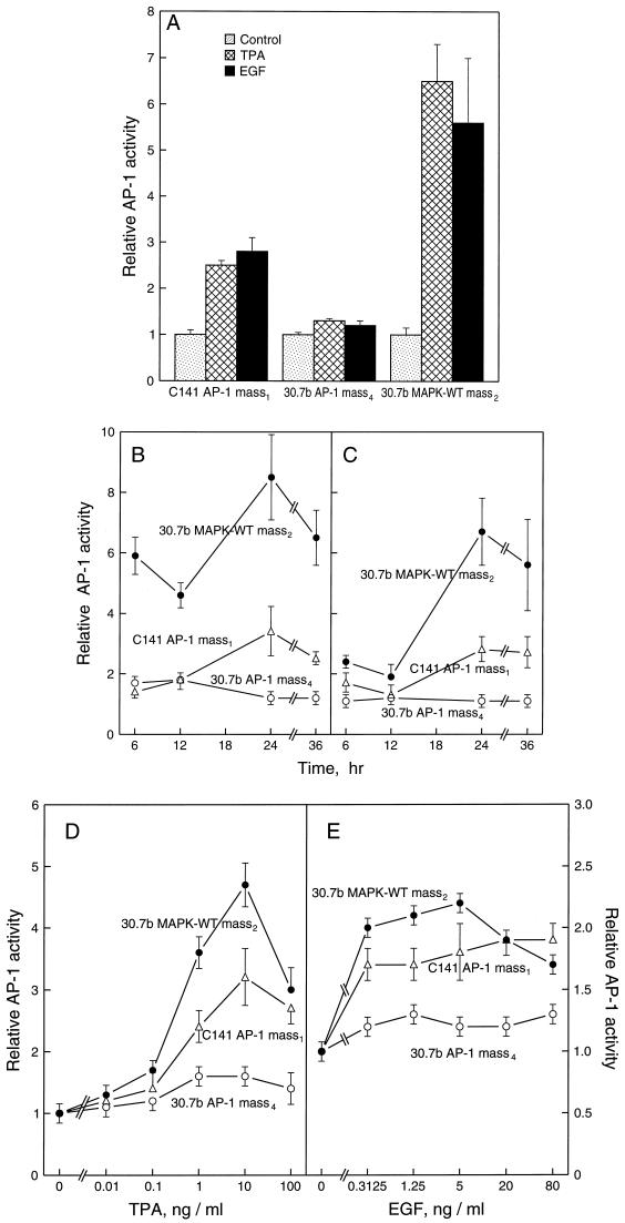
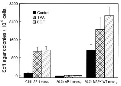
The JB6 mouse epidermal cell system, which includes tumor promotion-sensitive (P+) and tumor promotion-resistant (P-) cells, is a well-established and extensively used cell culture model for studying the mechanism of late-stage tumor promotion. Tumor promoters, such as 12-O-tetradecanoylphorbol 13-acetate (TPA) or epidermal growth factor (EGF), induce high levels of activator protein 1 (AP-1) activity and large, tumorigenic, anchorage-independent colonies in soft agar at a high frequency in JB6 P+ cells, but not in JB6 P- cells. We report here a molecular explanation for the defect in the AP-1 activation and promotion-resistant phenotype of P- cells. We demonstrate that the lack of AP-1 activation and cell transformation responses to TPA and EGF in P- cells appears attributable to the low level of mitogen-activated protein kinase (MAPK) (extracellular signal-regulated protein kinase, Erk) in these cells. TPA and EGF induce transactivation of AP-1 activity in P+ cells but not in P- cells. Nonphosphorylated forms and TPA- or EGF-induced phosphorylated forms of Erks (Erk1 and Erk2) in P- cells were much lower than those in P+ cells. Stable transfection of wild-type MAPK (Erk2) into P- cells restored its response to TPA and EGF for both AP-1 activation and cell transformation. These results suggest that the shortage of MAPK (Erk1 and Erk2) appears to be an important contributor to the tumor promotion-resistant phenotype in JB6 cells.


Figures

Figure 1
Figure 1
TPA- and EGF-induced AP-1 activation in JB6 P+ cells but not P cells. A total of 8 × 10³ of JB6 P+, C1 41 AP-1 mass1 or P, Cl 30.7b AP-1 mass3 suspended in 5% FBS MEM was seeded into each well of 96-well plates. After overnight culture at 37°C, the cells were starved for 24 hr by changing medium with 0.1% FBS MEM. Then, (A) the cells were treated with TPA (10 ng/ml) or EGF (10 ng/ml) for 24 hr before assaying for luciferase activity; (B) for dose response study, the cells were treated with the indicated dose of TPA or EGF and incubated for 24 hr, then luciferase activity was determined; and (C) for the time course study, the cells were exposed to TPA (10 ng/ml) or EGF (10 ng/ml). The luciferase activity was measured at time points as indicated. The results were expressed as relative AP-1 activity. The relative AP-1 activity was presented as the luciferase activity relative to the medium control.
Figure 2
Figure 2
Low level of MAPK (Erk1 or Erk2) expression and activation in JB6 P Cl 30.7b cells. A total of 8 × 10⁴ of JB6 P+ C1 41 or P Cl 30.7b cells was seeded into each well of 6-well plates. After culture at 37°C for 24 hr, the cells were starved for 48 hr by replacing medium with 0.1% FBS MEM. The medium was changed with fresh 0.1% FBS MEM and cultured for 4 hr. The cells were exposed to TPA (10 ng/ml) or EGF (10 ng/ml) for 30 min, then extracted and analyzed by Western blot analysis by using the PhosphoPlus MAPK antibody kit by New England Biolabs and visualized by chemiluminescence (ECL, Amersham). (A) Phosphorylated Erk proteins detected with phospho-specific MAPK antibody. (B) Erk1 and Erk2 proteins detected with MAPK antibody.
Figure 3
Figure 3
Overexpression of Erk2 in JB6 P Cl 30.7b cells. A total of 8 × 10⁴ of Cl 30.7b AP-1 mass4, Cl 30.7b MAPK-WT mass2 or Cl 41 AP-1 mass1 was treated, extracted, and analyzed as described in Fig. 2. (A) Erk1 and Erk2 proteins detected with MAPK antibody. To visualize the conversion of unphosphorylated Erk to phosphorylated Erk in these transfectants, extracts were separated on an 8% polyacrylamide gel, transferred, and blotted as above. Under these conditions the gel mobility shift due to phosphorylation of Erks could be visualized. (B) Phosphorylated Erk proteins detected with phospho-specific MAPK antibody.
Figure 4
Figure 4
Restoration of AP-1 response to TPA or EGF stimulation in wild-type Erk2-transfected JB6 P cells. A total of 8 × 10³ of Cl 30.7b AP-1 mass4, Cl 30.7b MAPK-WT mass2, or Cl 41 AP-1 mass1 was seeded into each well of 96-well plates. After overnight culture at 37°C, the cells were starved for 24 hr by replacing medium with 0.1% FBS MEM. Then, (A) the cells were treated with TPA (10 ng/ml) or EGF (10 ng/ml) for 24 h, the luciferase activity was measured as described (9, 24). (B and C) For time course study, the cells were exposed to TPA (10 ng/ml) (B) or EGF (10 ng/ml) (C) for the times as indicated. (D and E) For the dose response study, the cells were treated with the indicated doses of TPA or EGF and incubated for 24 hr before assaying luciferase activity. The luciferase activity was measured and the results were presented as relative AP-1 activity.
Figure 5
Figure 5
Overexpression of wild-type Erk2 converts the P cell to P+ phenotype. A total of 1 × 10⁴ of C1 41 AP-1 mass1, Cl 30.7b AP-1 mass3 or Cl 30.7b MAPK-WT mass2 was or was not exposed to TPA (10 ng/ml) or EGF (10 ng/ml) in 1 ml of 0.33% BME agar containing 10% FBS laid over 3.5 ml of 0.5% BME agar containing 10% FBS in each well of a 60-mm-diameter dish. The cultures are maintained in a 37°C, 5% CO2 incubator for 14–16 days, and the cell colonies were scored as described (11). The results were presented as soft agar colonies per 104 cells.

Similar articles

Cited by

References

    1. Boutwell R K. Prog Exp Tumor Res. 1964;4:207–250. - PubMed
    1. Drinkwater N R. In: Genes and Signal Transduction in Multistage Carcinogenesis. Colburn N, editor. New York: Dekker; 1989. pp. 3–17.
    1. Dong Z, Jeffrey A M. Cancer Invest. 1990;8:523–533. - PubMed
    1. Weinstein I B. Cancer Res. 1988;48:4135–4143. - PubMed
    1. Colburn N H, Former B F, Nelson K A, Yuspa S H. Nature (London) 1979;281:589–591. - PubMed

Publication types

MeSH terms

Substances