. 1998 Jan 6;95(1):258-63.
doi: 10.1073/pnas.95.1.258.

A chemokine expressed in lymphoid high endothelial venules promotes the adhesion and chemotaxis of naive T lymphocytes

Affiliations

A chemokine expressed in lymphoid high endothelial venules promotes the adhesion and chemotaxis of naive T lymphocytes

M D Gunn et al. Proc Natl Acad Sci U S A. .

Abstract

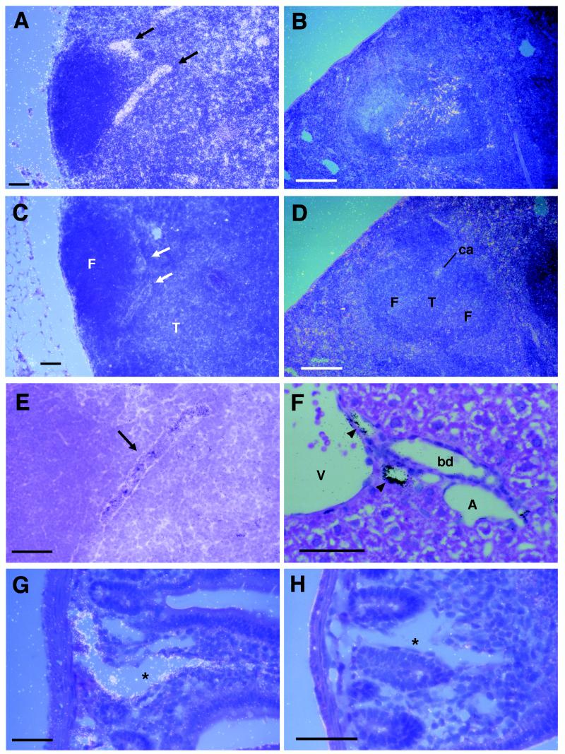
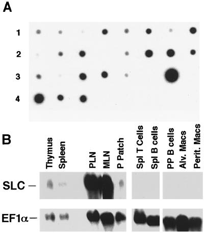
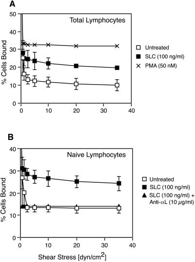
Preferential homing of naive lymphocytes to secondary lymphoid organs is thought to involve the action of chemokines, yet no chemokine has been shown to have either the expression pattern or the activities required to mediate this process. Here we show that a chemokine represented in the EST database, secondary lymphoid-tissue chemokine (SLC), is expressed in the high endothelial venules of lymph nodes and Peyer's patches, in the T cell areas of spleen, lymph nodes, and Peyer's patches, and in the lymphatic endothelium of multiple organs. SLC is a highly efficacious chemoattractant for lymphocytes with preferential activity toward naive T cells. Moreover, SLC induces firm adhesion of naive T lymphocytes via beta2 integrin binding to the counter receptor, intercellular adhesion molecule-1, a necessary step for lymphocyte recruitment. SLC is the first chemokine demonstrated to have the characteristics required to mediate homing of lymphocytes to secondary lymphoid organs. In addition, the expression of SLC in lymphatic endothelium suggests that the migration of lymphocytes from tissues into efferent lymphatics may be an active process mediated by this molecule.


Figures

Figure 1
Figure 1
Detection of SLC mRNA in mouse tissues by in situ hybridization. Sections were hybridized with antisense (A, B, E–G) or sense (C, D, H) 35S-labeled SLC riboprobe. (A) Dark field micrograph of SLC antisense probe hybridization in lymph node. Signal is seen as white dots. HEV are indicated by arrows. (B) SLC antisense probe hybridization in spleen. SLC sense probe does not hybridize to lymph node (C) or spleen (D). (E) High power view of HEV from A shown in bright field demonstrating typical morphology. Signal is seen as black dots. (F) Hybridization of SLC to the endothelium of small lymphatics (indicated by arrowheads) in liver. (G) Hybridization of SLC to the endothelial cells lining a central lactile (indicated by asterisks) in small intestine. (H) Lack of hybridization to SLC sense probe in small intestine. Identified structures are: F, follicle; T, T cell area; ca, central arteriole; A, artery; V, vein; bd, bile duct. [Bars = 100 μm (A–D) and 50 μm (E–H).]
Figure 2
Figure 2
Detection of SLC mRNA in tissues and purified cells by Northern analysis. (A) Autoradiograph of hSLC 32P-probe hybridization to a human multitissue dot blot. From left to right, tissues shown are: Row 1, heart, aorta, skeletal muscle, colon, bladder, uterus, prostate, stomach. Row 2, testis, ovary, pancreas, pituitary, adrenal, thyroid, salivary gland, breast. Row 3, kidney, liver, small intestine, spleen, thymus, peripheral leukocyte, lymph node, bone marrow. Row 4, appendix, lung, trachea, placenta. (B) Autoradiograph of mSLC 32P-probe hybridization to mouse northern blot. EF-1α hybridization is shown to indicate amounts of mRNA loaded in each lane.
Figure 3
Figure 3
Chemotactic activity of SLC on mouse lymphocyte subtypes. Murine lymphocytes were subjected to chemotaxis through 5-μm pore Transwell filters. The number of input and migrating cells of each subtype were determined by immunostaining and flow cytometry. Results are expressed as the percentage of input cells of each subtype that migrated to the lower chamber. (A) Migration of B cells, CD4+ T cells, and CD8+ T cells to SLC. Peak migration of same cells to SDF-1α is shown for comparison. (B) Comparison of naive vs. memory cell migration in response to SLC. Naive CD4+ cells are defined as those expressing L-selectin (LSEL+) and having low levels of CD44 (CD44 lo). Memory cells are the converse (LSEL and CD44 hi). (C) Inhibition of SLC-induced migration by pretreatment of cells for 2 hr with pertussis toxin (PTX). (D) Migration of mouse peripheral blood lymphocytes. Data points represent the mean ± SD for experiments performed in triplicate.
Figure 4
Figure 4
SLC induces shear-resistant binding of total and naive T lymphocytes to immobilized ICAM-1. T Lymphocytes treated as indicated were allowed to attach to ICAM-1 for 6 min under static conditions in a parallel plate flow chamber. Flow was initiated and increased in 2-fold increments every 10 sec. Results are expressed as the percentage of input cells remaining bound at each flow rate. (A) Adhesion of SLC-treated, untreated and phorbol 12-myristate 13-acetate-stimulated T lymphocytes. (B) Effect of SLC on naive (CD45RA+) T lymphocytes compared with untreated cells and cells treated with SLC and an anti-αL antibody. Data points represent the mean ± SD for at least three separate experiments. Analysis by using a paired two-tailed Student’s t test showed that SLC induced statistically significant binding of both total T lymphocytes (P < 0.01) and naive T lymphocytes (P < 0.05) to ICAM-1.

References

    1. Girard J-P, Springer T A. Immunol Today. 1995;16:449–457. - PubMed
    1. van Ewijk W, Nieuwenhuis P. Experientia. 1985;41:199–208. - PubMed
    1. Springer T A. Annu Rev Physiol. 1995;57:827–872. - PubMed
    1. Butcher E C, Picker L J. Science. 1996;272:60–66. - PubMed
    1. Butcher E C. Cell. 1991;67:1033–1036. - PubMed

Publication types

MeSH terms