. 1998 Jan 6;95(1):340-5.
doi: 10.1073/pnas.95.1.340.

Melanopsin: An opsin in melanophores, brain, and eye

Affiliations

Melanopsin: An opsin in melanophores, brain, and eye

I Provencio et al. Proc Natl Acad Sci U S A. .

Abstract

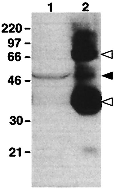
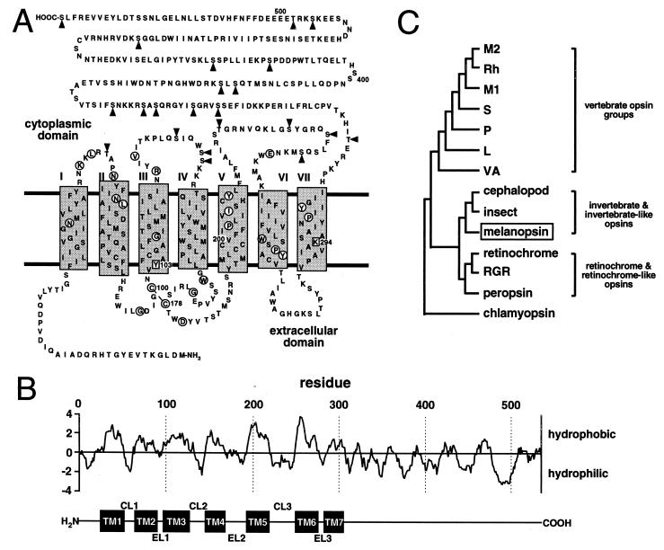
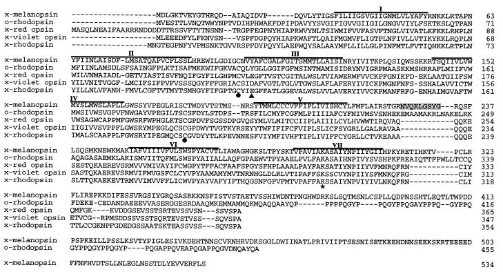
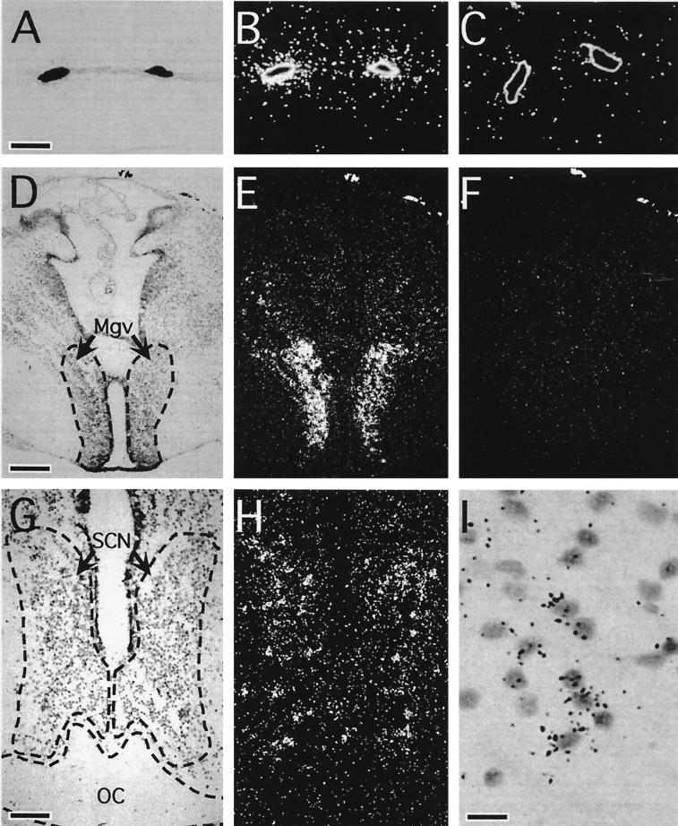
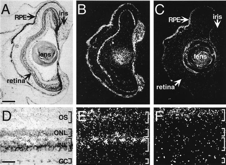
We have identified an opsin, melanopsin, in photosensitive dermal melanophores of Xenopus laevis. Its deduced amino acid sequence shares greatest homology with cephalopod opsins. The predicted secondary structure of melanopsin indicates the presence of a long cytoplasmic tail with multiple putative phosphorylation sites, suggesting that this opsin's function may be finely regulated. Melanopsin mRNA is expressed in hypothalamic sites thought to contain deep brain photoreceptors and in the iris, a structure known to be directly photosensitive in amphibians. Melanopsin message is also localized in retinal cells residing in the outermost lamina of the inner nuclear layer where horizontal cells are typically found. Its expression in retinal and nonretinal tissues suggests a role in vision and nonvisual photoreceptive tasks, such as photic control of skin pigmentation, pupillary aperture, and circadian and photoperiodic physiology.


Figures

Figure 1
Figure 1
Western blot of protein extracts from dermal melanophores and whole eye probed with an antiserum raised against bovine rhodopsin. Indicated molecular masses are in kDa. Lanes: 1, total protein from cultured melanophores; 2, 1% of total protein from a whole early postmetamorphic adult eye. A 50-kDa immunoreactive band is present in both lanes (solid arrowhead). The 35- and 70-kDa bands in lane 2 are monomeric and dimeric rhodopsin, respectively (open arrowheads).
Figure 2
Figure 2
Melanopsin structure and phylogeny. (A) Deduced amino acid sequence and predicted secondary structure of melanopsin. Transmembrane domains are designated by shaded rectangles and circled residues are identical to at least 65 of 67 aligned vertebrate and invertebrate opsins (ftp://swift.embl-heidelberg.de/tm7/align/opsins.ALIGN). Lys-294 is a putative site for chromophore attachment via a Schiff’s base linkage, and Tyr-103 is the aromatic residue conserved in all invertebrates and possibly involved in stabilizing the chromophore. Arrowheads indicate potential intracellular phosphorylation sites. (B) Hydropathy analysis [Kyte–Doolittle algorithm (15); window, 10 amino acids] predicts a secondary structure consistent with the tmpred algorithm. The corresponding transmembrane domains (TM1 to TM7), cytoplasmic loops (CL1 to CL3), and extracellular loops (EL1 to EL3) are indicated below the plot. (C) A phylogenetic tree was constructed by aligning melanopsin against representatives of the vertebrate opsin groups (L, long-wavelength-sensitive opsin; M1, blue-like middle-wavelength-sensitive opsin; M2, green-like middle-wavelength-sensitive opsin; P, pineal opsin; Rh, rhodopsin; S, short-wavelength-sensitive—all from chicken; VA, vertebrate ancient opsin—from Atlantic salmon), representatives of the retinochrome-like opsins (retinochrome from squid and RGR and peropsin from human), and representatives from cephalopods (octopus rhodopsin), insects (Drosophila Rh1 opsin), and unicellular eukaryotes (Chlamydomonas chlamyopsin). All sequences are available through GenBank.
Figure 3
Figure 3
Alignment of melanopsin amino acid sequence with those of octopus rhodopsin, Xenopus red opsin, Xenopus violet opsin, and Xenopus rhodopsin. Predicted transmembrane domains are overlined. Conserved sites of chromophore linkage (★) and disulfide bridge formation (•) are indicated. The residues corresponding to the acidic counterion in the vertebrate visual pigments are also indicated (▴). The peptide insertion within melanopsin’s third cytoplasmic loop has been shaded.
Figure 4
Figure 4
Bright-field and dark-field photomicrographs of melanopsin transcript distribution within extraocular structures. Melanopsin message was localized to tadpole (stages 56 and 57) dermal melanophores (A and B), the ventral aspect of the magnocellular preoptic nucleus (Mgv) (D and E), and the SCN (G and H) of the tadpole brain (stages 56 and 57). Cell-specific antisense hybridization was restricted to a subset of SCN cells (I). In contrast, incubation with the sense control probe yielded no hybridization above background (C and F). (Corresponding bright-field views are not shown.) OC, optic chiasm. [Bars = 20 μm (A), 150 μm (D), 75 μm (G), and 15 μm (I).]
Figure 5
Figure 5
Bright-field and dark-field photomicrographs of melanopsin transcript distribution within ocular structures. In sections of tadpole (stages 56 and 57) (A and B) and adult (D and E) eye, melanopsin transcripts were identified in retinal cells within the outer lamina of the inner nuclear layer (INL) (A, B, D, and E). In contrast, incubation of adjacent sections with the sense RNA probe (C and F) resulted in no labeling above background. (Corresponding bright-field views are not shown.) Intense cell-specific hybridization was also observed in the iris and less intensely within the RPE. GC, ganglion cell layer; ONL, outer nuclear layer; OS, photoreceptor outer segments. [Bars = 150 μm (A) and 35 μm (D).]

References

    1. Bagnara J T, Obika M. Experientia. 1967;23:155–157. - PubMed
    1. Daniolos A, Lerner A B, Lerner M R. Pigment Cell Res. 1990;3:38–43. - PubMed
    1. McClintock T S, Rising J P, Lerner M R. J Cell Physiol. 1996;167:1–7. - PubMed
    1. Lerner M R. Trends Neurosci. 1994;17:142–146. - PubMed
    1. Quillan J M, Jayawickreme C K, Lerner M R. Proc Natl Acad Sci USA. 1995;92:2894–2898. - PMC - PubMed

Publication types

Associated data

LinkOut - more resources