. 1998 Jan 6;95(1):352-7.
doi: 10.1073/pnas.95.1.352.

Controlling signaling with a specifically designed Gi-coupled receptor

Affiliations

Controlling signaling with a specifically designed Gi-coupled receptor

P Coward et al. Proc Natl Acad Sci U S A. .

Abstract

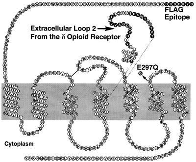
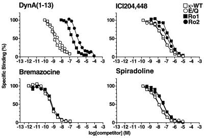
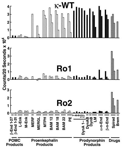
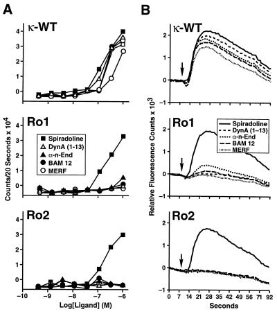
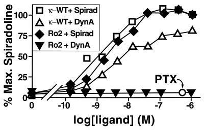
We are developing a system to control G protein signaling in vivo to regulate a broad range of physiologic responses. Our system utilizes G protein-coupled peptide receptors engineered to respond exclusively to synthetic small molecule ligands and not to their natural ligand(s). These engineered receptors are designated RASSLs (receptor activated solely by a synthetic ligand). We have made two prototype RASSLs that are based on the human kappa opioid receptor. Small molecule drugs that activate the kappa receptor are nonaddictive and safe to administer in vivo. Binding and signaling assays reveal 200-2000-fold reductions in the ability of our RASSLs to bind or be activated by dynorphin, an endogenous peptide ligand of the kappa opioid receptor. In a high-throughput signaling assay, these prototype RASSLs expressed in Chinese hamster ovary K1 cells showed little or no response to a panel of 21 opioid peptides but still signaled normally in response to small molecule drugs such as spiradoline. Activation of a RASSL by spiradoline also caused proliferation of rat-1a tissue culture cells. These data provide evidence that G protein-coupled receptors can be made into RASSLs. The potential in vivo applications for RASSLs include the positive enrichment of transfected cells and the development of new animal models of disease.


Figures

Figure 1
Figure 1
Modifying the human κ receptor to make the Ro1, E/Q, and Ro2 receptors. The wild-type sequence is shown as ○; residues that have been changed are shown as •. The EL2 loop exchange is included in Ro1 and Ro2 constructs. The E297Q mutation is included in the E/Q and Ro2 constructs. The FLAG epitope is included in all the constructs in this study.
Figure 2
Figure 2
Competition binding assay. Membranes prepared from transfected COS-7 cells were incubated in the presence of 1.5 nM [3H]EKC and various concentrations of dynorphin A(1–13), bremazocine, spiradoline, or ICI204,448. The specific transfected receptor cDNAs are indicated in the figure. Ki values are listed in Table 1. The amount of radioligand bound in the absence of competing ligand was assigned a value of 100%.
Figure 3
Figure 3
Agonist activities of 21 natural opioid peptides on κ-WT, Ro1, and Ro2. Agonist-mediated changes in intracellular calcium were measured by the FLIPR as described in Materials and Methods. For dose responses (indicated by three bars/ligand), agonist concentrations are 1.0, 0.1, and 0.01 μM. For single doses (indicated by a single bar/ligand), the agonist concentration is 1.0 μM. Spirad, spiradoline; Brem, bremazocine. See Table 2 for peptide ligand amino acid sequences. Although abbreviations are used here, the order of the peptide ligands is the same as in Table 2. Data in Figs. 3 and Fig. 4A are expressed as mean of duplicate determinations in a single experiment; two additional experiments gave similar results.
Figure 4
Figure 4
Agonist activities of selected ligands on κ-WT, Ro1, and Ro2. Agonist-mediated changes in intracellular calcium were measured by the FLIPR as described in Materials and Methods. (A) Dose responses on κ-WT, Ro1, and Ro2. (B) Sample tracings of the actual calcium fluorescence curves from the 1 μM dose. The arrow indicates the addition of agonist.
Figure 5
Figure 5
[3H]Thymidine incorporation assay. Amount of [3H]thymidine incorporation in rat-1a cell clones expressing κ-WT or Ro2 in the presence of dynorphin A(1–13) (DynA) or spiradoline (Spirad) as indicated. Ro2 plus spiradoline, plus pertussis toxin (PTX) 200 ng/ml is indicated by ○. The value for 100% is the value for the highest dose of spiradoline for each receptor. The basal point is untreated cells. Data in are expressed as mean ± SD of duplicate determinations in a single experiment; three additional experiments gave similar results.

References

    1. Strader C D, Fong T M, Tota M R, Underwood D. Annu Rev Biochem. 1994;63:101–132. - PubMed
    1. Spiegel A M, Shenker A, Weinstein L S. Endocr Rev. 1992;13:536–565. - PubMed
    1. Baldwin J M. Curr Opin Cell Biol. 1994;6:180–190. - PubMed
    1. Conklin B R, Bourne H R. Cell. 1993;73:631–641. - PubMed
    1. Bourne H R. Curr Opin Cell Biol. 1997;9:134–142. - PubMed

Publication types

LinkOut - more resources