. 1998 Jan;72(1):32-41.
doi: 10.1128/JVI.72.1.32-41.1998.

Quantitative disassembly and reassembly of human papillomavirus type 11 viruslike particles in vitro

Affiliations

Quantitative disassembly and reassembly of human papillomavirus type 11 viruslike particles in vitro

M P McCarthy et al. J Virol. 1998 Jan.

Abstract

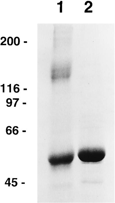
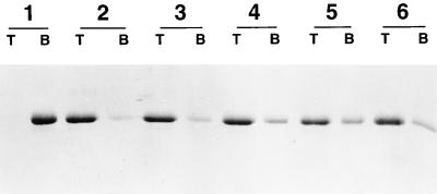
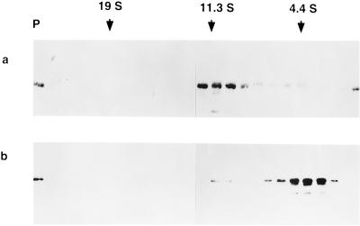
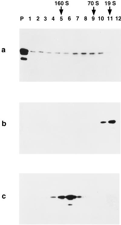
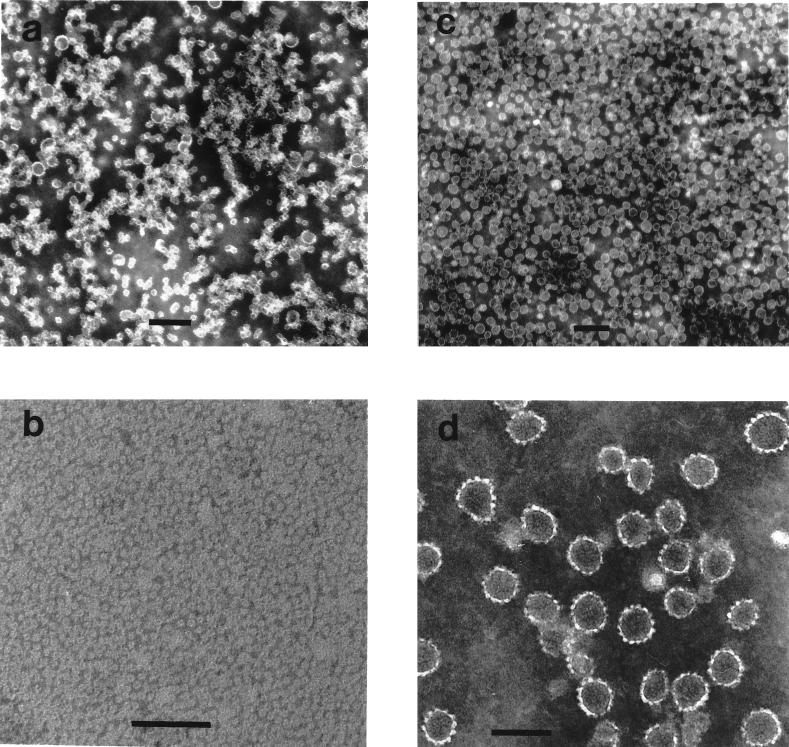
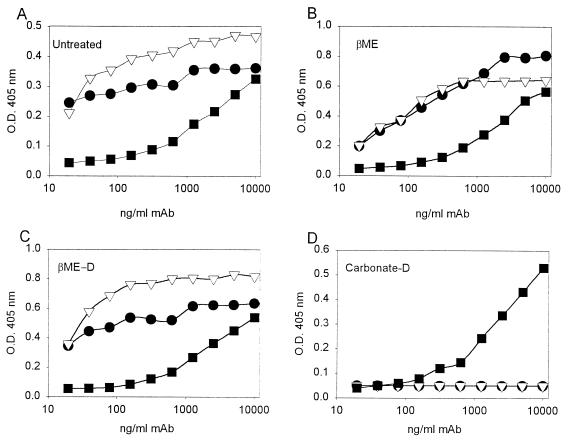
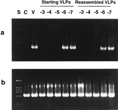
The human papillomavirus (HPV) capsid is primarily composed of a structural protein denoted L1, which forms both pentameric capsomeres and capsids composed of 72 capsomeres. The L1 protein alone is capable of self-assembly in vivo into capsidlike structures referred to as viruslike particles (VLPs). We have determined conditions for the quantitative disassembly of purified HPV-11 L1 VLPs to the level of capsomeres, demonstrating that disulfide bonds alone are essential to maintaining long-term HPV-11 L1 VLP structure at physiological ionic strength. The ionic strength of the disassembly reaction was also important, as increased NaCl concentrations inhibited disassembly. Conversely, chelation of cations had no effect on disassembly. Quantitative reassembly to a homogeneous population of 55-nm, 150S VLPs was reliably achieved by the re-formation of disulfide linkages following removal of reducing agent at near-neutral pH and moderate NaCl concentration. HPV-11 L1 VLPs could also be dissociated by treatment with carbonate buffer at pH 9.6, but VLPs could not be regenerated following carbonate treatment. When probed with conformationally sensitive and/or neutralizing monoclonal antibodies, both capsomeres generated by disulfide reduction of purified VLPs and reassembled VLPs formed from capsomeres upon removal of reducing agents exhibited epitopes found on the surface of authentic HPV-11 virions. Antisera raised against either purified VLP starting material or reassembled VLPs similarly neutralized infectious HPV-11 virions. The ability to disassemble and reassemble VLPs in vitro and in bulk allows basic features of capsid assembly to be studied and also opens the possibility of packaging selected exogenous compounds within the reassembled VLPs.


Figures

FIG. 1
FIG. 1
SDS-PAGE analysis of purified HPV-11 L1 protein. The protein was mixed with sample preparation buffer in the absence (lane 1) or presence (lane 2) of 2 mM DTT and boiled for 2 min prior to gel electrophoresis. Shown on the left are the positions at which molecular weight standards (in kilodaltons) migrated.
FIG. 2
FIG. 2
Thirty percent sucrose cushion analysis of HPV-11 VLP disassembly. HPV-11 preparations were treated at 4°C as described in the text, and samples were taken at the top (T) or bottom (B) of the sucrose cushion prior to gel electrophoresis. Group 1, untreated, purified HPV-11 VLP starting material in PBS; group 2, VLPs incubated with 5% βME for 16 h; group 3, VLPs incubated with 5% βME for 1 h; group 4, VLPs incubated with 2% βME for 16 h; group 5, VLPs incubated with 0.5% βME for 16 h; group 6, VLPs incubated with 10 mM DTT–5 mM EDTA for 16 h.
FIG. 3
FIG. 3
Five to 20% linear sucrose gradient analysis of disassembled HPV-11 VLPs. VLPs in PBS were incubated with 5% βME (a) or 200 mM NaHCO3 (pH 9.6) (b) for 16 h at 4°C and then centrifuged on a 5 to 20% linear sucrose gradient as described in the text. The gradient was collected in 25 fractions (0.5 ml), and the pellet (P) was resuspended in 0.5 ml of PBS. Shown is an immunoblot demonstrating positions of the L1 protein across the gradient. Also indicated are the peak positions at which sedimentation standards migrated when run on separate gradients.
FIG. 4
FIG. 4
Ten to 65% linear sucrose gradient analysis of HPV-11 VLPs in various states of assembly. An aliquot of purified VLP starting material (a) was incubated with 5% βME for 16 h at 4°C (b). A portion of βME-treated VLPs were then reassembled by dialysis into PBS–0.5 NaCl to remove reducing agent (c). The samples were then centrifuged on 10 to 65% linear sucrose gradients as described in the text. Each gradient was collected in 12 fractions (1 ml), and the pellet (P) was resuspended in 1 ml of PBS. Shown are immunoblots demonstrating the positions at which the L1 protein migrated on the different gradients. Also indicated are the peak positions at which sedimentation standards migrated, as in Fig. 3.
FIG. 5
FIG. 5
Electron micrographs of HPV-11 VLPs in various states of assembly. VLPs, treated as described below, were stained with 2% phosphotungstic acid, applied to grids, and photographed at magnifications of ×15,000 to ×25,000. (a) Purified VLP starting material; (b) VLPs disassembled to the level of capsomeres by incubation with 5% βME for 16 h at 4°C; (c) VLPs reassembled from disassembled VLPs by dialysis into PBS–0.5 M NaCl; (d) the central region of image c at greater magnification. Scale bars: a and c, 200 nm; b and d, 100 nm.
FIG. 6
FIG. 6
Reactions of intact and disassembled VLPs with HPV-11 structure-specific monoclonal antibodies. HPV-11 L1 VLP starting material (A), VLPs disassembled by treatment with 5% βME either without (B) or with (C) subsequent dialysis into PBS–0.5 M NaCl to remove reducing agent, and VLPs disassembled in the presence of 200 mM carbonate (pH 9.6) and then dialyzed into PBS–0.5 M NaCl (D) were attached to the wells of microtiter plates. HPV-11 structure-specific monoclonal antibodies H11.F1 (HPV-11 neutralizing; ▿) and H11.A3 (HPV-11 nonneutralizing; •) were tested for immunoreactivity to the bound antigens in an ELISA as described in Materials and Methods. Reactivity with monoclonal antibody AU1 (▪), which recognizes a linear epitope found on HPV-11 L1, was used as a control to demonstrate antigen attachment to the microtiter wells.
FIG. 7
FIG. 7
Comparison of the abilities of antisera raised against initial purified HPV-11 VLPs and against reassembled VLPs to neutralize HPV-11 virus. Serial log10 dilutions of anti-HPV-11 antiserum (10−3 to 10−7) raised against initial purified HPV-11 VLPs or reassembled VLPs were incubated with HPV-11 virions before addition to HaCaT cells. Control experiments without added virions (lane C) or virions added to cells without preincubation with serum (lane V) were also run. Six days postinfection, the cells were harvested and the presence of the E1∧E4 spliced message, diagnostic of HPV-11 infection, was determined by RT-PCR. PCR products were separated on 2% agarose gels, stained with ethidium bromide, and examined under UV light for the presence of the ∼0.6-kb E1∧E4 band (a). PCR amplification of β-actin was performed on all cDNA samples as an internal control (b). The expected size of the β-actin band is ∼0.6 kb. Lane S contains molecular size markers.

References

    1. Baker T S, Newcombe W N, Olson B H, Cowsert L M, Olson C, Brown J C. Structure of bovine and human papillomaviruses. Analysis by cryoelectron microscopy and three-dimensional image reconstruction. Biophys J. 1991;60:1445–1456. - PMC - PubMed
    1. Belnap D V, Olson N H, Cladel N M, Newcomb W N, Brown J C, Kreider J W, Christensen N D, Baker T S. Conserved features in papillomavirus and polyomavirus capsids. J Mol Biol. 1996;259:249–263. - PMC - PubMed
    1. Berger B, Shor P W, Tucker-Kellogg L, King J. Local rule-based theory of virus shell assembly. Proc Natl Acad Sci USA. 1994;91:7732–7736. - PMC - PubMed
    1. Boukamp P, Petrussekvka R T, Breifreutz D, Hornung J, Markham A, Fusenig N E. Normal keratinization in a spontaneously immortalized aneuploid human keratinocyte cell line. J Cell Biol. 1988;106:761–771. - PMC - PubMed
    1. Bradford M M. A rapid and sensitive method for the quantitation of microgram quantities of protein utilizing the principle of protein-dye binding. Anal Biochem. 1976;72:248–254. - PubMed

LinkOut - more resources