. 1998 Jan;72(1):65-72.
doi: 10.1128/JVI.72.1.65-72.1998.

The gH-gL complex of herpes simplex virus (HSV) stimulates neutralizing antibody and protects mice against HSV type 1 challenge

Affiliations

The gH-gL complex of herpes simplex virus (HSV) stimulates neutralizing antibody and protects mice against HSV type 1 challenge

T Peng et al. J Virol. 1998 Jan.

Abstract

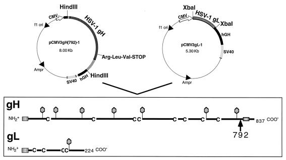
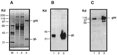
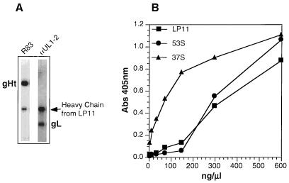
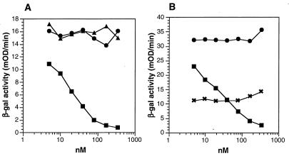
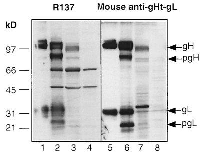
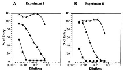
The herpes simplex virus type 1 (HSV-1) gH-gL complex which is found in the virion envelope is essential for virus infectivity and is a major antigen for the host immune system. However, little is known about the precise role of gH-gL in virus entry, and attempts to demonstrate the immunologic or vaccine efficacy of gH and gL separately or as the gH-gL complex have not succeeded. We constructed a recombinant mammalian cell line (HL-7) which secretes a soluble gH-gL complex, consisting of gH truncated at amino acid 792 (gHt) and full-length gL. Purified gHt-gL reacted with gH- and gL-specific monoclonal antibodies, including LP11, which indicates that it retains its proper antigenic structure. Soluble forms of gD (gDt) block HSV infection by interacting with specific cellular receptors. Unlike soluble gD, gHt-gL did not block HSV-1 entry into cells, nor did it enhance the blocking capacity of gD. However, polyclonal antibodies to the complex did block entry even when added after virus attachment. In addition, these antibodies exhibited high titers of complement-independent neutralizing activity against HSV-1. These sera also cross-neutralized HSV-2, albeit at low titers, and cross-reacted with gH-2 present in extracts of HSV-2-infected cells. To test the potential for gHt-gL to function as a vaccine, BALB/c mice were immunized with the complex. As controls, other mice were immunized with gD purified from HSV-infected cells or were sham immunized. Sera from the gD- or gHt-gL-immunized mice exhibited high titers of virus neutralizing activity. Using a zosteriform model of infection, we challenged mice with HSV-1. All animals showed some evidence of infection at the site of virus challenge. Mice immunized with either gD or gHt-gL showed reduced primary lesions and exhibited no secondary zosteriform lesions. The sham-immunized control animals exhibited extensive secondary lesions. Furthermore, mice immunized with either gD or gHt-gL survived virus challenge, while many control animals died. These results suggest that gHt-gL is biologically active and may be a candidate for use as a subunit vaccine.


Figures

FIG. 1
FIG. 1
Plasmids used to construct the HL-7 cell line and diagrammatic representations of gHt and gL. HL-7 cells were obtained by cotransfecting mouse L cells with pCMV3gH(792)-1, pCMV3gL-1 (9), and pX343, which confers resistance to hygromycin B (1) (not shown). HL-7 was one of four separate clones which expressed and secreted gHt-gL as a complex. The stick diagrams illustrate major structural features of full-length gH-1 and gL-1. An arrow indicates the location of the truncation of gH at amino acid 792. Balloons indicate positions of predicted N-linked oligosaccharides, and C’s indicate positions of cysteine residues. The predicted signal peptide and transmembrane anchor regions are indicated with shaded boxes. SV40, simian virus 40; hGH, human growth hormone.
FIG. 2
FIG. 2
Extracellular expression and purification of gHt-gL complex from HL-7 cells. (A) Twenty microliters of HL-7 cell supernatant (lane 1), 20 μl of flowthrough (lane 2), and 1 μg of protein eluted from the 53S immunoabsorbent column (lane 3) were analyzed by electrophoresis on an SDS–10% polyacrylamide gel. The gel was stained for protein with silver stain. (B) The proteins were electrophoresed on an SDS–10% polyacrylamide gel, transferred to nitrocellulose, and probed with anti-gL ascites 8H4. (C) The proteins were electrophoresed on an SDS–10% polyacrylamide gel, transferred to nitrocellulose, and probed with anti-gH serum R83.
FIG. 3
FIG. 3
Reactivity of purified gHt-gL with gH-specific antibodies. (A) Purified gHt-gL was immunoprecipitated with MAb LP11 and then electrophoresed on an SDS–10% polyacrylamide gel. The proteins were transferred to nitrocellulose and probed with R83 (anti-gH serum) (lane 1) or with αUL1-2 (anti-gL serum) (lane 2) (B) Various concentrations of gHt-gL were coated onto an ELISA plate for 2 h at RT. Wells were reacted with anti-gH MAb LP11, 53S, or 37S. Binding of these antibodies was detected with horseradish peroxidase-labeled goat anti-mouse antibody and ABTS substrate. Abs, absorbance.
FIG. 4
FIG. 4
Effect of gHt-gL on HSV cell entry. Various concentrations of purified proteins gC1(457t) (gCt), gD-1(306t) (gDt), and gHt-gL were added to Vero cell monolayers in 96-well plates for 90 min at 4°C. HSV-1(hrR3) was added at an MOI of 0.5, and the plate was incubated for another 90 min at 4°C. Plates were then shifted to 37°C for 5 h. Cells were lysed, and β-galactosidase (β-gal) activity was measured on aliquots of the cytoplasmic extract using the substrate CPRG and measuring the increase in absorption at 570 nm (expressed as milli-optical density units [mOD]). (A) Blocking of virus entry with purified gCt (▴), gDt (▪), or gHt-gL (•); (B) blocking of virus entry with gDt alone (▪), gHt-gL (•), or a mixture of 40 nM gD (concentration which gave 50% inhibition of entry) with various concentrations of gHt-gL (×).
FIG. 5
FIG. 5
Immunoblot (Western blot) analysis of serum samples from rabbits immunized with gHt-gL. (A) Purified gHt-gL was electrophoresed on a denaturing SDS–10% polyacrylamide gel, transferred to nitrocellulose, and reacted with R136 (lane 1), R137 (lane 2), R138 (lane 3), or R139 (lane 4). (B) Purified gHt-gL was electrophoresed on a nondenaturing (native) SDS–10% polyacrylamide gel, transferred to nitrocellulose, and reacted with R136 (lane 1), R137 (lane 2), R138 (lane 3), or R139 (lane 4).
FIG. 6
FIG. 6
Blocking of HSV entry by rabbit antibodies to gHt-gL. (A) HSV-1(hrR3) was incubated for 90 min at 37°C with various concentrations of rabbit anti-gHt-gL serum R136, R137, R138, or R139, and the serum-virus mixture was added to Vero cell monolayers in a 96-well plate, incubated at 4°C for 90 min, and then incubated at 37°C for 5 h. Virus entry was assayed as an increase in β-galactosidase activity in cytoplasmic extracts from each well and expressed as percentages of control values obtained with virus alone. (B) HSV-1(hrR3) was added to Vero cell monolayers at 4°C for 90 min. The medium was removed, various dilutions of either R83, R137, or LP11 were added, and monolayers were incubated at 4°C for 90 min and then at 37°C for 5 h. Virus entry was assayed as described for panel A.
FIG. 7
FIG. 7
Immunoblot (Western blot) analysis of cytoplasmic extracts of HSV-1 or HSV-2-infected Vero cells. Samples of purified gHt-gL or cytoplasmic extracts were electrophoresed on a denaturing SDS–10% polyacrylamide gel, transferred to nitrocellulose, and reacted with R137 (lanes 1 to 4) or mouse anti-gHt-gL (lanes 5 to 8). The mouse serum was pooled from nine animals immunized with gHt-gL (Table 2, experiment I). gHt-gL purified from HL-7 cells (lanes 1 and 5) was included on the gel as a control. Cytoplasmic extracts were prepared from cells infected with HSV-1(NS) (lanes 2 and 6) or HSV-2(333) (lanes 3 and 7) or from uninfected cells (lanes 4 and 8).
FIG. 8
FIG. 8
Blocking of HSV entry by mouse antibodies to gD or to gHt-gL. (A) HSV-1(hrR3) was incubated for 90 min at 37°C with various concentrations of antisera from mice immunized either with full-length gD (from HSV-1-infected cells; ▪), with gHt-gL (from HL-7 cells; •), or with PBS (▴) according to experiment I (Table 2). The serum-virus mixture was added to Vero cell monolayers in a 96-well plate, incubated at 4°C for 90 min, and then incubated at 37°C for 5 h. Virus entry was assayed as an increase in β-galactosidase activity in cytoplasmic extracts from each well and expressed as percentages of control values obtained with virus alone. (B) Same as panel A except that the sera were from mice immunized as part of experiment II (Table 2). Each of the sera from both experiments was assayed, and only one representative curve for each experimental group is shown. All of the sera for each group gave similar curves.

Similar articles

Cited by

References

    1. Blochlinger K, Diggelmann H. Hygromycin B phosphotransferase as a selectable marker for DNA transfer experiments with higher eucaryotic cells. Mol Cell Biol. 1984;4:2929–2931. - PMC - PubMed
    1. Boursnell, M. E. Personal communication.
    1. Browne H, Baxter V, Minson T. Analysis of protective immune responses to the glycoprotein H-glycoprotein L complex of herpes simplex virus type 1. J Gen Virol. 1993;74:2813–2817. - PubMed
    1. Buckmaster E A, Gompels U, Minson A. Characterisation and physical mapping of an HSV-1 glycoprotein of approximately 115 × 103 molecular weight. Virology. 1984;139:408–413. - PubMed
    1. Burke R L. HSV vaccine development. In: Roizman B, Lopez C, editors. The herpesviruses. Vol. 5. New York, N.Y: Raven Press; 1993. pp. 367–380.

Publication types

MeSH terms