. 1998 Jan;72(1):142-50.
doi: 10.1128/JVI.72.1.142-150.1998.

The papillomavirus minor capsid protein, L2, induces localization of the major capsid protein, L1, and the viral transcription/replication protein, E2, to PML oncogenic domains

Affiliations

The papillomavirus minor capsid protein, L2, induces localization of the major capsid protein, L1, and the viral transcription/replication protein, E2, to PML oncogenic domains

P M Day et al. J Virol. 1998 Jan.

Abstract

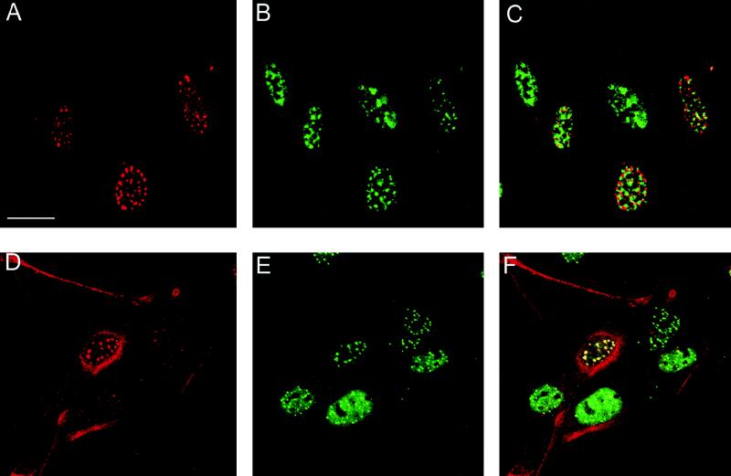
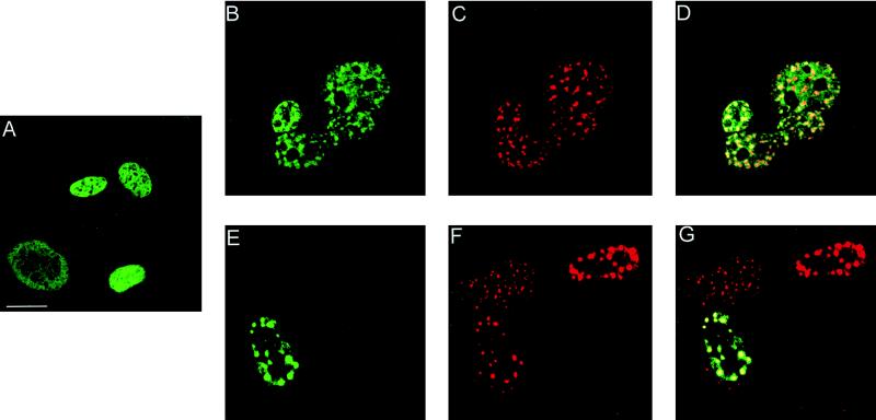
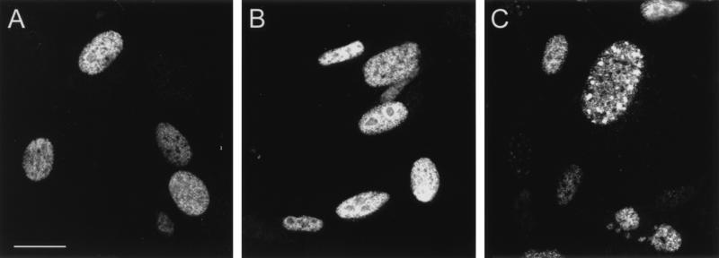
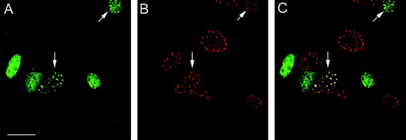
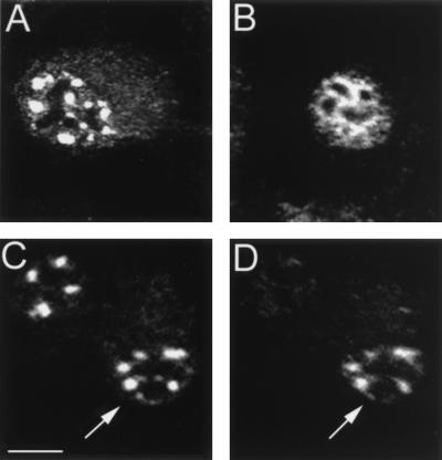
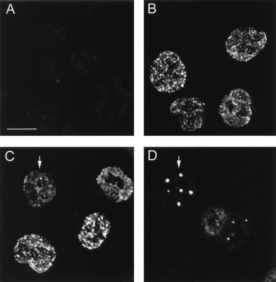
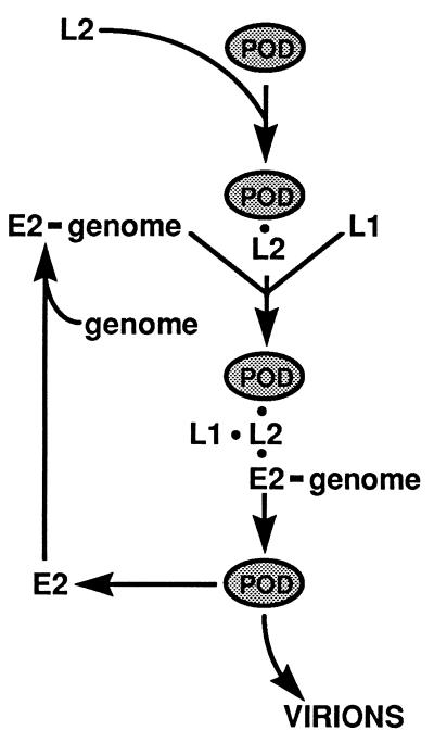
We have used immunofluorescent staining and confocal microscopy to examine the subcellular localization of structural and nonstructural bovine papillomavirus (BPV) proteins in cultured cells that produce infectious virions. When expressed separately, L1, the major capsid protein, showed a diffuse nuclear distribution while L2, the minor capsid protein, was found to localize to punctate nuclear regions identified as promonocytic leukemia protein (PML) oncogenic domains (PODs). Coexpression of L1 and L2 induced a relocation of L1 into the PODs, leading to the colocalization of L1 and L2. The effect of L2 expression on the distribution of the nonstructural viral proteins E1 and E2, which are required for maintenance of the genome and viral DNA synthesis, was also examined. The localization of the E1 protein was unaffected by L2 expression. However, the pattern of anti-E2 staining was dramatically altered in L2-expressing cells. Similar to L1, E2 was shifted from a dispersed nuclear locality into the PODs and colocalized with L2. The recruitment of full-length E2 by L2 occurred in the absence of other viral components. L2 was shown previously to be essential for the generation of infectious BPV. Our present results provide evidence for a role for L2 in the organization of virion components by recruiting them to a distinct nuclear domain. This L2-dependent colocalization probably serves as a mechanism to promote the assembly of papillomaviruses either by increasing the local concentration of virion constituents or by providing the physical architecture necessary for efficient packaging and assembly. The data also suggest a role for a nonstructural viral protein, E2, in virion assembly, specifically the recruitment of the viral genome to the sites of assembly, through its high-affinity interaction with specific sequences in the viral DNA.


Figures

FIG. 1
FIG. 1
Colocalization of L2 with PML protein. BPHE-1 cells were infected with the L2-SFV recombinant. The cells were fixed, and double-staining immunolocalization against the L2 protein and either SC35 or PML was performed. (A and D) The L2 protein was detected with rabbit polyclonal antiserum 17/28 and Texas red-conjugated goat anti-rabbit IgG. (B) SC35 was detected with a mouse monoclonal antibody (Sigma) and FITC-conjugated goat anti-mouse IgG. The same field is shown in panels A and B. (C) Digital superimposition of the two images. Coincidence of staining would be depicted in yellow in the merged panel. (E) PML localization. PML was detected with mouse monoclonal antibody 5E10 and FITC-conjugated goat anti-mouse IgG. Panel E shows the same field as the anti-L2 staining in panel D. (F) The overlap in the distribution of the patterns is evident in the merged image. Note that in panels D to F, the L2-PML colocalization is independent of the level of expression of L2. Bar, 10 mm.
FIG. 2
FIG. 2
L2 expression affects the cellular distribution of L1. BPHE-1 cells were either infected with the L1-SFV recombinant (A) or coinfected with the L1-SFV recombinant and the L2-SFV recombinant, and the proteins were detected by double immunofluorescence (B to G). (A) Staining with the anti-L1 antibody, 5B6, detected with FITC-conjugated goat anti-mouse IgG. Panels B to D show the same field of cells as do panels E to G. (B and E) Optically gated images showing the unique fluorescence of the 5B6 antibody; (C and F) optically gated images showing the unique fluorescence of the anti-L2 antiserum, 17/28, detected with Texas-red conjugated goat anti-rabbit IgG. The digital merge of the images is shown in panels D and G. Coincidence of staining appears yellow in the merged image. Bar, 10 μm.
FIG. 3
FIG. 3
E2 distribution in BPHE-1 cells that are uninfected or infected with either the L1-SFV or L2-SFV recombinant. The E2 protein was detected with monoclonal antibody B201 and FITC-conjugated goat anti-mouse IgG. (A) E2 distribution in uninfected cells. (B) E2 distribution in L1-SFV-infected cells. (C) E2 distribution in L2-SFV-infected cells. All panels were collected with identical image settings. Bar, 10 μm.
FIG. 4
FIG. 4
L2 recruits the E2 protein into PODs. BPHE-1 cells were infected with the L2-SFV recombinant, and the L2 and E2 proteins were detected by double immunofluorescence. Panels A and B show the same field of cells. (A) Localization of E2 detected with B201 and FITC-conjugated goat anti-mouse IgG. (B) Localization of L2 detected with 17/28 and Texas red-conjugated goat anti-rabbit IgG. (C) Digital merge of the images in panels A and B. Yellow areas indicate regions of colocalized staining. Bar, 10 μm.
FIG. 5
FIG. 5
The full-length form of E2 relocates to PODs. BHK-21 cells were infected with either the L2-SFV recombinant or the E2-SFV recombinant or coinfected to simultaneously express both proteins. (A) In cells that were infected with L2-SFV, the L2 protein was detected with 17/28 and Texas red-conjugated goat anti-rabbit IgG. (B) Cells that were infected with E2-SFV were stained with anti-E2 monoclonal antibody B201 and FITC conjugated goat anti-mouse IgG. The proteins in the coinfected cells were detected by double staining with the same primary and secondary antibodies. (C) Anti-L2 staining, detected in the red channel. (D) Anti-E2 staining of the same field, detected in the green channel. A cell that is coinfected is indicated by the arrows in these panels. Bar, 20 μm.
FIG. 6
FIG. 6
E1 localization is unaffected by L2 expression. BHK-21 cells were coinfected with the E1-SFV and L2-SFV recombinants. E1 was detected with rabbit polyclonal antiserum 150-1 and Texas-red conjugated goat anti-rabbit IgG. (A) Binding of these antisera to uninfected cells. (B) 150-1 staining of cells infected with E1-SFV. (C) E1 localization after coinfection with E1-SFV and L2-SFV (arrow). Panels A through C were collected with identical image settings. (D) The L2 protein was detected in the coinfected cells by staining with monoclonal antibody 6A8-E6H6 and FITC-conjugated goat anti-mouse IgG. Panels C and D show identical fields; a coexpressing cell is indicated by the arrow. Bar, 20 μm.
FIG. 7
FIG. 7
Model for L2-mediated assembly of papillomavirus virions. It is proposed that L2 acts to mediate papillomavirus assembly by causing the concentration of the virion components within the PODs. L2 localizes to PODs independently of other viral proteins. The L2 localization will cause the subsequent recruitment of E2 with the bound genome and L1. These events are independent of each other. This L2-L1-E2-genome association within the PODs confers an appropriate environment and/or concentration for virion assembly. See the text for details.

References

    1. Androphy A J, Lowy D R, Schiller J T. Bovine papillomavirus E2 trans activating gene product binds to specific sites in papillomavirus DNA. Nature. 1987;325:70–73. - PubMed
    1. Ascoli C, Maul G J. Identification of a novel nuclear domain. J Cell Biol. 1991;112:785–795. - PMC - PubMed
    1. Boddy M N, Howe K, Etkin L D, Solomon E, Freemont P S. Pic 1, a novel ubiquitin-like protein which interacts with the PML component of a multiprotein complex that is disrupted in acute promyelocytic leukemia. Oncogene. 1996;13:971–982. - PubMed
    1. Carrasco L. The inhibition of cell functions after viral infection: a proposed general mechanism. FEBS Lett. 1977;76:11–15. - PubMed
    1. Carvalho T, Seeler J S, Ohman K, Jordan P, Petterson U, Akusjarvi G, Carmo-Fonseca M, Dejean A. Targeting of adenovirus E1A and E4-ORF3 proteins to nuclear matrix-associated PML bodies. J Cell Biol. 1995;131:45–56. - PMC - PubMed

MeSH terms

LinkOut - more resources