Inactivation of the RNase activity of glycoprotein E(rns) of classical swine fever virus results in a cytopathogenic virus
- PMID: 9420210
- PMCID: PMC109359
- DOI: 10.1128/JVI.72.1.151-157.1998
Inactivation of the RNase activity of glycoprotein E(rns) of classical swine fever virus results in a cytopathogenic virus
Abstract
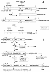
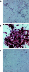
Envelope glycoprotein E(rns) of classical swine fever virus (CSFV) has been shown to contain RNase activity and is involved in virus infection. Two short regions of amino acids in the sequence of E(rns) are responsible for RNase activity. In both regions, histidine residues appear to be essential for catalysis. They were replaced by lysine residues to inactivate the RNase activity. The mutated sequence of E(rns) was inserted into the p10 locus of a baculovirus vector and expressed in insect cells. Compared to intact E(rns), the mutated proteins had lost their RNase activity. The mutated proteins reacted with E(rns)-specific neutralizing monoclonal and polyclonal antibodies and were still able to inhibit infection of swine kidney cells (SK6) with CSFV, but at a concentration higher than that measured for intact E(rns). This result indicated that the conformation of the mutated proteins was not severely affected by the inactivation. To study the effect of these mutations on virus infection and replication, a CSFV mutant with an inactivated E(rns) (FLc13) was generated with an infectious DNA copy of CSFV strain C. The mutant virus showed the same growth kinetics as the parent virus in cell culture. However, in contrast to the parent virus, the RNase-negative virus induced a cytopathic effect in swine kidney cells. This effect could be neutralized by rescue of the inactivated E(rns) gene and by neutralizing polyclonal antibodies directed against E(rns), indicating that this effect was an inherent property of the RNase-negative virus. Analyses of cellular DNA of swine kidney cells showed that the RNase-negative CSFV induced apoptosis. We conclude that the RNase activity of envelope protein E(rns) plays an important role in the replication of pestiviruses and speculate that this RNase activity might be responsible for the persistence of these viruses in their natural host.
Figures


Similar articles
-
Dimerization of glycoprotein E(rns) of classical swine fever virus is not essential for viral replication and infection.Arch Virol. 2005 Nov;150(11):2271-86. doi: 10.1007/s00705-005-0569-y. Epub 2005 Jun 28. Arch Virol. 2005. PMID: 15986175
-
Chimeric classical swine fever viruses containing envelope protein E(RNS) or E2 of bovine viral diarrhoea virus protect pigs against challenge with CSFV and induce a distinguishable antibody response.Vaccine. 2000 Oct 15;19(4-5):447-59. doi: 10.1016/s0264-410x(00)00198-5. Vaccine. 2000. PMID: 11027808
-
Mutations abrogating the RNase activity in glycoprotein E(rns) of the pestivirus classical swine fever virus lead to virus attenuation.J Virol. 1999 Dec;73(12):10224-35. doi: 10.1128/JVI.73.12.10224-10235.1999. J Virol. 1999. PMID: 10559339 Free PMC article.
-
Comparison of the effects of RNase-negative and wild-type classical swine fever virus on peripheral blood cells of infected pigs.J Gen Virol. 2004 Jul;85(Pt 7):1899-1908. doi: 10.1099/vir.0.79988-0. J Gen Virol. 2004. PMID: 15218175
-
Laboratory diagnosis, epizootiology, and efficacy of marker vaccines in classical swine fever: a review.Vet Q. 2000 Oct;22(4):182-8. doi: 10.1080/01652176.2000.9695054. Vet Q. 2000. PMID: 11087126 Review.
Cited by
-
Abrogation of the RNase activity of Erns in a low virulence classical swine fever virus enhances the humoral immune response and reduces virulence, transmissibility, and persistence in pigs.Virulence. 2021 Dec;12(1):2037-2049. doi: 10.1080/21505594.2021.1959715. Virulence. 2021. PMID: 34339338 Free PMC article.
-
Characteristics of Classical Swine Fever Virus Variants Derived from Live Attenuated GPE- Vaccine Seed.Viruses. 2021 Aug 23;13(8):1672. doi: 10.3390/v13081672. Viruses. 2021. PMID: 34452536 Free PMC article.
-
Characterization of helper virus-independent cytopathogenic classical swine fever virus generated by an in vivo RNA recombination system.J Virol. 2005 Feb;79(4):2440-8. doi: 10.1128/JVI.79.4.2440-2448.2005. J Virol. 2005. PMID: 15681445 Free PMC article.
-
Recovery of virulent and RNase-negative attenuated type 2 bovine viral diarrhea viruses from infectious cDNA clones.J Virol. 2002 Aug;76(16):8494-503. doi: 10.1128/jvi.76.16.8494-8503.2002. J Virol. 2002. PMID: 12134054 Free PMC article.
-
Structure-function analysis of Rny1 in tRNA cleavage and growth inhibition.PLoS One. 2012;7(7):e41111. doi: 10.1371/journal.pone.0041111. Epub 2012 Jul 19. PLoS One. 2012. PMID: 22829915 Free PMC article.
References
-
- Becher P, Shannon A D, Tautz N, Thiel H-J. Molecular characterization of border disease virus, a pestivirus from sheep. Virology. 1994;198:542–551. - PubMed
-
- Bolin S R, McClurkin A W, Cutlip R C, Coria M F. Severe clinical disease induced in cattle persistently infected with noncytopathic bovine viral diarrhea virus by super-infection with cytopathic bovine viral diarrhea virus. Am J Vet Res. 1985;46:573–576. - PubMed
-
- Brownlie J, Clarke M C, Howard C J. Experimental production of fatal mucosal disease in cattle. Vet Rec. 1984;114:535–536. - PubMed
MeSH terms
Substances
LinkOut - more resources
Full Text Sources
Other Literature Sources

