. 1998 Jan;72(1):266-72.
doi: 10.1128/JVI.72.1.266-272.1998.

Hepatitis B virus X protein interferes with cellular DNA repair

Affiliations

Hepatitis B virus X protein interferes with cellular DNA repair

S A Becker et al. J Virol. 1998 Jan.

Abstract

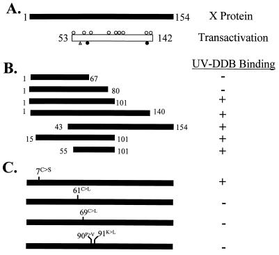
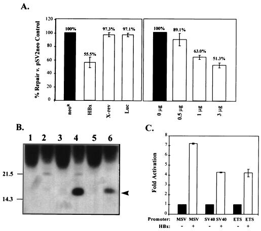
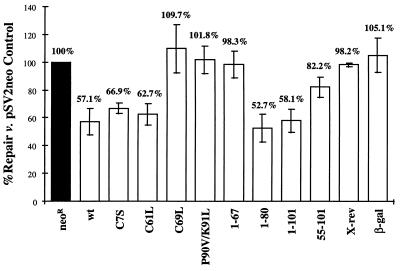
The hepatitis B virus X protein (HBx) is a broadly acting transactivator implicated in the development of liver cancer. Recently, HBx has been reported to interact with several different cellular proteins, including our report of its binding to XAP-1, the human homolog of the simian repair protein UVDDB. In the present study, several HBx mutants were used to localize the minimal domain of HBx required for binding to XAP-1/UVDDB to amino acids 55 to 101. The normal function of XAP-1/UVDDB is thought to involve binding to damaged DNA, the first step in nucleotide excision repair (NER); therefore, we hypothesized that this interaction may affect the cell's capacity to correct lesions in the genome. When tested in two independent assays that measure NER (unscheduled DNA synthesis and host cell reactivation), the expression of HBx significantly inhibited the ability of cells to repair damaged DNA. Under the assay conditions, HBx was expressed at a level similar to that previously observed during natural viral infection and was able to transactivate several target reporter genes. These results are consistent with a model in which HBx acts as a cofactor in hepatocarcinogenesis by preventing the cell from efficiently repairing damaged DNA, thus leading to an accumulation of DNA mutations and, eventually, cancer. An adverse effect on cellular DNA repair processes suggests a new mechanism by which a tumor-associated virus might contribute to carcinogenesis.


Figures

FIG. 1
FIG. 1
Mutant X proteins used for identifying regions of HBx required for interaction with XAP-1/UVDDB. (A) The 154-amino-acid HBx protein (black bar). The region known to be important for HBx transactivation of the SV40 early promoter in HepG2 cells (10) is shown as an open box. Open circles above the transactivation domain bar indicate point mutants (codons 58, 64, 74, 82, 107, 111, 114, 126, and 134) whose alteration has no effect on transactivation by HBx in HepG2 cells; an open triangle indicates the point mutation (residue 61) whose alteration reduces or completely abolishes transactivation, depending on the amino acid change; and filled circles below the bar indicate point mutations (codons 69 and 132) whose alteration completely abolishes HBx activity (4, 24, 33). (B) Deletion mutants of HBx. Black bars represent regions of HBx retained in the deletion mutants. Numbers represent amino acids retained in the mutant. XAP-1 binding was measured in the yeast two-hybrid system, with a positive interaction indicated by β-galactosidase activity as described in Materials and Methods. (C) HBx proteins containing point mutations. Mutations were introduced into pAS1-X by site-directed mutagenesis and mutated oligonucleotides. Mutations included a conserved Cys (position 7) changed to Ser; Cys at positions 61 and 69 each converted to Leu; and amino acids at positions 90 and 91 changed to eliminate a Pro and a charged (Lys) residue.
FIG. 2
FIG. 2
The UDS assay measures repair ability. (A) DNA repair in fibroblasts. Normal (CCD27Sk), XPE (XP2RO), and XPC (GOR DO) fibroblasts were plated in 60-mm-diameter dishes and subjected to the UDS protocol as described in Materials and Methods. Repair seen in the normal cells was set to 100%, and repair for the XPC and XPE cells was compared to this value (numbers above the bars). (B) XAP-1/UVDDB is not detected in XP2RO cells. HeLa cells and XP fibroblasts were extracted and immunoprecipitated with either preimmune rabbit antiserum or antiserum produced against a carboxy-terminal peptide of XAP-1/UVDDB (37). Following fractionation by sodium dodecyl sulfate–8% polyacrylamide gel electrophoresis and Western transfer, the proteins were detected with antiserum produced against an amino-terminal peptide of XAP-1/UVDDB (37) and enhanced chemiluminescence (Amersham, Arlington Heights, Ill.). The 127-kDa protein (arrowhead at right) was detected in HeLa cells (lane 2), XPC cells (lane 3), and normal fibroblasts (data not shown) but not in XPE fibroblasts (lane 4) or HeLa cells immunoprecipitated with preimmune rabbit serum (lane 1). The original autoradiogram was photographed with the U.V.P. documentation system (SW2000; U.V.P. Inc., San Gabriel, Calif.).
FIG. 3
FIG. 3
Expression of HBx protein interferes with cellular UDS. (A) Repair of damaged DNA in HBx-expressing cells. HepG2 cells were cotransfected with the pSV plasmid including the gene indicated below each bar and a luciferase reporter to control for transfection efficiency. Neor, neomycin resistance gene; HBx, X gene from HBV subtype adw2; X-rev, X in the reverse orientation; Luc, luciferase reporter gene alone. The number of micrograms of pSV-X transfected is shown below each bar in the righthand graph. Control pSV2neo plasmid was used to standardize the total amount of DNA introduced into the cells to 6 μg per plate. Repair for cells transfected with pSV2neo was set to 100%, and the repair measured for other transfected cells was compared to that value (numbers above each bar). Values shown are the means of at least three independent experiments; the error bars represent the corresponding standard deviations. (B) HBx expression. Duplicate 60-mm-diameter plates of cells transfected with pSV2neo (lanes 1 and 2) or pSV-X (lanes 3 and 4) were harvested 48 h posttransfection, pooled, and immunoprecipitated with anti-p28 negative control (odd-numbered lanes) or anti-X (even-numbered lanes). Liver extracts (25%, wt/vol) from ATX transgenic mice (38) were diluted 150-fold and then similarly immunoprecipitated and used as controls (lanes 5 and 6). Recovered proteins were transferred to nitrocellulose membrane for detection with anti-X by Western blotting. Migration of the 17-kDa HBx protein is shown at the right (arrowhead). (C) Transactivation by HBx. Cells were cotransfected with 5 μg of pSV-X and 1 μg of luciferase reporter under the control of one of several regulatory elements, noted below each bar: MSV, murine sarcoma virus; SV40, SV40 early promoter; ETS, regulatory element for the ets oncogene. The activation of each luciferase construct in the absence of HBx was normalized to 1.0 (black bars), and the fold activation of that basal level in the presence of HBx was calculated. Error bars represent the standard errors of the means from triplicate samples.
FIG. 4
FIG. 4
Interference with repair by HBx and mutant derivatives. HepG2 cells were assayed by the HCR assay described in Materials and Methods with the test plasmid construct listed under each bar: Neor, neomycin-resistance; wt, wild-type HBx subtype adw2; amino acid numbers, residues mutagenized (point mutants with the changed codon indicated by the standard single-letter abbreviation) or retained (deletion constructs); X-rev, X gene in the reverse orientation; β-gal, β-galactosidase reporter. Repair for the pSV2neo control (black bar) was set to 100%, and repair for cells transfected with the other constructs was compared to that value (numbers above each bar). Values shown are the means of at least three independent experiments, and the error bars represent the standard deviations.

References

    1. Aboussekhra A, Biggerstaff M, Shivji M K, Vilpo J A, Moncollin V, Podust V N, Protic M, Hubscher U, Egly J M, Wood R D. Mammalian DNA nucleotide excision repair reconstituted with purified protein components. Cell. 1995;80:859–868. - PubMed
    1. Aden D P, Fogel A, Plotkin S, Damjanov I, Knowles B B. Controlled synthesis of HBsAg in a differentiated human liver carcinoma-derived cell line. Nature (London) 1979;282:615–616. - PubMed
    1. Anonymous. Statistical Package for the Social Sciences (SPSS) for Windows, version 7. Chicago, Ill: SPSS, Inc.; 1995.
    1. Arii M, Takada S, Koike K. Identification of three essential regions of hepatitis B virus X protein for trans-activation function. Oncogene. 1992;7:397–403. - PubMed
    1. Athas W F, Hedayati M A, Matanoski G M, Farmer E R, Grossman L. Development and field-test validation of an assay for DNA repair in circulating human lymphocytes. Cancer Res. 1991;51:5786–5793. - PubMed

Publication types

MeSH terms

LinkOut - more resources