. 1998 Jan;72(1):273-8.
doi: 10.1128/JVI.72.1.273-278.1998.

An avirulent mutant of rabies virus is unable to infect motoneurons in vivo and in vitro

Affiliations

An avirulent mutant of rabies virus is unable to infect motoneurons in vivo and in vitro

P Coulon et al. J Virol. 1998 Jan.

Abstract

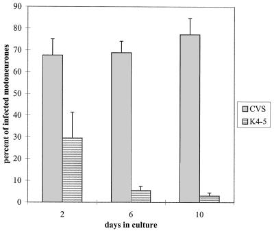
An antigenic double mutant of rabies virus (challenge virus standard [CVS] strain) was selected by successive use of two neutralizing antiglycoprotein monoclonal antibodies, both specific for antigenic site III. This mutant differed from the original virus strain by two amino acid substitutions in the ectodomain of the glycoprotein. The lysine in position 330 and the arginine in position 333 were replaced by asparagine and methionine, respectively. This double mutant was not pathogenic for adult mice. When injected intramuscularly into the forelimbs of adult mice, this virus could not penetrate the nervous system, either by the motor or by the sensory route, while respective single mutants infected motoneurons in the spinal cord and sensory neurons in the dorsal root ganglia. In vitro experiments showed that the double mutant was able to infect BHK cells, neuroblastoma cells, and freshly prepared embryonic motoneurons, albeit with a lower efficiency than the CVS strain. Upon further incubation at 37 degrees C, the motoneurons became resistant to infection by the mutant while remaining permissive to CVS infection. These results suggest that rabies virus uses different types of receptors: a molecule which is ubiquitously expressed at the surface of continuous cell lines and which is recognized by both CVS and the double mutant and a neuron-specific molecule which is not recognized by the double mutant.


Figures

FIG. 1
FIG. 1
Conservation of antigenic site III in laboratory and wild-type strains of rabies virus as well as Mokola virus (26), a rabies-related virus. Sequences are shown for fixed strains CVS (32), ERA (1), SAD (5), and Pasteur virus (PV) (27) and for wild-type strains CGX89-1 (2), COSRV (14), T5-A1 (16), and Algiers (4). The K4-5 sequence was determined in this study. Positively charged amino acids are in bold and are underlined. The glycosylation site and the proline are boxed.
FIG. 2
FIG. 2
One-step growth curves for CVS (⧫) and K4-5 (•) on BSR (——) and NG108-15 (–––) cells. The MOIs were 5 (CVS) and 6 (K4-5). After 1 h of adsorption at room temperature, the inoculum was removed and cell layers were washed before the addition of the medium. Supernatants were changed after 4 h in order to remove desorbed virus. Incubation was done at 37°C. p.i., postinfection.
FIG. 3
FIG. 3
Purified rat embryonic spinal motoneurons in cultures. (a) Cultures of dissociated motoneurons were grown on glass coverslips coated with poly-l-lysine (0.005%) and laminin (0.001%) in the presence of DMEM-F12 medium with additives. Living cells were observed by phase-contrast microscopy 6 days after plating. (b) Immunoperoxidase detection of acetylcholine in the same culture of motoneurons after fixation with paraformaldehyde. Bar, 15 μm.
FIG. 4
FIG. 4
Infection of spinal motoneurons from rat embryos after 10 days in cultures with CVS or antigenic double mutant K4-5. Cells were plated on glass coverslips treated with poly-l-lysine and laminin, kept for 10 days, and then infected for 24 h with CVS (A and B) or K4-5 (C and D). Cultures were fixed and permeabilized before simultaneous detection of neurons (A and C) and viral antigens (B and D) by double-labeling immunofluorescence. Bar, 30 μm.
FIG. 5
FIG. 5
Rat embryonic spinal motoneurons acquire progressive resistance to infection with mutant K4-5 in vitro. Motoneurons were seeded on poly-l-lysine- and laminin-treated glass coverslips at a density of 3 × 10⁴ to 3 × 10⁵ cells per coverslip, depending on the culture time. They were infected at day 2, 6, or 10 with CVS or K4-5 for 24 h at 37°C. After fixation and permeabilization, neurons were identified with an anti-MAP2 mouse MAb, and infected cells were detected with a rabbit anti-rabies virus nucleocapsid antibody. The percentage of infected motoneurons was estimated as the ratio of the number of doubly-labeled cells to the number of MAP2-labeled cells (value estimated from 103 MAP2+ cells counted). The experiment was performed in quadruplicate.

References

    1. Anilionis A, Wunner W H, Curtis P J. Structure of the glycoprotein gene in rabies virus. Nature (London) 1981;294:275–278. - PubMed
    1. Bai X H, Warner C K, Fekadu M. Comparisons of nucleotide and deduced amino acid sequences of the glycoprotein genes of a Chinese street strain (CGX89-1) and a Chinese vaccine strain (3aG) of rabies virus. Virus Res. 1993;27:101–112. - PubMed
    1. Bataillé, S., P. Portalier, P. Coulon, and J. P. Ternaux. Influence of acetylcholinesterase on embryonic spinal rat motoneuron growth in culture: a quantitative morphometric study. Eur. J. Neurosci., in press. - PubMed
    1. Benmansour A, Brahimi M, Tuffereau C, Coulon P, Lafay F, Flamand A. Rapid sequence evolution of street rabies glycoprotein is related to the highly heterogeneous nature of the viral population. Virology. 1992;187:33–45. - PubMed
    1. Benmansour A, Leblois H, Coulon P, Tuffereau C, Gaudin Y, Flamand A, Lafay F. Antigenicity of rabies virus glycoprotein. J Virol. 1991;65:4198–4203. - PMC - PubMed

Publication types

MeSH terms

LinkOut - more resources