

doi: 10.1128/JVI.72.1.303-308.1998.
Cells with high cyclophilin A content support replication of human immunodeficiency virus type 1 Gag mutants with decreased ability to incorporate cyclophilin A
Affiliations
- PMID: 9420228
- PMCID: PMC109377
- DOI: 10.1128/JVI.72.1.303-308.1998
Item in Clipboard
Cells with high cyclophilin A content support replication of human immunodeficiency virus type 1 Gag mutants with decreased ability to incorporate cyclophilin A
J Virol.
1998 Jan.
Abstract
Gag polyprotein-mediated incorporation of cellular cyclophilin A (CyPA) into virions is essential for the formation of infectious human immunodeficiency virus type 1 (HIV-1) virions. Either a point mutation in Gag (P222A) or drugs which bind CyPA decrease virion incorporation of CyPA and interfere with HIV-1 replication. We have found that lymphoid cells varied greatly in their CyPA content and that cells with high CyPA content supported the replication of P222A HIV-1 Gag mutants. These experiments demonstrated that a higher cellular CyPA content of some cells was able to compensate for the decreased binding affinity of P222A mutant Gag for CyPA, allowing virus replication to occur.
Figures
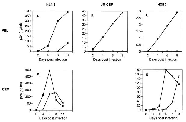
Effect of Gag P222A mutation on HIV-1 replication in PBLs and CEM cells. Following infection of 2 × 10⁶ CEM cells or PHA-stimulated PBLs with HIV-1 virus stock containing 20 ng of p24, viral replication was monitored by measuring the amount of p24 in the viral culture supernatant (ordinate) as a function of days postinfection (abscissa). CEM cells were split 1:3 at 72 hpi and every 48 h thereafter. ▪, wild type; □, P222A mutant.
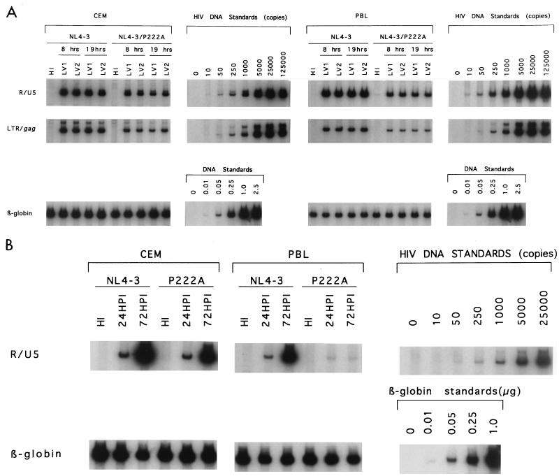
PCR analysis of HIV-1 DNA in HIV-1NL4-3- or HIV-1NL4-3/P222A-infected CEM cells or PHA-stimulated PBLs. CEM cells or PHA-stimulated PBLs were infected with HIV-1NL4-3 or HIV-1NL4-3/P222A (20 ng of p24/2 × 106), and cells were harvested at 8 and 19 hpi (A) or 24 and 72 hpi (B). DNA was purified and subjected to PCR analysis with the R/U5 (AA55/667), LTR (long terminal repeat)/gag (M661/667), or β-globin (LA1/LA2) primers (42). LV1 and LV2, live virus stock infections done in duplicate; HI, infections done with heat-inactivated virus stock (42). HIV-1 DNA corresponds to the number of copies per lane, while β-globin DNA is given as micrograms per lane.

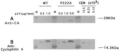
CyPA content of HIV-1NL4-3, HIV-1NL4-3/P217A, and HIV- 1NL4-3/P222A virions. Sucrose gradient-purified HIV-1NL4-3, HIV-1NL4-3/P217A, and HIV-1NL4-3/P222A virions containing 230 ng of p24 collected from CEM cells infected with virus stocks from supernatants of COS-7 cell transfections were acetone precipitated, separated by SDS-PAGE, and transferred to nitrocellulose. Membranes were labelled with a mixture of human monoclonal antibodies against capsid (CA) (A) or rabbit polyclonal antibody against CyPA (B). Bound antibody was detected with horseradish peroxidase-conjugated secondary antibody and a chemoluminescent detection system. Serially diluted human CyPA was used for standards. M, molecular mass standards; WT, wild type.
CyPA content of HIV-1NL4-3 and HIV-1NL4-3/P222A virions produced by COS-7 cells. Different amounts (measured by p24 ELISA) of sucrose gradient-purified HIV-1NL4-3 and HIV-1NL4-3/P222A virions from COS-7 cell transfections were precipitated by centrifugation, and their proteins were separated by SDS-PAGE and transferred to nitrocellulose membranes. Membranes were labelled with human monoclonal antibody against CA (A) or rabbit polyclonal antibody against human CyPA (B). Bound antibody was detected with alkaline phosphatase-conjugated secondary antibody with a precipitation substrate. Serially diluted CEM cells were analyzed similarly as standards. M, molecular mass standards; WT, wild type.
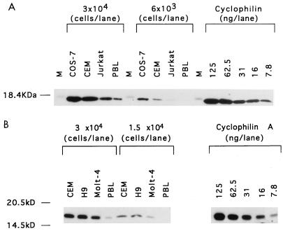
CyPA content of different cell types. Proteins from 3 × 10⁴ or 6 × 10³ COS-7, CEM, and Jurkat cells and PBLs (A) or 3 × 10⁴ or 1.5 × 10⁴ CEM, H9, and Molt-4 cells and PBLs (B) were separated by SDS-PAGE and transferred to nitrocellulose membranes. Membranes were decorated with rabbit polyclonal antibody against human CyPA. Horseradish peroxidase-conjugated goat anti-rabbit immunoglobulin was used as a secondary antibody. Serially diluted human CyPA was used for standards. M, molecular mass standards.
References
-
- Bess J W, Jr, Gorelick R J, Bosche W J, Henderson L E, Arthur L O. Microvesicles are a source of contaminating cellular proteins found in purified HIV-1 preparations. Virology. 1997;230:134–144. - PubMed
-
- Billich A, Hammerschmid F, Peichl P, Wenger R, Zenke G, Quesniaux V, Rosenwirth B. Mode of action of SDZ NIM 811, a nonimmunosuppressive cyclosporin A analog with activity against human immunodeficiency virus (HIV) type 1: interference with protein-cyclophilin A interactions. J Virol. 1995;69:2451–2461. - PMC - PubMed
Publication types
MeSH terms
Substances
Grants and funding
LinkOut - more resources
Full Text Sources
Other Literature Sources
