. 1998 Jan;72(1):309-19.
doi: 10.1128/JVI.72.1.309-319.1998.

Infectious clones and vectors derived from adeno-associated virus (AAV) serotypes other than AAV type 2

Affiliations

Infectious clones and vectors derived from adeno-associated virus (AAV) serotypes other than AAV type 2

E A Rutledge et al. J Virol. 1998 Jan.

Abstract

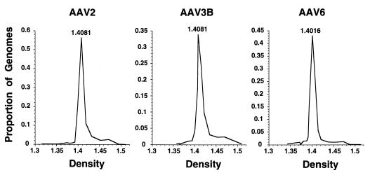
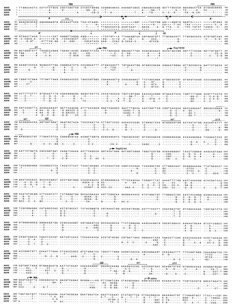
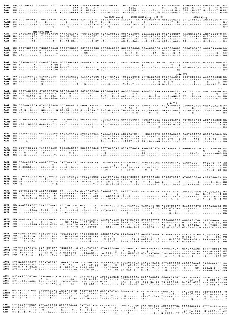
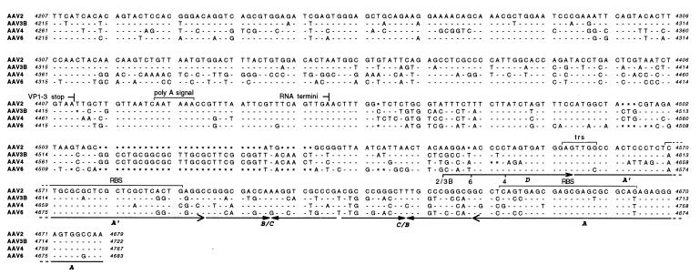
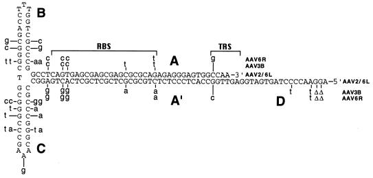
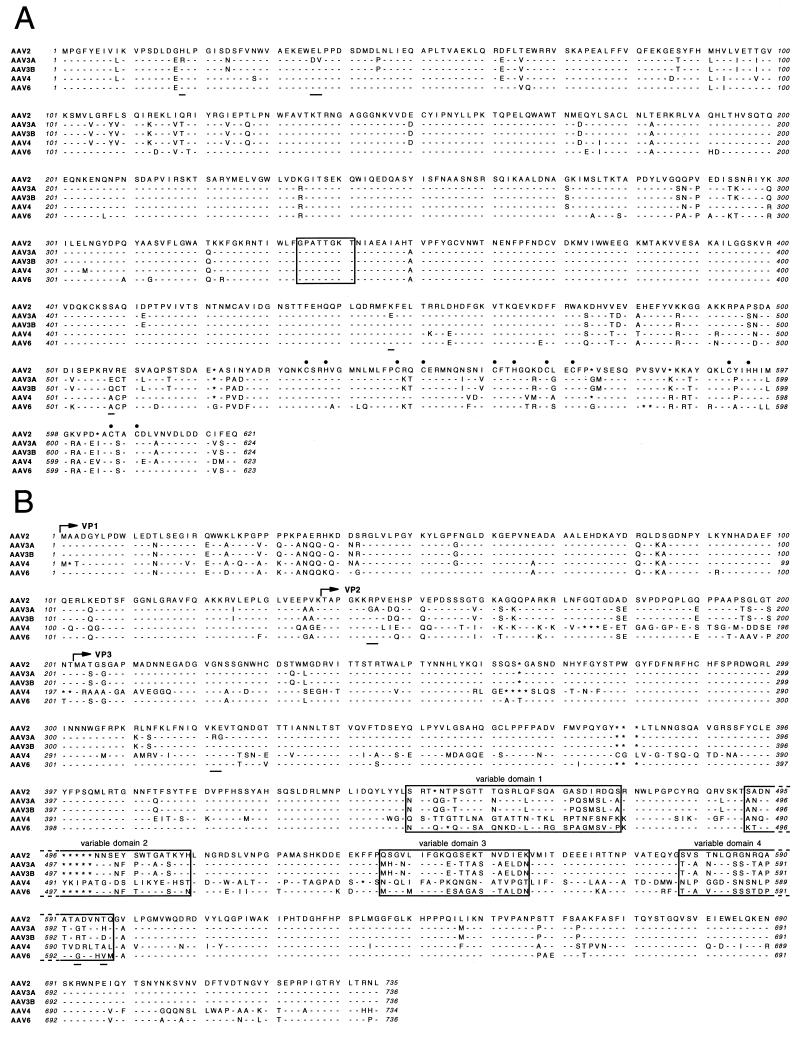
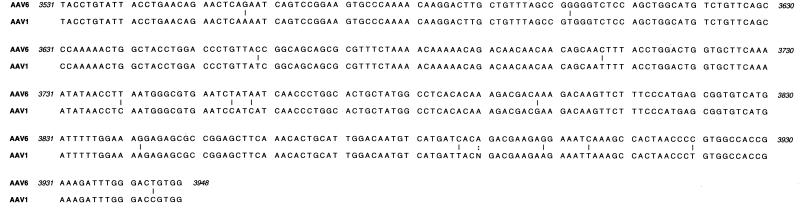
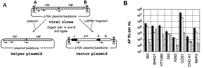
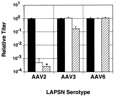
Adeno-associated viruses (AAVs) are single-stranded dependent parvoviruses being developed as transducing vectors. Although at least five serotypes exist (AAV types 1 to 5 [AAV1 to -5]), only AAV2, AAV3, and AAV4 have been sequenced, and the vectors in use were almost all derived from AAV2. Here we report the cloning and sequencing of a second AAV3 genome and a new AAV serotype designated AAV6 that is related to AAV1. AAV2, AAV3, and AAV6 were 82% identical at the nucleotide sequence level, and AAV4 was 75 to 78% identical to these AAVs. Significant sequence variation was noted in portions of the capsid proteins that presumably are responsible for serotype-specific functions. Vectors produced from AAV3 and AAV6 differed from AAV2 vectors in host range and serologic reactivity. The AAV3 and AAV6 vector serotypes were able to transduce cells in the presence of serum from animals previously exposed to AAV2 vectors. Our results suggest that vectors based on alternative AAV serotypes will have advantages over existing AAV2 vectors, including the transduction of different cell types, and resistance to neutralizing antibodies against AAV2. This could be especially important for gene therapy, as significant immunity against AAV2 exists in human populations and many protocols will likely require multiple vector doses.


Figures

FIG. 1
FIG. 1
Cesium chloride gradient fractionation of AAV serotypes. AAV2, AAV3B, and AAV6 virus stocks produced from infectious clones were purified on CsCl gradients. The number of AAV genomes in each gradient fraction was determined by Southern analysis, and the density of each fraction was determined by refractometry. Each fraction genome number was calculated as a proportion of the total present in the gradient and plotted against the density of the fraction. The density of the fraction with the greatest AAV signal is indicated.
FIG. 2
FIG. 2
Sequences of infectious AAV clones. The DNA sequences of AAV2, AAV3B, AAV4 (14), and AAV6 are shown aligned, with nucleotides identical to those of AAV2 indicated by dashes and gaps indicated by asterisks. The positions of conserved genetic elements are shown, including the p5, p19, and p40 promoters, transcription start and stop sites, intron splice sites, polyadenylation signal (poly A signal), translation start and stop sites for the Rep (Rep 78, 68, 52, and 40) and capsid (VP1, VP2, and VP3) proteins, sp1 and sp1-like (GGT) binding sites (42, 43), Rep protein binding site (RBS) (13, 49), terminal resolution site (trs) (56), and the terminal repeat A, B, C, A′, and D domains. The AAV2 sequence (57) includes published corrections (11, 46).
FIG. 2
FIG. 2
Sequences of infectious AAV clones. The DNA sequences of AAV2, AAV3B, AAV4 (14), and AAV6 are shown aligned, with nucleotides identical to those of AAV2 indicated by dashes and gaps indicated by asterisks. The positions of conserved genetic elements are shown, including the p5, p19, and p40 promoters, transcription start and stop sites, intron splice sites, polyadenylation signal (poly A signal), translation start and stop sites for the Rep (Rep 78, 68, 52, and 40) and capsid (VP1, VP2, and VP3) proteins, sp1 and sp1-like (GGT) binding sites (42, 43), Rep protein binding site (RBS) (13, 49), terminal resolution site (trs) (56), and the terminal repeat A, B, C, A′, and D domains. The AAV2 sequence (57) includes published corrections (11, 46).
FIG. 3
FIG. 3
Terminal repeat secondary structure. The sequences of the terminal repeats of AAV2, AAV3B, and AAV6 are shown in the secondary structure predicted to exist in single-stranded vector genomes. The AAV2 and AAV6 left (AAV6L) sequences are shown in capital letters. The nucleotide differences in the AAV3B and AAV6 right (AAV6R) terminal repeats are shown in lowercase letters above or below the repeat structure. Nucleotide deletions are indicated by Δ. The A, B, C, A′, and D repeat domains, Rep binding site (RBS) (13, 49), and terminal resolution site (TRS) (56) are indicated.
FIG. 4
FIG. 4
Translated reading frames. The predicted amino acid sequences of the Rep78 (A) and capsid VP1, VP2, and VP3 proteins (B) are aligned for AAV2, AAV3A, AAV3B, AAV4, and AAV6. Amino acid identities with AAV2 sequences are indicated by dashes. Gaps are indicated by asterisks. Differences between AAV3B and AAV3A are also indicated by underlining. (A) The consensus ATP binding site (54, 58) is boxed, and the cysteine and histidine residues of the zinc finger binding motif (10, 28) are indicated by filled circles. (B) The start sites for each of the capsid proteins, VP1, VP2, and VP3, are indicated by arrows, and four variable domains (regions with significant sequence differences among all 4 serotypes, as determined by inspection) are boxed.
FIG. 5
FIG. 5
Sequence comparison of AAV6 and AAV1. The variable region of the cap gene in AAV1 DNA was amplified by PCR and sequenced and is shown aligned with the corresponding sequence of AAV6. The vertical lines indicate nucleotide differences. Dots indicate an uncertainty. Numbers refer to the AAV6 genomic sequence in Fig. 2.
FIG. 6
FIG. 6
Transduction by AAV vector serotypes. (A) Strategy for construction of helper and vector plasmids from the proviral clone. The viral clone was digested at the engineered restriction sites (shown as A and B) inside the terminal repeats (TR). The rep and cap genes were ligated into another plasmid backbone to create the helper plasmid, and the LAPSN fragment was inserted into the viral clone backbone inside the terminal repeats to create the vector plasmid. The positions of the murine leukemia virus long terminal repeat promoter (L), alkaline phosphatase gene (AP), SV40 early promoter (S), and neomycin phosphotransferase gene (N) are indicated. (B) AAV2-LAPSN (filled bars), AAV3-LAPSN (open bars), and AAV6-LAPSN (hatched bars) vector stocks were adjusted to contain 5 × 10⁷ particles/μl and used to infect a panel of cell lines, and the numbers of cell foci expressing alkaline phosphatase (AP ffu) per ml of vector stock were determined (see Materials and Methods). Results are means ± standard deviations of three measurements. The asterisk indicates that the value was lower than the indicated amount (no stained cells were detected).
FIG. 7
FIG. 7
Inactivation by anti-AAV2 serum. Vectors were left untreated (filled bars) or treated with 1:100 (open bars) or 1:20 (hatched bars) rabbit anti-AAV2 serum dilutions before infection of BHK21 cells. Two days after infection, the numbers of cell foci expressing alkaline phosphatase were determined by histochemical staining. The results (means ± standard deviations) are expressed as relative titers for each vector serotype after normalization of the untreated titers to 1.0 (mean ± standard deviation). The asterisk indicates a value lower than the indicated amount (no stained cells were detected).

References

    1. Afione S A, Conrad C K, Kearns W G, Chunduru S, Adams R, Reynolds T C, Guggino W B, Cutting G R, Carter B J, Flotte T R. In vivo model of adeno-associated virus vector persistence and rescue. J Virol. 1996;70:3235–3241. - PMC - PubMed
    1. Alexander I E, Russell D W, Spence A M, Miller A D. Effects of gamma irradiation on the transduction of dividing and nondividing cells in brain and muscle of rats by adeno-associated virus vectors. Hum Gene Ther. 1996;7:841–850. - PubMed
    1. Atchison R W, Casto B C, Hammon W M. Adenovirus-associated defective virus particles. Science. 1965;149:754–756. - PubMed
    1. Bantel Schaal U, zur Hausen H. Characterization of the DNA of a defective human parvovirus isolated from a genital site. Virology. 1984;134:52–63. - PubMed
    1. Bertran J, Miller J L, Yang Y, Fenimore-Justman A, Rueda F, Vanin E F, Nienhuis A W. Recombinant adeno-associated virus-mediated high-efficiency, transient expression of the murine cationic amino acid transporter (ecotropic retroviral receptor) permits stable transduction of human HeLa cells by ecotropic retroviral vectors. J Virol. 1996;70:6759–6766. - PMC - PubMed

Publication types

MeSH terms

Associated data