. 1998 Jan;72(1):758-66.
doi: 10.1128/JVI.72.1.758-766.1998.

An arenavirus RING (zinc-binding) protein binds the oncoprotein promyelocyte leukemia protein (PML) and relocates PML nuclear bodies to the cytoplasm

Affiliations

An arenavirus RING (zinc-binding) protein binds the oncoprotein promyelocyte leukemia protein (PML) and relocates PML nuclear bodies to the cytoplasm

K L Borden et al. J Virol. 1998 Jan.

Abstract

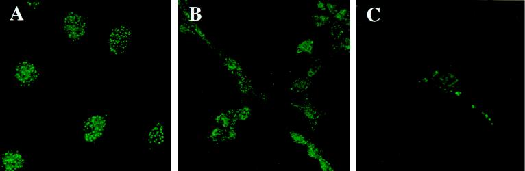
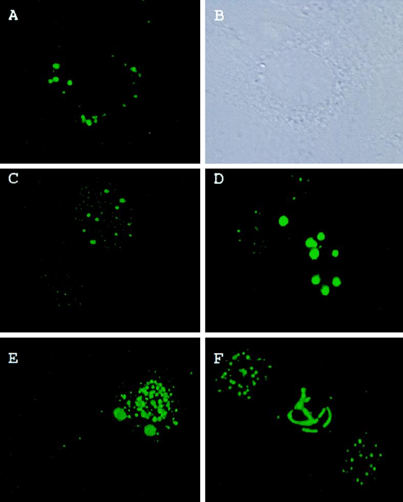
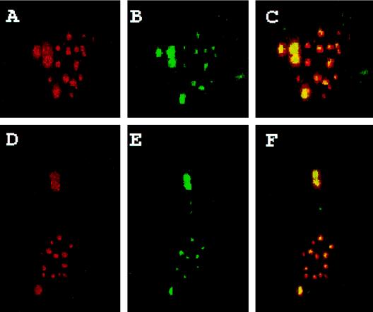
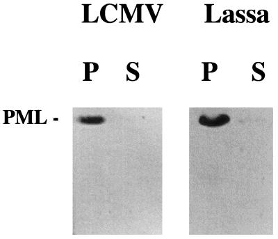
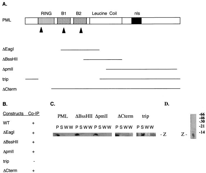
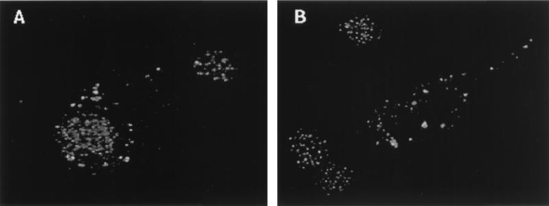
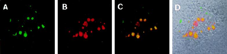
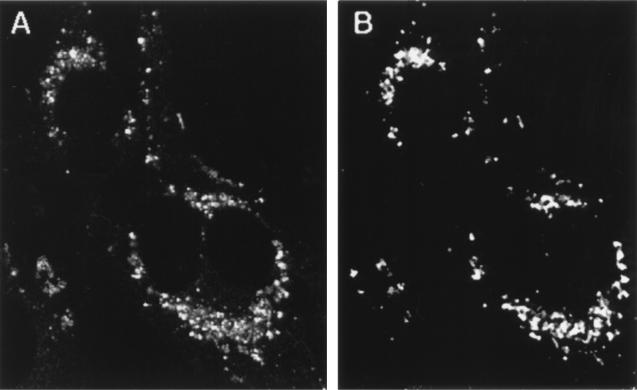
The promyelocytic leukemia protein (PML) forms nuclear bodies which are altered in some disease conditions. We report that the cytoplasmic RNA virus lymphocytic choriomeningitis virus (LCMV) influences the distribution of PML bodies. In cells infected with LCMV, the Z protein and PML form large bodies primarily in the cytoplasm. Transient transfection studies indicate that Z alone is sufficient to redistribute PML to the cytoplasm and that PML and Z colocalize. Coimmunoprecipitation studies show specific interaction between PML and Z proteins. A similar result was observed with a Z protein from another arenavirus, Lassa virus, suggesting that this is a general feature of the Arenaviridae. Genetically engineered mutations in PML were used to show that the Z protein binds the N-terminal region of PML and does not need the PML RING or the nuclear localization signal to colocalize. The Z protein acts dominantly to overcome the diffuse phenotype observed in several PML mutants. The interaction between PML and Z may influence certain unique characteristics of arenavirus infection.


Figures

FIG. 1
FIG. 1
Effect of LCMV infection on PML nuclear bodies. (A) Uninfected NIH 3T3 cells; (B) 70-h LCMV-infected NIH 3T3 cells; (C) 90-h LCMV-infected cells. LCMV infection is described in Materials and Methods. Cells were stained with the PML polyclonal antibody and FITC-conjugated secondary antibody as described in Materials and Methods. Immunofluorescence was observed by confocal laser microscopy. Magnification, ×40 objective with zooms of 2.6 (A), 1.7 (B), and 3.9 (C).
FIG. 2
FIG. 2
Subcellular localization of the Z protein, PML, and the effect of Z protein expression on PML subcellular distribution. Experiments were carried out as described in Materials and Methods. (A) Cell transfected only with the Z construct and stained with the affinity-purified Z antibody followed by FITC; (B) phase-contrast view of the same cell; (C) two cells, one of which was transfected with the PML construct to show the normal PML phenotype for the 69-kDa isoform which is found in the nucleus (see reference and references therein). See text for further details. (D to F) Cells transfected with both the expression constructs for Z and PML but stained only with the PML polyclonal antibody followed by FITC. Immunofluorescence was observed with confocal laser microscopy. FITC was excited at 488 nm. Magnifications: ×40 objective with zooms of 3.5 (A and B), 3.5 (C), 3.0 (D), and 3.0 (E) and ×25 objective with a zoom of 6.0 (F).
FIG. 3
FIG. 3
Colocalization studies of the Z protein and PML. Cells were transfected as described in Materials and Methods with equivalent amounts of the Z- and PML-containing mammalian expression vectors. Upper panels correspond with lower panels. (A and D) Cells stained with the PML MAb 5E10 with Texas Red-labeled anti-mouse secondary antibodies; (B and E) cells stained with affinity-purified Z antibody with FITC-labeled anti-rabbit secondary antibodies; (C and F) overlay (colocalization is in yellow). FITC was excited at 488 nm, and Texas Red was excited at 568 nm. The two channels were recorded independently. Images were overlaid in Photoshop. Magnification, ×40 objective with a zoom of 2. Images were further enlarged in Photoshop for presentation.
FIG. 4
FIG. 4
Direct protein-protein interaction between PML and Z shown by coimmunoprecipitation. Z proteins from both LCMV and Lassa virus were studied. The respective Z-GST fusion proteins were mixed with the PML protein and coimmunoprecipitated with an anti-GST antibody as described in Materials and Methods. Samples were run on SDS-20% PAGE gels and then blotted onto PVDF membranes. The Western analysis was carried out with the enhanced chemiluminescence detection system (ECL; Amersham) and MAb 5E10 to detect any PML that was immunoprecipitated. S, supernatant; P, precipitate. Lane P of the autoradiograph indicates that PML coprecipitates with LCMV Z and Lassa virus Z fusion proteins. Lane S shows that supernatant after coimmunoprecipitation retains only trace amounts of PML.
FIG. 5
FIG. 5
Mapping the interaction between PML and Z. (A) Summary of the constructs used in these studies. The boxes indicate the various motifs found in PML; B1 and B2, the respective B boxes; nls, nuclear localization signal (see text). Lines indicate the deleted regions of PML. See Materials and Methods for details of constructs. (B) Summary of the coimmunoprecipitation (Co-IP) results with PML and Z. The Z protein and not Z-GST was used for these studies. + and −, PML and Z did and did not immunoprecipitate, respectively. Mutations are as described for panel A. WT, wild type. (C) The relevant autoradiographs used for the data in panel B. P, precipitate; S, supernatant; W, wash. Two washes were done for each immunoprecipitation. Experiments were carried out as described in Materials and Methods. (D) Specificity of the affinity-purified Z antibody. The S100 fraction of the cell lysate was immunoblotted and probed with the affinity-purified Z antibody. Molecular weight markers are shown. See text for further details.
FIG. 6
FIG. 6
PML mutants cotransfected with LCMV Z gene. Mutants stained with the PML polyclonal antibody with FITC secondary antibody. Images were collected by confocal laser microscopy. (A) PML RING mutant cotransfected with Z; (B) PML B1 B box mutant cotransfected with Z. See Materials and Methods for description of mutations. Magnification, ×40 with zooms of 1.7 (A) and 2.8 (B).
FIG. 7
FIG. 7
Cotransfection of wild-type PML and the Z gene with a double-point mutation in the RING region. The cells were stained with affinity-purified anti-Z antibody with an FITC secondary (A) or with the PML MAb 5E10 with a Texas Red secondary antibody (B). (C) Overlay showing colocalization in yellow; (D) the phase view and fluorescence overlay. Images were collected on a confocal laser microscope exciting each channel independently. Magnification, ×40 with a zoom of 1.9.
FIG. 8
FIG. 8
Effects of transient transfection when both PML and Z have mutations in the RING domain. Cells were stained with the PML MAb 5E10 with Texas Red (A) or the affinity-purified Z antibody with an FITC secondary antibody (B). See Materials and Methods for description of mutants. Images were collected on a confocal laser microscope by exciting each channel independently. Magnification, ×100.

References

    1. Ascoli C A, Maul G G. Identification of a novel nuclear domain. J Cell Biol. 1991;112:785–795. - PMC - PubMed
    1. Banerjee S N, Buchmeier M, Rawls W E. Requirement of a cell nucleus for the replication of an arenavirus. Intervirology. 1976;6:190–196. - PubMed
    1. Barlow P N, Luisi B, Milner A, Elliot M, Everett R. Structure of the C3HC4 domain by 1H-nuclear magnetic resonance spectroscopy. J Mol Biol. 1994;237:201–211. - PubMed
    1. Boddy M N, Howe K, Etkin L D, Solomon E, Freemont P S. PIC1, a novel ubiquitin-like protein which interacts with the PML component of a multiprotein complex that is disrupted in acute promyelocytic leukaemia. Oncogene. 1996;13:971–982. - PubMed
    1. Borden K L B, Freemont P S. The RING finger: an example of a sequence structure family. Curr Opin Struc Biol. 1996;6:395–401. - PubMed

Publication types

MeSH terms

LinkOut - more resources