. 1998 Jan 1;12(1):34-44.
doi: 10.1101/gad.12.1.34.

New core promoter element in RNA polymerase II-dependent transcription: sequence-specific DNA binding by transcription factor IIB

Affiliations

New core promoter element in RNA polymerase II-dependent transcription: sequence-specific DNA binding by transcription factor IIB

T Lagrange et al. Genes Dev. .

Abstract

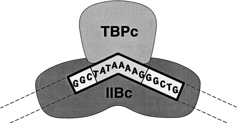
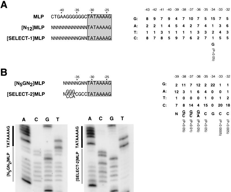
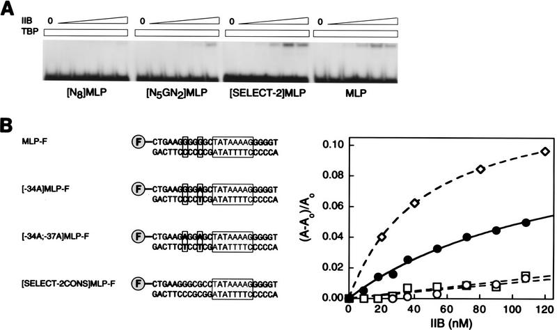
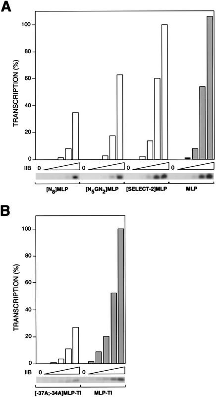
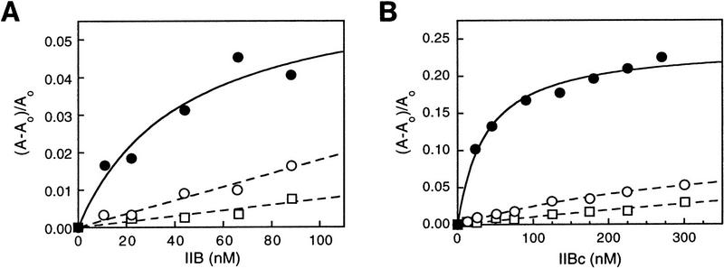
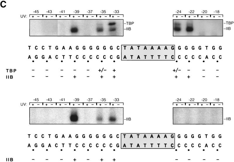
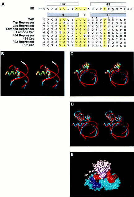
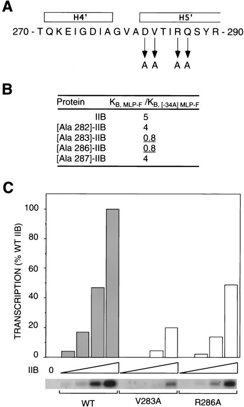
A sequence element located immediately upstream of the TATA element, and having the consensus sequence 5'-G/C-G/C-G/A-C-G-C-C-3', affects the ability of transcription factor IIB to enter transcription complexes and support transcription initiation. The sequence element is recognized directly by the transcription factor IIB. Recognition involves alpha-helices 4' and 5' of IIB, which comprise a helix-turn-helix DNA-binding motif. These observations establish that transcription initiation involves a fourth core promoter element, the IIB recognition element (BRE), in addition to the TATA element, the initiator element, and the downstream promoter element, and involves a second sequence-specific general transcription factor, IIB, in addition to transcription factor IID.


Figures

Figure 1
Figure 1
Structure of the IIB–TBP–TATA complex. Schematic representation of the crystallographic structure of a ternary complex of transcription factor IIB core domain (IIBc), TBP core domain (TBPc), and a 16-bp DNA fragment containing the TATA element (Nikolov et al. 1995). (TATAAAAG) TATA element; (GGC and GGCTG) DNA segments upstream and downstream of the TATA element in the 16-bp DNA fragment; (dashed lines) projected paths of distal upstream and downstream DNA segments.
Figure 2
Figure 2
New sequence element. (A) Binding site selection, round 1. (Left) Positions −43 to −24 of the wild-type, N12-randomized, and consensus round-1-selected adenovirus major late promoter sequences (TATA element boxed and shaded). (Right) Summary of 19 round-1-selected sequences. (B) Binding site selection, round 2. (Left) Positions −39 to −24 of the N5GN2- randomized and consensus round-2-selected adenovirus major late promoter sequences (TATA element boxed and shaded). (Right) Summary of 22 round-2-selected sequences. (Bottom) Sequences of pooled N5GN2-randomized and round-2-selected DNA fragments. Probabilities in A and B are from χ2 analysis (Statistix v. 2.1).
Figure 3
Figure 3
New sequence element is important for TBP–IIB–DNA complex formation. (A) Electrophoretic mobility shift analysis of IIB–TBP–DNA complex formation. Experiments were performed with DNA fragments containing N8-randomized, N5GN2-randomized, pooled round-2-selected, and wild-type adenovirus major late promoter sequences. TBP was at 30 nm; IIB was at 0, 0.0625, 0.25, 1, 4, and 16 nm. The upper and lower bands correspond to the IIB–TBP–DNA complex and free DNA, respectively; under the conditions used, the TBP–DNA complex is not stable to electrophoresis (see Maldonado et al. 1990). (B) Fluorescence anisotropy analysis of IIB–TBPc–DNA complex formation. (Left) DNA fragments used. The DNA fragments contain fluorescein and wild-type, −34A, −37A;−34A, and consensus round-2-selected adenovirus major late promoter sequences. Fluorescein is indicated as “F”; positions −37 and −34 are boxed; the TATA element is boxed and shaded. (Right) Data. (•) DNA fragment MLP-F; (○) DNA fragment [−34A]MLP-F; (□) DNA fragment [−37A;−34A]MLP-F; (⋄) DNA fragment [SELECT-2CONS]MLP-F.
Figure 4
Figure 4
New sequence element is important for transcription initiation. (A) Transcription initiation at the N8-randomized, N5GN2-randomized, pooled round-2-selected, and wild-type adenovirus major late promoters. IIB was at 0, 15, 60, 240, 960, and 3800 pm. (B) Transcription initiation at −37A;−34A and wild-type adenovirus major late promoter derivatives. IIB was at 0, 20, 40, 80, 160, and 320 pm.
Figure 5
Figure 5
IIB recognizes the new sequence element. (A) Fluorescence anisotropy analysis of IIB–DNA complex formation. (•) DNA fragment MLP-F; (○) DNA fragment [−34A]MLP-F; (□) DNA fragment [−37A; −34A]MLP-F (sequences in Fig. 3B). (B) Fluorescence anisotropy analysis of IIBc–DNA complex formation (symbols as in A). (C) Site-specific protein–DNA photo-cross-linking analysis of IIB–TBP–DNA complex formation (top) and IIB–DNA complex formation (bottom). The TATA element is boxed and shaded. (•) Phosphates at which a phenyl-azide photoactivatible cross-linking agent was incorporated; (+, ±) sites at which strong and weak cross-linking, respectively, were observed. The autoradiographs present results of reactions with UV irradiation and control reactions without UV irradiation.
Figure 5
Figure 5
IIB recognizes the new sequence element. (A) Fluorescence anisotropy analysis of IIB–DNA complex formation. (•) DNA fragment MLP-F; (○) DNA fragment [−34A]MLP-F; (□) DNA fragment [−37A; −34A]MLP-F (sequences in Fig. 3B). (B) Fluorescence anisotropy analysis of IIBc–DNA complex formation (symbols as in A). (C) Site-specific protein–DNA photo-cross-linking analysis of IIB–TBP–DNA complex formation (top) and IIB–DNA complex formation (bottom). The TATA element is boxed and shaded. (•) Phosphates at which a phenyl-azide photoactivatible cross-linking agent was incorporated; (+, ±) sites at which strong and weak cross-linking, respectively, were observed. The autoradiographs present results of reactions with UV irradiation and control reactions without UV irradiation.
Figure 6
Figure 6
IIB recognizes the new sequence element through a helix–turn–helix motif: structural evidence. (A) Amino acid sequences of helices 4′ and 5′ of IIB (residues 270–290 of IIB; Bagby et al. 1995; Nikolov et al. 1995) and known HTHs (residues −1–20 of each HTH; Pabo and Sauer 1992). Amino acids conserved among known HTHs are highlighted in yellow; positions of amino acids that contact DNA base pairs in known structures of HTH–DNA complex structures are marked with filled circles. (B,C) Superimposition of helices 4′ and 5′ of IIB (blue; Nikolov et al. 1995; coordinates kindly supplied by S. Burley, Rockefeller University, New York, NY) on the HTHs of CAP (yellow; Parkinson et al. 1996), λ repressor (green; Beamer and Pabo 1992; coordinates obtained from Brookhaven Protein Data Bank, accession code 1LMB), and Trp repressor (red; Otwinowski et al. 1988; coordinates obtained from Brookhaven Protein Data Bank, accession code 1TRO) (two orthogonal views). The 3 nucleotide pairs upstream of the TATA element present in the crystallographic structure of the IIBc–TBPc–TATA complex are shown in ribbon representation and colored blue; these nucleotide pairs correspond to positions −34 to −32 of the adenovirus major late promoter (position −32 at bottom in B). Nine nucleotide pairs in the Trp repressor–DNA complex are shown in ribbon representation and colored red. (D) Superimposition of helices 3′–5′ of IIB (blue) on helices C–E of Trp repressor (red). (E) Structure of the IIBc–TBPc–TATA complex (Nikolov et al. 1995; Fig. 1) showing helices 4′ and 5′ of IIB (dark blue), the nucleotide pair corresponding to position −34 of the adenovirus major late promoter (arrow), and the projected location of the nucleotide pair corresponding to position −37 of the adenovirus major late promoter (arrow). IIBc is blue, TBP is white, the DNA nontemplate strand is bright red, and the DNA template strand is dark red.
Figure 7
Figure 7
IIB recognizes the new sequence element through a HTH motif: functional evidence. (A) Substitutions of amino acids of IIB that correspond to base-pair-contacting amino acids of HTH DNA-binding proteins (see Fig. 6A). (B) Effects of substitutions on specificity between G:C and A:T at position −34 of the adenovirus major late promoter. Data are from fluorescence anisotropy assays of IIB–TBPc–DNA complex formation. (C) Effects on transcription initiation at the adenovirus major late promoter. Data are from run-off transcription assays. IIB and IIB derivatives were at 1, 2, 4, and 8 nm.

Similar articles

Cited by

References

    1. Bagby S, Kim S, Maldonado E, Tonk K, Reinberg D, Ikura M. Solution structure of the carboxy-terminal core domain of human TFIIB: Similarity to cyclin A and interaction with TATA-binding protein. Cell. 1995;82:857–867. - PubMed
    1. Beamer L, Pabo C. Refined 1.8 Å crystal structure of the lambda repressor-operator complex. J Mol Biol. 1992;227:177–196. - PubMed
    1. Blackwell T, Weintraub H. Differences and similarities in DNA-binding preferences of MyoD and E2A protein complexes revealed by binding site selection. Science. 1990;250:1104–1110. - PubMed
    1. Blackwell T, Kretzner L, Blackwood E, Eisenman R, Weintraub H. Sequence-specific DNA binding by the c-Myc protein. Science. 1990;250:1149–1151. - PubMed
    1. Bucher P. The Eukaryotic Promoter Database EPD, EMBL, nucleotide sequence data library, release 48. Cambridge, UK: European Bioinformatics Institute; 1996.

Publication types