. 1998 Jan 1;12(1):45-54.
doi: 10.1101/gad.12.1.45.

The Med proteins of yeast and their function through the RNA polymerase II carboxy-terminal domain

Affiliations

The Med proteins of yeast and their function through the RNA polymerase II carboxy-terminal domain

L C Myers et al. Genes Dev. .

Abstract

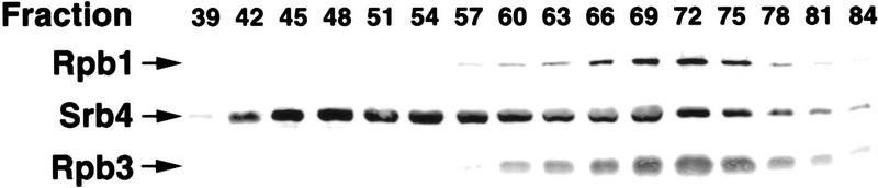
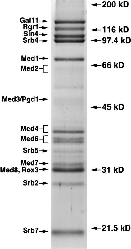
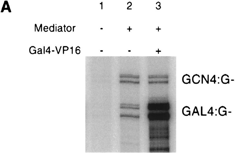
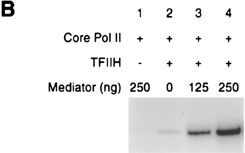
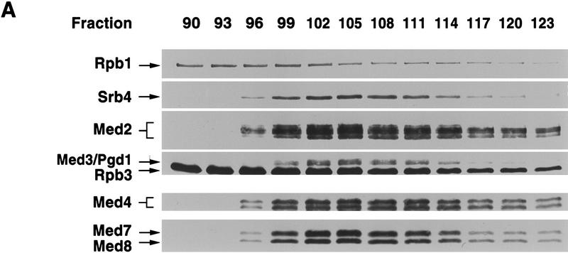
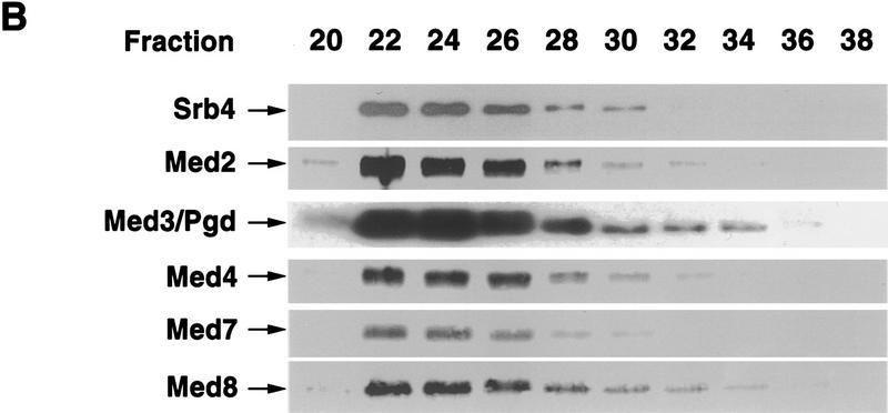
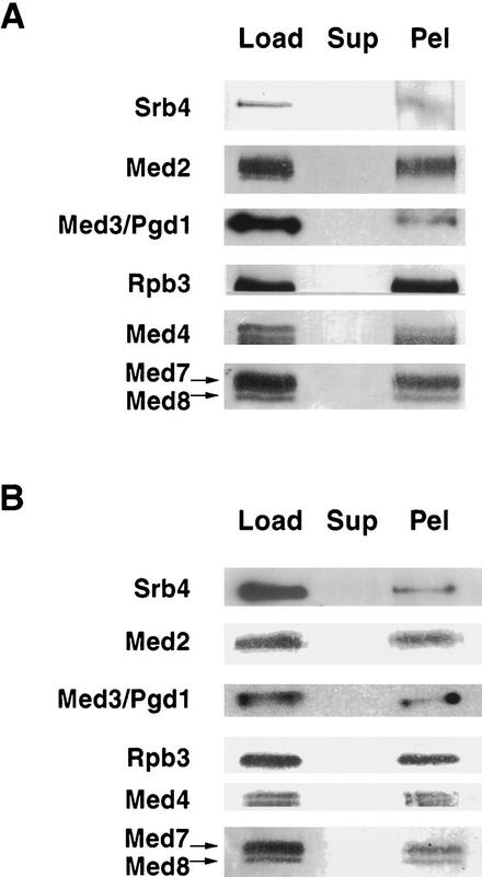
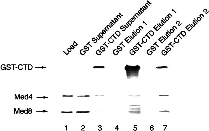
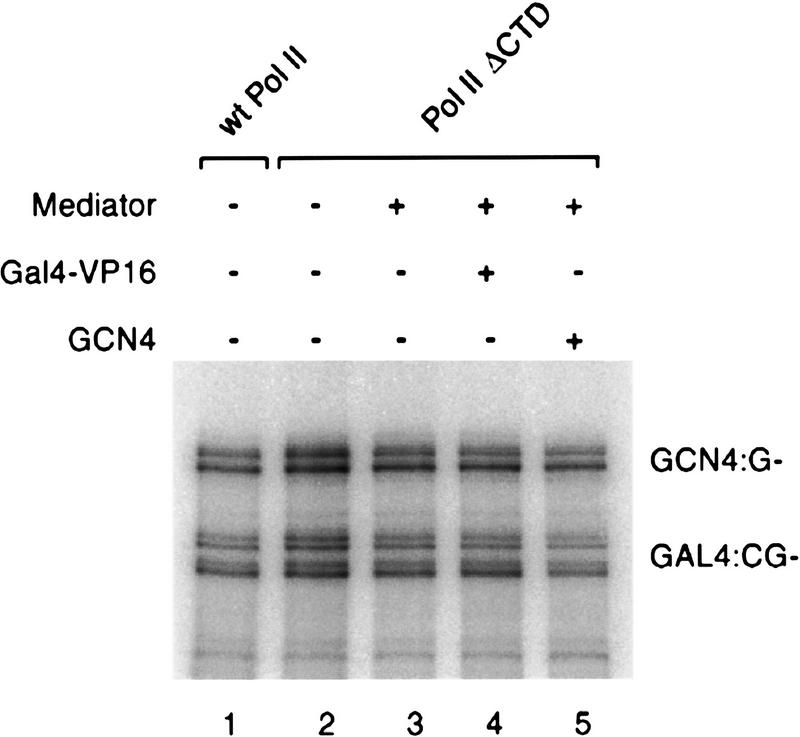
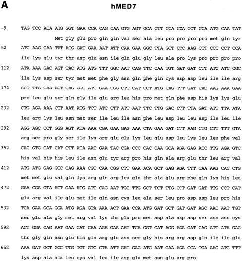
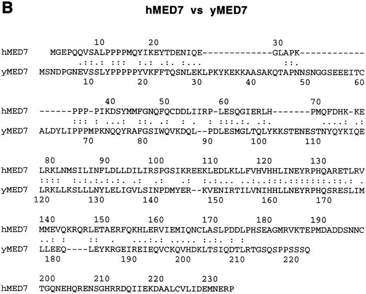
Mediator was resolved from yeast as a multiprotein complex on the basis of its requirement for transcriptional activation in a fully defined system. Three groups of mediator polypeptides could be distinguished: the products of five SRB genes, identified as suppressors of carboxy-terminal domain (CTD)-truncation mutants; products of four genes identified as global repressors; and six members of a new protein family, termed Med, thought to be primarily responsible for transcriptional activation. Notably absent from the purified mediator were Srbs 8, 9, 10, and 11, as well as members of the SWI/SNF complex. The CTD was required for function of mediator in vitro, in keeping with previous indications of involvement of the CTD in transcriptional activation in vivo. Evidence for human homologs of several mediator proteins, including Med7, points to similar mechanisms in higher cells.


Figures

Figure 1
Figure 1
Separation of free mediator and RNA polymerase II holoenzyme by chromatography on Mono Q. Hydroxyapatite fractions containing both mediator and polymerase II were applied to Mono Q and eluted with a linear gradient of potassium acetate. Fractions were analyzed by immunoblotting with antibodies against the mediator subunit Srb4 and against the polymerase II subunits Rpb1 and Rpb3. Mediator, in the free form, peaked at 600 mm potassium acetate and, in the holoenzyme form, peaked at 800 mm.
Figure 2
Figure 2
Polypeptide composition of free mediator. Peak fractions from Bio-Sil SEC 400 were pooled and analyzed by SDS-PAGE in an 11% gel. Proteins were revealed by staining with Coomassie blue. Srb6 was present, but was too small to be detected in the gel system used. Positions of molecular mass markers are indicated on the right.
Figure 3
Figure 3
Functional characterization of free mediator. (A) Activities of pure mediator (heparin fraction) in transcription. The final gel filtration fraction used to assess the stoichiometry of mediator polypeptides (see text) was functionally indistinguishable from the heparin fraction used here and of comparable purity (data not shown). Transcription was performed with templates containing binding sites for Gal4 (GAL4:G−) and Gcn4 (GCN4:G−) upstream of the S. cerevisiae CYC1 promoter fused to a G-less cassette (Kim et al. 1994). Reaction mixtures contained 2 μg of RNA polymerase II, 0.5 μg of mediator (lanes 2,3), and 10 ng of Gal4–VP16 (lane 3), in addition to basal transcription factors as described (Kim et al. 1994). Enhancement of basal transcription (lane 2) was 20.5-fold and could vary between 5- and 60-fold depending on reaction conditions. Activation of transcription by Gal4–VP16 (lane 3) was 18.4-fold, and could vary between 10- and 20-fold depending on reaction conditions. (B) Free mediator stimulates CTD phosphorylation by TFIIH. RNA polymerase II (Core Pol II, 100 ng) was phosphorylated by holoTFIIH (20 ng) in the presence of increasing amounts of a free mediator (heparin fraction). Stimulation of CTD kinase activity was 17.3-fold (lane 3) and 36.3-fold (lane 4).
Figure 3
Figure 3
Functional characterization of free mediator. (A) Activities of pure mediator (heparin fraction) in transcription. The final gel filtration fraction used to assess the stoichiometry of mediator polypeptides (see text) was functionally indistinguishable from the heparin fraction used here and of comparable purity (data not shown). Transcription was performed with templates containing binding sites for Gal4 (GAL4:G−) and Gcn4 (GCN4:G−) upstream of the S. cerevisiae CYC1 promoter fused to a G-less cassette (Kim et al. 1994). Reaction mixtures contained 2 μg of RNA polymerase II, 0.5 μg of mediator (lanes 2,3), and 10 ng of Gal4–VP16 (lane 3), in addition to basal transcription factors as described (Kim et al. 1994). Enhancement of basal transcription (lane 2) was 20.5-fold and could vary between 5- and 60-fold depending on reaction conditions. Activation of transcription by Gal4–VP16 (lane 3) was 18.4-fold, and could vary between 10- and 20-fold depending on reaction conditions. (B) Free mediator stimulates CTD phosphorylation by TFIIH. RNA polymerase II (Core Pol II, 100 ng) was phosphorylated by holoTFIIH (20 ng) in the presence of increasing amounts of a free mediator (heparin fraction). Stimulation of CTD kinase activity was 17.3-fold (lane 3) and 36.3-fold (lane 4).
Figure 4
Figure 4
Med proteins comigrate during hydroxyapatite chromatography and gel filtration. (A) The peak of RNA polymerase II holopolymerase from DEAE-Sephacel was applied to a 20 ml hydroxyapatite column and eluted with a 200 ml gradient of 0.01–0.2 mm potassium phosphate. Fractions were analyzed by SDS-PAGE in a 10% gel and immunoblotted with antibodies directed against the proteins indicated. (B) The mediator peak from Mono Q was subjected to gel filtration through Bio-Sil SEC 400. Fractions were analyzed by SDS-PAGE in a 10% gel and immunoblotted with antibodies directed against the proteins indicated.
Figure 4
Figure 4
Med proteins comigrate during hydroxyapatite chromatography and gel filtration. (A) The peak of RNA polymerase II holopolymerase from DEAE-Sephacel was applied to a 20 ml hydroxyapatite column and eluted with a 200 ml gradient of 0.01–0.2 mm potassium phosphate. Fractions were analyzed by SDS-PAGE in a 10% gel and immunoblotted with antibodies directed against the proteins indicated. (B) The mediator peak from Mono Q was subjected to gel filtration through Bio-Sil SEC 400. Fractions were analyzed by SDS-PAGE in a 10% gel and immunoblotted with antibodies directed against the proteins indicated.
Figure 5
Figure 5
Co-immunoprecipitation of Med proteins with RNA polymerase II holoenzyme. (A) Immunoprecipitation by anti-Med2 antibody coupled to protein A–Sepharose. An RNA polymerase II holopolymerase fraction from Mono Q (Load, 5 μg) was incubated with anti-Med2 antibody beads. The supernatant (Sup) was removed, the beads were washed, and immunoprecipitated protein was eluted (Pel). Equal amounts of the load, supernatant, and eluted proteins were analyzed by immunoblotting with antibodies against the proteins indicated. (B) Immunoprecipitation with anti-Med4 antibody coupled to protein A–Sepharose, as in A.
Figure 6
Figure 6
Mediator binds directly to the CTD. Free mediator was applied to GST–CTD and GST (control) resins and eluted with glutathione (Elution 1) and SDS (Elution 2). Proteins were analyzed by immunoblotting with antibodies directed against Med4, Med8, and the CTD (to reveal the presence of GST–CTD).
Figure 7
Figure 7
CTD requirement for mediator function in vitro. Transcription was performed as described with wild type polymerase (1 μg) or Pol II ΔCTD (1 μg) in the presence of mediator (500 ng), Gal4–VP16 (10 ng), and GCN4 (10 ng) as indicated.
Figure 8
Figure 8
Human homolog of yeast Med7 protein. (A) Human Med7 homolog sequence (GenBank accession no. AF031383). (B) Human and yeast Med7 alignment. Colons indicate identity and dots similarity by the program FASTA (Pearson and Lipman 1988). FASTA aligned the sequences using the BLOSUM50 matrix resulting in a Smith–Waterman score of 291, and a 31.8% identity and 59.2% similarity in a 211-amino-acid region of overlap.
Figure 8
Figure 8
Human homolog of yeast Med7 protein. (A) Human Med7 homolog sequence (GenBank accession no. AF031383). (B) Human and yeast Med7 alignment. Colons indicate identity and dots similarity by the program FASTA (Pearson and Lipman 1988). FASTA aligned the sequences using the BLOSUM50 matrix resulting in a Smith–Waterman score of 291, and a 31.8% identity and 59.2% similarity in a 211-amino-acid region of overlap.

References

    1. Boeke J, Truehart J, Natsoulis B, Fink GR. 5-Fluoroorotic acid as a selective agent in yeast molecular genetics. Methods Enzymol. 1987;154:164–175. - PubMed
    1. Bröhl S, Lisowsky T, Riemen G, Michaelis G. A new nuclear suppressor system for a mitochondrial RNA polymerase mutant identifies an unusual zinc-finger protein and a polyglutamine domain protein in Saccharomyces cerevisiae. Yeast. 1994;10:719–731. - PubMed
    1. Burley SK, Roeder RG. Biochemistry and structural biology of transcription factor IID (TFIID) Annu Rev Biochem. 1996;65:769–799. - PubMed
    1. Cairns BR, Kim YJ, Sayre MH, Laurent BC, Kornberg RD. A multisubunit complex containing the SWI1-ADR6, SWI2-SNF2, SWI3, SNF5, and SNF6 gene products isolated from yeast. Proc Natl Acad Sci. 1994;91:1950–1954. - PMC - PubMed
    1. Chao DM, Gadbois EL, Murray PJ, Anderson SF, Sonu MS, Parvin JD, Young RA. A mammalian SRB protein associated with an RNA polymerase II holoenzyme. Nature. 1996;380:82–85. - PubMed

Publication types

MeSH terms

Associated data