. 1998 Jan 1;12(1):55-66.
doi: 10.1101/gad.12.1.55.

A specific subset of SR proteins shuttles continuously between the nucleus and the cytoplasm

Affiliations

A specific subset of SR proteins shuttles continuously between the nucleus and the cytoplasm

J F Cáceres et al. Genes Dev. .

Abstract

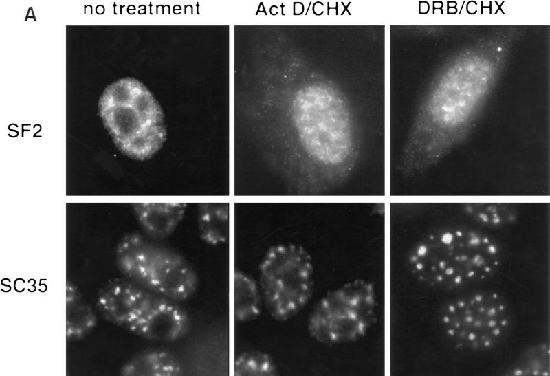
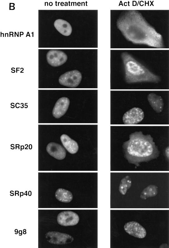
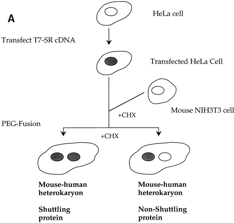
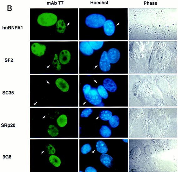
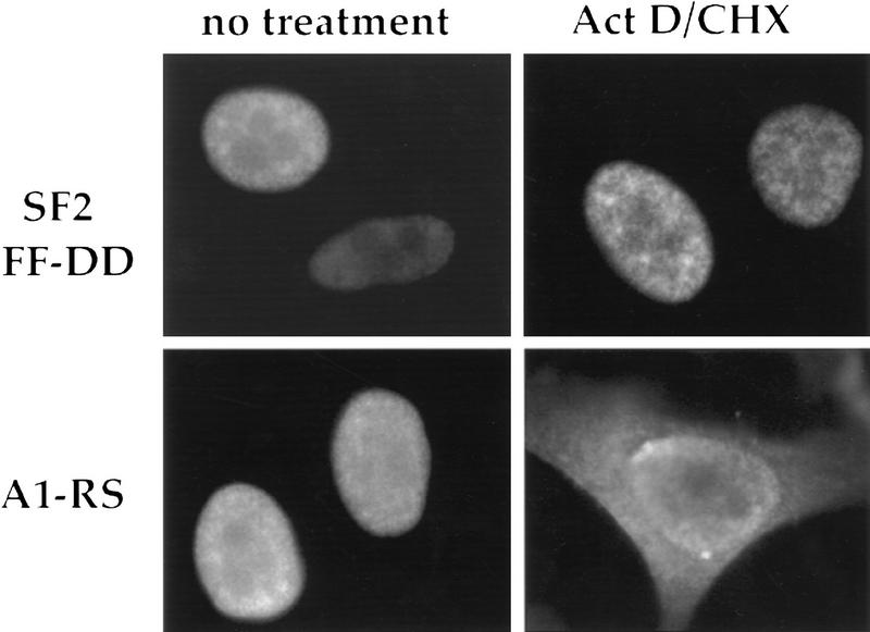
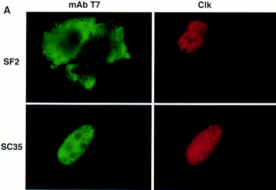
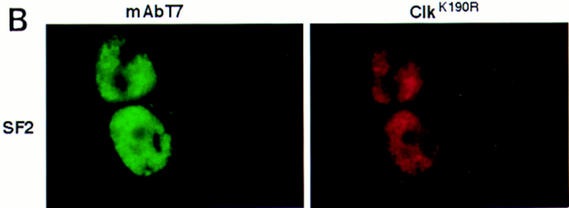
The SR proteins constitute a large family of nuclear phosphoproteins required for constitutive pre-mRNA splicing. These factors also have global, concentration-dependent effects on alternative splicing regulation and this activity is antagonized by members of the hnRNP A/B family of proteins. We show here that whereas some human SR proteins are confined to the nucleus, three of them-SF2/ASF, SRp20, and 9G8-shuttle rapidly and continuously between the nucleus and the cytoplasm. By swapping the corresponding domains between shuttling and nonshuttling SR proteins, we show that the carboxy-terminal arginine/serine-rich (RS) domain is required for shuttling. This domain, however, is not sufficient to promote shuttling of an unrelated protein reporter, suggesting that stable RNA binding mediated by the RNA-recognition motifs may be required for shuttling. Consistent with such a requirement, a double point-mutation in RRM1 of SF2/ASF that impairs RNA binding prevents the protein from shuttling. In addition, we show that phosphorylation of the RS domain affects the shuttling properties of SR proteins. These findings show that different SR proteins have unique intracellular transport properties and suggest that the family members that shuttle may have roles not only in nuclear pre-mRNA splicing but also in mRNA transport, cytoplasmic events, and/or processes that involve communication between the nucleus and the cytoplasm.


Figures

Figure 1
Figure 1
Effect of transcription inhibition on subcellular localization of SR proteins. (A) Endogenous SF2/ASF and SC35. HeLa cells were incubated alone (left panels), or in the presence of actinomycin D plus cycloheximide (middle panels), or of DRB plus cycloheximide (right panels) for 3 hr. The cells were fixed and the localization of SF2/ASF and of SC35 was determined by indirect immunofluorescence with an anti-SF2/ASF monoclonal antibody (top panels) or an SC35 monoclonal antibody (bottom panels). Cycloheximide alone had no effect, and was included together with the transcription inhibitors to prevent further protein synthesis from pre-existing mRNA. (B) Transiently expressed SR proteins. HeLa cells were transfected with expression plasmids encoding the indicated T7 epitope-tagged SR proteins, or hnRNP A1 as a control. At 24 hr post-transfection, the cells were incubated with actinomycin D plus cycloheximide (right panels) or mock treated (left panels) for 3 hr and subsequently fixed. The localization of the tagged proteins was determined by indirect immunofluorescence with anti-T7 monoclonal antibody and FITC-conjugated secondary antibody.
Figure 1
Figure 1
Effect of transcription inhibition on subcellular localization of SR proteins. (A) Endogenous SF2/ASF and SC35. HeLa cells were incubated alone (left panels), or in the presence of actinomycin D plus cycloheximide (middle panels), or of DRB plus cycloheximide (right panels) for 3 hr. The cells were fixed and the localization of SF2/ASF and of SC35 was determined by indirect immunofluorescence with an anti-SF2/ASF monoclonal antibody (top panels) or an SC35 monoclonal antibody (bottom panels). Cycloheximide alone had no effect, and was included together with the transcription inhibitors to prevent further protein synthesis from pre-existing mRNA. (B) Transiently expressed SR proteins. HeLa cells were transfected with expression plasmids encoding the indicated T7 epitope-tagged SR proteins, or hnRNP A1 as a control. At 24 hr post-transfection, the cells were incubated with actinomycin D plus cycloheximide (right panels) or mock treated (left panels) for 3 hr and subsequently fixed. The localization of the tagged proteins was determined by indirect immunofluorescence with anti-T7 monoclonal antibody and FITC-conjugated secondary antibody.
Figure 2
Figure 2
Analysis of nucleocytoplasmic shuttling of SR proteins by transient expression in interspecies heterokaryons. (A) Diagram of the experimental approach. The shaded nuclei indicate localization of the transiently expressed human SR protein. Cycloheximide (CHX) was added before fusion to prevent further protein synthesis from the human mRNA in the heterokaryons. (B) Detection of the transiently expressed SR proteins in interspecies heterokaryons. HeLa cells were transfected with expression plasmids encoding the indicated T7 epitope-tagged proteins, or hnRNP A1 as a control. At 24 hr post-transfection, the HeLa cells were treated with cycloheximide and subsequently fused with mouse NIH 3T3 cells in the presence of polyethylene glycol to form heterokaryons. The cells were incubated further for 2 hr in the presence of cycloheximide, followed by fixation. The localization of the expressed proteins was determined by indirect immunofluorescence with anti-T7 monoclonal antibody and FITC-conjugated secondary antibody (left panels). The cells were simultaneously incubated with Hoechst 33258 for differential staining of human and mouse nuclei within heterokaryons (middle panels). The arrows indicate the mouse nuclei within human–mouse heterokaryons. Phase-contrast images of the same heterokaryons are shown (right panels).
Figure 2
Figure 2
Analysis of nucleocytoplasmic shuttling of SR proteins by transient expression in interspecies heterokaryons. (A) Diagram of the experimental approach. The shaded nuclei indicate localization of the transiently expressed human SR protein. Cycloheximide (CHX) was added before fusion to prevent further protein synthesis from the human mRNA in the heterokaryons. (B) Detection of the transiently expressed SR proteins in interspecies heterokaryons. HeLa cells were transfected with expression plasmids encoding the indicated T7 epitope-tagged proteins, or hnRNP A1 as a control. At 24 hr post-transfection, the HeLa cells were treated with cycloheximide and subsequently fused with mouse NIH 3T3 cells in the presence of polyethylene glycol to form heterokaryons. The cells were incubated further for 2 hr in the presence of cycloheximide, followed by fixation. The localization of the expressed proteins was determined by indirect immunofluorescence with anti-T7 monoclonal antibody and FITC-conjugated secondary antibody (left panels). The cells were simultaneously incubated with Hoechst 33258 for differential staining of human and mouse nuclei within heterokaryons (middle panels). The arrows indicate the mouse nuclei within human–mouse heterokaryons. Phase-contrast images of the same heterokaryons are shown (right panels).
Figure 3
Figure 3
Domain requirements for SR protein shuttling. (A) Analysis of shuttling of SF2/ASF–SRp40 chimeras by transient expression and transcription inhibition. HeLa cells transfected with epitope-tagged constructs and either mock treated (left panels), or treated with actinomycin D and cycloheximide (right panels), were analyzed as in Fig. 1B. SF2/ASF (top row) and SRp40 (second row) are composed of two RRMs and a carboxy-terminal RS domain. The names of the chimeras (bottom four rows) denote the origin of each domain, with RRM1 and RRM2 indicated by roman numerals and the subscript indicating whether the domain derives from SF2/ASF or from SRp40. (B) Shuttling by a chimera with the RRM of SC35 and the RS domain of SF2/ASF. Cells were transfected, treated, and analyzed as in A, using either epitope-tagged SC35 (top row) or a construct consisting of the single RRM from SC35 (denoted by I35) and the RS domain from SF2/ASF (denoted by RSSF2) replacing the SC35 carboxy-terminal RS domain. (C) Analysis of shuttling of SR protein chimeras by the heterokaryon assay. SR protein chimeras were transiently expressed in HeLa cells, which were then treated with cycloheximide and fused to NIH 3T3 cells. The localization of the epitope-tagged proteins in the heterokaryons was analyzed as in Fig. 2B. The chimeras are named as in A and B, and the arrows indicate the mouse nuclei in the human–mouse heterokaryons. The same heterokaryons were analyzed for localization of the epitope-tagged proteins (left panels), staining with Hoechst 33258 (middle panels), and by phase-contrast (right panels).
Figure 3
Figure 3
Domain requirements for SR protein shuttling. (A) Analysis of shuttling of SF2/ASF–SRp40 chimeras by transient expression and transcription inhibition. HeLa cells transfected with epitope-tagged constructs and either mock treated (left panels), or treated with actinomycin D and cycloheximide (right panels), were analyzed as in Fig. 1B. SF2/ASF (top row) and SRp40 (second row) are composed of two RRMs and a carboxy-terminal RS domain. The names of the chimeras (bottom four rows) denote the origin of each domain, with RRM1 and RRM2 indicated by roman numerals and the subscript indicating whether the domain derives from SF2/ASF or from SRp40. (B) Shuttling by a chimera with the RRM of SC35 and the RS domain of SF2/ASF. Cells were transfected, treated, and analyzed as in A, using either epitope-tagged SC35 (top row) or a construct consisting of the single RRM from SC35 (denoted by I35) and the RS domain from SF2/ASF (denoted by RSSF2) replacing the SC35 carboxy-terminal RS domain. (C) Analysis of shuttling of SR protein chimeras by the heterokaryon assay. SR protein chimeras were transiently expressed in HeLa cells, which were then treated with cycloheximide and fused to NIH 3T3 cells. The localization of the epitope-tagged proteins in the heterokaryons was analyzed as in Fig. 2B. The chimeras are named as in A and B, and the arrows indicate the mouse nuclei in the human–mouse heterokaryons. The same heterokaryons were analyzed for localization of the epitope-tagged proteins (left panels), staining with Hoechst 33258 (middle panels), and by phase-contrast (right panels).
Figure 4
Figure 4
The RS domain of shuttling SR proteins is not sufficient for shuttling. Interspecies heterokaryon analysis of transiently expressed fusions of the nucleoplasmin core domain reporter (NPc) and the RS domain of SF2/ASF or SRp20. HeLa cells were transfected with plasmids encoding the indicated epitope-tagged proteins, fused to mouse cells, and the heterokaryons were analyzed as in Fig. 2B.
Figure 5
Figure 5
RNA binding is required for shuttling. HeLa cells were transfected with expression plasmids encoding the indicated T7 epitope-tagged SR proteins. At 24 hr post-transfection, the cells were incubated with actinomycin D plus cycloheximide (right panels) or mock treated (left panels) for 3 hr and subsequently fixed. The localization of the tagged proteins was determined by indirect immunofluorescence with anti-T7 monoclonal antibody and FITC-conjugated secondary antibody.
Figure 6
Figure 6
Effect of expression of the Clk/Sty kinase on SF2/ASF shuttling. (A) HeLa cells were cotransfected with plasmids encoding either T7-tagged SF2/ASF (top panels) or SC35 (bottom panels), together with another plasmid expressing a myc-tagged Clk/Sty kinase. The localization of the transiently expressed SR proteins was determined by indirect immunofluorescence with anti-T7 monoclonal antibody and FITC-conjugated secondary antibody (left panels), and that of Clk/Sty with a rabbit polyclonal anti-myc antibody and Texas-Red conjugated secondary antibody (right panels). (B) Same as in A, except that a mutant Clk/Sty kinase, Clk/StyK190R, was cotransfected with T7-tagged SF2/ASF.
Figure 6
Figure 6
Effect of expression of the Clk/Sty kinase on SF2/ASF shuttling. (A) HeLa cells were cotransfected with plasmids encoding either T7-tagged SF2/ASF (top panels) or SC35 (bottom panels), together with another plasmid expressing a myc-tagged Clk/Sty kinase. The localization of the transiently expressed SR proteins was determined by indirect immunofluorescence with anti-T7 monoclonal antibody and FITC-conjugated secondary antibody (left panels), and that of Clk/Sty with a rabbit polyclonal anti-myc antibody and Texas-Red conjugated secondary antibody (right panels). (B) Same as in A, except that a mutant Clk/Sty kinase, Clk/StyK190R, was cotransfected with T7-tagged SF2/ASF.

References

    1. Alzhanova-Ericsson AT, Sun X, Visa N, Kiseleva E, Wurtz T, Daneholt B. A protein of the SR family of splicing factors binds extensively to exonic Balbiani ring pre-mRNA and accompanies the RNA from the gene to the nuclear pore. Genes & Dev. 1996;10:2881–2893. - PubMed
    1. Birney E, Kumar S, Krainer AR. Analysis of the RNA-recognition motif and RS and RGG domains: Conservation in metazoan pre-mRNA splicing factors. Nucleic Acids Res. 1993;21:5803–5816. - PMC - PubMed
    1. Blencowe BJ, Nickerson JA, Issner R, Penman S, Sharp PA. Association of nuclear antigens with exon-containing splicing complexes. J Cell Biol. 1994;127:593–607. - PMC - PubMed
    1. Blencowe BJ, Issner R, Kim J, McCaw P, Sharp PA. New proteins related to the Ser-Arg family of splicing factors. RNA. 1995;1:852–865. - PMC - PubMed
    1. Borer RA, Lehner CF, Eppenberger HM, Nigg EA. Major nucleolar proteins shuttle between nucleus and cytoplasm. Cell. 1989;56:379–390. - PubMed

Publication types

MeSH terms