. 1998 Jan 1;12(1):67-83.
doi: 10.1101/gad.12.1.67.

A prokaryotic-like mode of cytoplasmic eukaryotic ribosome binding to the initiation codon during internal translation initiation of hepatitis C and classical swine fever virus RNAs

Affiliations

A prokaryotic-like mode of cytoplasmic eukaryotic ribosome binding to the initiation codon during internal translation initiation of hepatitis C and classical swine fever virus RNAs

T V Pestova et al. Genes Dev. .

Abstract

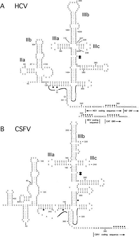
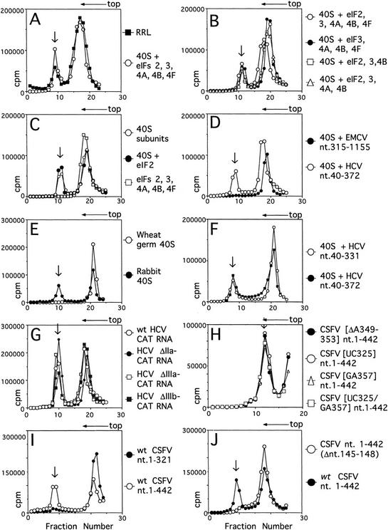
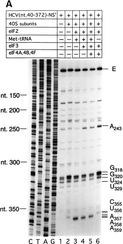
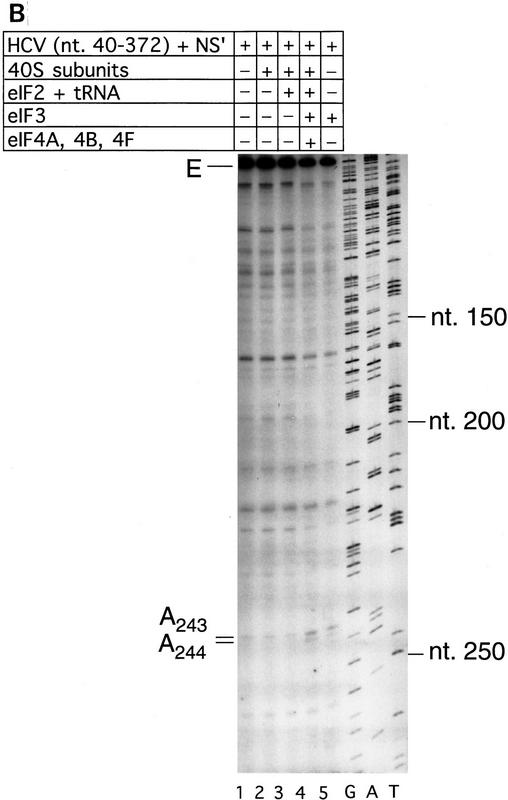
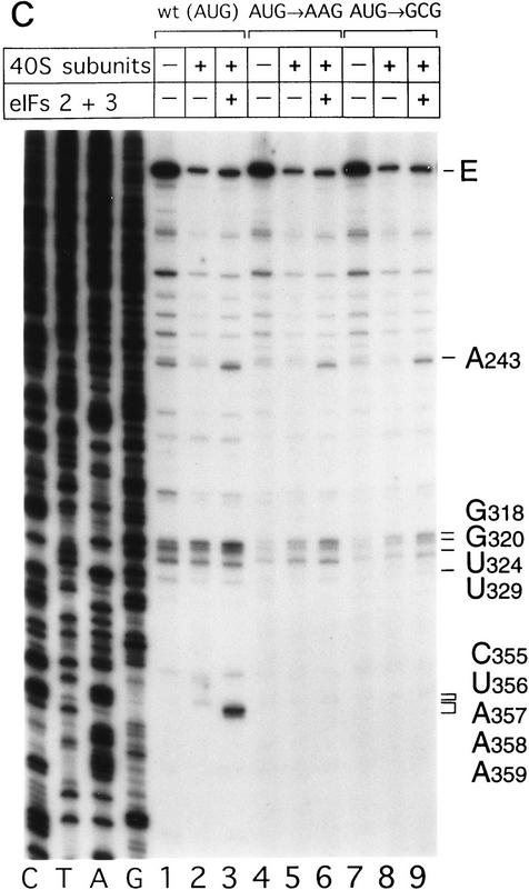
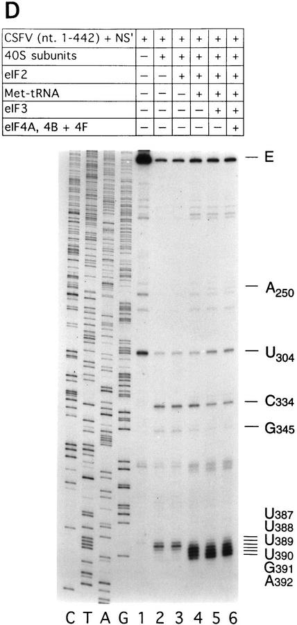
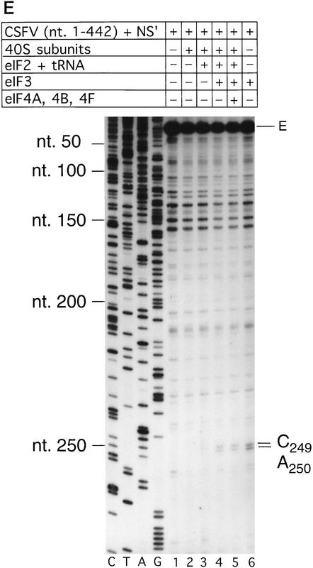
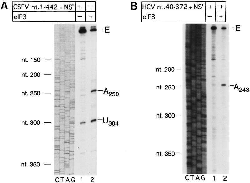
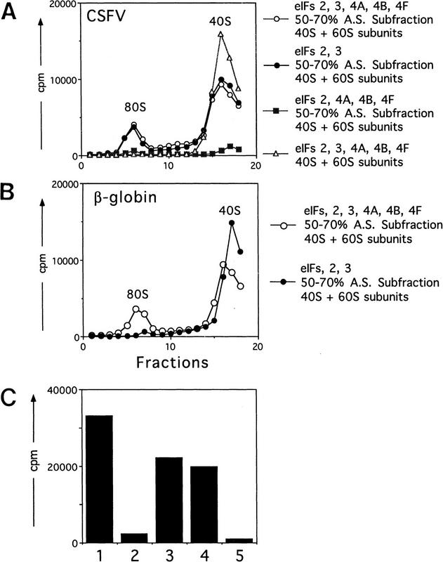
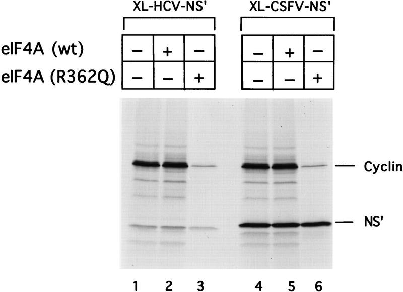
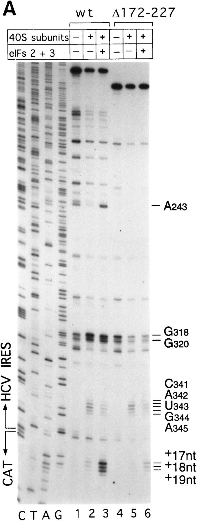
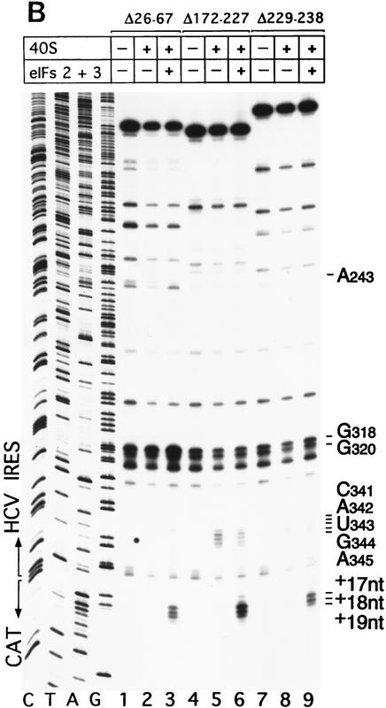
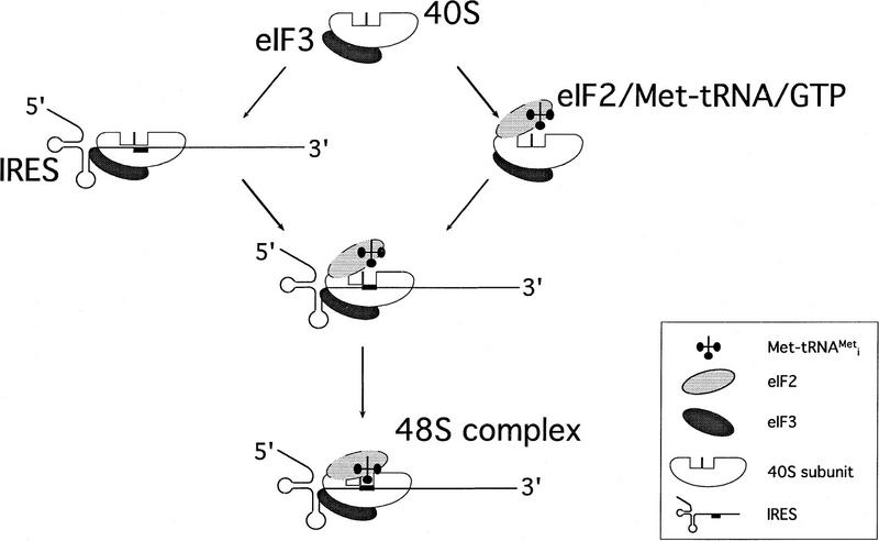
Initiation of translation of hepatitis C virus and classical swine fever virus mRNAs results from internal ribosomal entry. We reconstituted internal ribosomal entry in vitro from purified translation components and monitored assembly of 48S ribosomal preinitiation complexes by toe-printing. Ribosomal subunits (40S) formed stable binary complexes on both mRNAs. The complex structure of these RNAs determined the correct positioning of the initiation codon in the ribosomal "P" site in binary complexes. Ribosomal binding and positioning on these mRNAs did not require the initiation factors eIF3, eIF4A, eIF4B, and eIF4F and translation of these mRNAs was not inhibited by a trans-dominant eIF4A mutant. Addition of Met-tRNAiMet, eIF2, and GTP to these binary ribosomal complexes resulted in formation of 48S preinitiation complexes. The striking similarities between this eukaryotic initiation mechanism and the mechanism of translation initiation in prokaryotes are discussed.


Figures

Figure 1
Figure 1
Model secondary and tertiary RNA structures of the 5′NTRs of hepatitis C virus (A) and classical swine fever virus (B), based on proposals by Brown et al. (1992), Honda et al. (1996), and Wang et al. (1995). The nomenclature of domains is as described by Honda et al. (1996). HCV constructs were linked either to NS′ or CAT reporter cistrons–both sequences are shown. An NS′ reporter cistron (not shown) was linked 74 nucleotides downstream of the CSFV initiation codon in all CSFV constructs. HCV and CSFV initiation codons are underlined. Stop-sites where primer extension was arrested on naked RNA only are indicated by open circles; sites of RT arrest caused or enhanced by binding of eIF3 and of 40S subunits are indicated by solid diamonds and circles, respectively. The 3′ border of the HCV nucleotides 26–67 deletion and both borders of the HCV nucleotides 172–227 and HCV nucleotides 229–238 deletions are labeled and indicated by asterisks.
Figure 2
Figure 2
Ribosomal complex formation on HCV and CSFV IRESs. Assays were done using HCV nucleotides 40–372 RNA (A–F) and HCV, CSFV, and EMCV RNAs (D,F–J) as indicated and with RRL or rabbit 40S subunits and eIF2, eIF3, eIF4A, eIF4B, and eIF4F (A) with rabbit 40S subunits and combinations of eIF2, eIF3, eIF4A, eIF4B, and eIF4F (B,C) and with rabbit or (where indicated) wheat germ 40S subunits (D–J). ATP was included only in reactions that contained eIF4A, eIF4B, or eIF4F; Met–tRNAiMet and GMP–PNP were included only in reactions that contained eIF2. Other components described in Materials and Methods were present in all reactions. Sedimentation was from right to left. The positions of ribosomal complexes are indicated by arrows. (G) HCV ΔIIa–CAT, ΔIIIb–CAT, and ΔIIIc–CAT mRNAs are identical to wild-type HCV–CAT mRNA except for deletions of HCV nucleotides 28–67, 172–277, and 229–238, respectively. Sucrose gradients shown in H were centrifuged for a shorter time than others shown here. Fractions from upper parts of the sucrose gradient have been omitted from graphs in H for greater clarity.
Figure 3
Figure 3
Toeprint analysis of 48S complex formation on HCV and CSFV IRESs. (A) Wild-type HCV (nucleotides 40–372)–NS′ RNA was incubated with 40S ribosomal subunits (lanes 2–6), eIF2 (lanes 3–6), Met–tRNAiMet (lanes 3–5), eIF3 (lanes 4–6), and eIF4A, eIF4B, and eIF4F (lanes 5,6); (B) wild-type HCV (nucleotides 40–372)–NS′ RNA was incubated with 40S ribosomal subunits (lanes 2–4), eIF2, Met–tRNAiMet, and GMP–PNP (lanes 3,4), eIF3 (lanes 4,5), and eIF4A, eIF4B, and eIF4F (lane 4); (C) wildtype HCV (nucleotides 40–372)–NS′ (lanes 1–3), (AUG → AAG) mutant HCV (nucleotides 40–372)–NS′ (lanes 4–6), and (AUG → GCG) mutant HCV (nucleotides 40–372)–NS′ RNAs (lanes 7–9) were incubated with 40S ribosomal subunits (lanes 2,3,5,6,8,9) and with eIF2 and eIF3, Met–tRNAiMet and GMP–PNP (lanes 3,6,9); (D) wild-type CSFV (nucleotides 1–442)–NS′ RNA was incubated with 40S ribosomal subunits (lanes 2–6), eIF2 (lanes 3–6), Met–tRNAiMet (lanes 4–6), eIF3 (lanes 5–6), and eIF4A, eIF4B, and eIF4F (lane 6); (E) wild-type CSFV (nucleotides 1–442)–NS′ RNA was incubated with 40S ribosomal subunits (lanes 2–5), eIF2, Met–tRNA iMet and GMP–PNP (lanes 3–5), eIF3 (lanes 4–6), and eIF4A, eIF4B, and eIF4F (lane 5). Reaction conditions are described in Materials and Methods. In A,C, and D, the primers (5′-GGGATTTCTGATCTCGGCG-3′) and (5′-CTCGTTTGCGGACATGCC-3′) were annealed to the NS′ cistron 130 nucleotides downstream of the HCV initiation codon and 110 nucleotides downstream of the CSFV initiation codon, respectively, and were extended with AMV–RT. In B and D, the primers 5′-CGCAAGCACCCTATC-3′ (complementary to HCV nucleotides 295–309) and 5′-CCTGATAGGGTGCTGCAG-3′ (complementary to CSFV nucleotides 309–326) were annealed to HCV and CSFV IRESs, as appropriate, and were extended with AMV–RT. Full-length cDNA is marked E. Other cDNA products terminated at the sites are indicated on the right. Reference lanes C,T,A, and G depict HCV or CSFV sequences, as appropriate.
Figure 3
Figure 3
Toeprint analysis of 48S complex formation on HCV and CSFV IRESs. (A) Wild-type HCV (nucleotides 40–372)–NS′ RNA was incubated with 40S ribosomal subunits (lanes 2–6), eIF2 (lanes 3–6), Met–tRNAiMet (lanes 3–5), eIF3 (lanes 4–6), and eIF4A, eIF4B, and eIF4F (lanes 5,6); (B) wild-type HCV (nucleotides 40–372)–NS′ RNA was incubated with 40S ribosomal subunits (lanes 2–4), eIF2, Met–tRNAiMet, and GMP–PNP (lanes 3,4), eIF3 (lanes 4,5), and eIF4A, eIF4B, and eIF4F (lane 4); (C) wildtype HCV (nucleotides 40–372)–NS′ (lanes 1–3), (AUG → AAG) mutant HCV (nucleotides 40–372)–NS′ (lanes 4–6), and (AUG → GCG) mutant HCV (nucleotides 40–372)–NS′ RNAs (lanes 7–9) were incubated with 40S ribosomal subunits (lanes 2,3,5,6,8,9) and with eIF2 and eIF3, Met–tRNAiMet and GMP–PNP (lanes 3,6,9); (D) wild-type CSFV (nucleotides 1–442)–NS′ RNA was incubated with 40S ribosomal subunits (lanes 2–6), eIF2 (lanes 3–6), Met–tRNAiMet (lanes 4–6), eIF3 (lanes 5–6), and eIF4A, eIF4B, and eIF4F (lane 6); (E) wild-type CSFV (nucleotides 1–442)–NS′ RNA was incubated with 40S ribosomal subunits (lanes 2–5), eIF2, Met–tRNA iMet and GMP–PNP (lanes 3–5), eIF3 (lanes 4–6), and eIF4A, eIF4B, and eIF4F (lane 5). Reaction conditions are described in Materials and Methods. In A,C, and D, the primers (5′-GGGATTTCTGATCTCGGCG-3′) and (5′-CTCGTTTGCGGACATGCC-3′) were annealed to the NS′ cistron 130 nucleotides downstream of the HCV initiation codon and 110 nucleotides downstream of the CSFV initiation codon, respectively, and were extended with AMV–RT. In B and D, the primers 5′-CGCAAGCACCCTATC-3′ (complementary to HCV nucleotides 295–309) and 5′-CCTGATAGGGTGCTGCAG-3′ (complementary to CSFV nucleotides 309–326) were annealed to HCV and CSFV IRESs, as appropriate, and were extended with AMV–RT. Full-length cDNA is marked E. Other cDNA products terminated at the sites are indicated on the right. Reference lanes C,T,A, and G depict HCV or CSFV sequences, as appropriate.
Figure 4
Figure 4
Primer extension analysis of RNP complexes formed on CSFV and HCV IRES elements as follows. (A) CSFV (nucleotides 1–442)–NS′ and (B) HCV (nucleotides 340–372)–NS′ RNAs were incubated with (lane 2) or without eIF3 (lane 1) under standard conditions. Primers were annealed to the NS′-coding sequences of these RNAs and extended with AMV–RT. The full-length cDNA extension product is marked E. The cDNA products labeled A250, U304, and A243 on the right terminated at these nucleotides. The reference lanes C, T, A, and G depict the CSFV sequence (A) and the HCV sequence (B). Positions of CSFV and HCV nucleotides are indicated on the left of the appropriate panel.
Figure 5
Figure 5
Assembly of 80S ribosomal initiation complexes on the CSFV IRES and on β-globin mRNA and analysis of their activity in stimulating methionylpuromycin synthesis. Assays were done using CSFV (nucleotides 1–442)–NS′ mRNA (A,C) and β-globin mRNA (B,C) and with initiation factors and other translation components as described in Materials and Methods. (A,B) Sedimentation was from right to left, and the formation of complexes was assayed by incorporation of [35S]methionine–tRNA. The positions of ribosomal complexes are indicated. Fractions from upper parts of sucrose gradients have been omitted from graphs in A and B for greater clarity. (C) The methionylpuromycin synthesis assay was done as described in Materials and Methods, using β-globin mRNA (columns 1 and 2); CSFV (nucleotides 1–442)–NS′ mRNA (columns 3–5); eIF2, eIF3, eIF4A, eIF4B, eIF4F (columns 1 and 3), eIF2 and eIF3 (columns 2 and 4); and eIF2, eIF4A, eIF4B, and eIF4F (column 5). All reactions also contained GTP, 40S and 60S ribosomal subunits, and a 50%–70% A.S. RSW subfraction as described in Materials and Methods. A background value determined with charged initiator tRNA alone was subtracted from all panels.
Figure 6
Figure 6
Dominant-negative effect of the eIF4A R362Q mutant protein on cap-mediated translation and its lack of effect on HCV and CSFV IRES-mediated translation. Reticulocyte lysate (10 μl) was preincubated alone (lanes 1,4), with eIF4A wild-type (1.2 μg) (lanes 2,5), or eIF4A mutant (1.2 μg) (lanes 3,6) for 5 min at 30°C, then incubated for 60 min at 30°C with dicistronic mRNAs (0.5 μg) as described in Materials and Methods. Translation products were analyzed by autoradiography after electrophoresis on SDS–17% polyacrylamide gel.
Figure 7
Figure 7
Binding of 40S subunits and assembly of 48S complexes on HCV and CSFV mutant IRESs. (A) Wild-type HCV (nucleotides 1–349)–CAT (lanes 1–3) and mutant HCV (nucleotides 1–349Δ172–227)–CAT (lanes 4–6) were incubated under standard conditions with 40S ribosomal subunits (lanes 2,3 and 5,6) and with eIF2, eIF3, Met–tRNAiMet and GMP–PNP (lanes 3,6). (B) Mutant HCV (nucleotides 1–380Δ26–67)–CAT (lanes 1–3), (nucleotides 1–380Δ172–227)–CAT (lanes 4–6) and (nucleotides 1–380Δ229–238) (lanes 7–9) RNAs were incubated with 40S subunits (lanes 2,3,5,6,8,9) and with eIF2, eIF3, Met–tRNAiMet, and GMP–PNP (lanes 3,6,9). Reaction conditions are described in Materials and Methods. A primer (5′-GCAACTGACTGAAATGCC-3′) was annealed to the CAT cistron ∼80 nucleotides downstream of the HCV initiation codon and was extended with AMV–RT. Primer extension was arrested at sites indicated on the right either as nucleotides in the HCV IRES or as positions relative to the A of the HCV initiation codon. Reference lanes C, T, A, and G depict the HCV–CAT sequence. The junction between the HCV IRES and the CAT cistron is indicated. This gel was exposed to x-ray film longer than that shown in A to show the differences in the intensity of stops on various mutant RNAs. (C) Model of the CSFV pseudoknot and adjacent nucleotides showing the substitutions present in the mutants UC325, GA357, and UC325GA357 and the deletions made in the mutants ΔA145–U148 and ΔA349–A353. In this model, helix I of the pseudoknot comprises nucleotides 129–142 and 331–346, and helix II comprises nucleotides 323–329 and 354–360. (D) CSFV (nucleotides 1–442)–NS′ RNAs containing the mutations UC325 (lanes 1–3), GA357 (lanes 4–6), UC325GA357 (lanes 7–9), and ΔA349–A353 (lanes 10–12) were incubated with 40S subunits (lanes 2,3,5,6,8,9,11,12) and with eIF2, eIF3, Met–tRNAiMet and GMP–PNP (lanes 3,6,9,12).
Figure 7
Figure 7
Binding of 40S subunits and assembly of 48S complexes on HCV and CSFV mutant IRESs. (A) Wild-type HCV (nucleotides 1–349)–CAT (lanes 1–3) and mutant HCV (nucleotides 1–349Δ172–227)–CAT (lanes 4–6) were incubated under standard conditions with 40S ribosomal subunits (lanes 2,3 and 5,6) and with eIF2, eIF3, Met–tRNAiMet and GMP–PNP (lanes 3,6). (B) Mutant HCV (nucleotides 1–380Δ26–67)–CAT (lanes 1–3), (nucleotides 1–380Δ172–227)–CAT (lanes 4–6) and (nucleotides 1–380Δ229–238) (lanes 7–9) RNAs were incubated with 40S subunits (lanes 2,3,5,6,8,9) and with eIF2, eIF3, Met–tRNAiMet, and GMP–PNP (lanes 3,6,9). Reaction conditions are described in Materials and Methods. A primer (5′-GCAACTGACTGAAATGCC-3′) was annealed to the CAT cistron ∼80 nucleotides downstream of the HCV initiation codon and was extended with AMV–RT. Primer extension was arrested at sites indicated on the right either as nucleotides in the HCV IRES or as positions relative to the A of the HCV initiation codon. Reference lanes C, T, A, and G depict the HCV–CAT sequence. The junction between the HCV IRES and the CAT cistron is indicated. This gel was exposed to x-ray film longer than that shown in A to show the differences in the intensity of stops on various mutant RNAs. (C) Model of the CSFV pseudoknot and adjacent nucleotides showing the substitutions present in the mutants UC325, GA357, and UC325GA357 and the deletions made in the mutants ΔA145–U148 and ΔA349–A353. In this model, helix I of the pseudoknot comprises nucleotides 129–142 and 331–346, and helix II comprises nucleotides 323–329 and 354–360. (D) CSFV (nucleotides 1–442)–NS′ RNAs containing the mutations UC325 (lanes 1–3), GA357 (lanes 4–6), UC325GA357 (lanes 7–9), and ΔA349–A353 (lanes 10–12) were incubated with 40S subunits (lanes 2,3,5,6,8,9,11,12) and with eIF2, eIF3, Met–tRNAiMet and GMP–PNP (lanes 3,6,9,12).
Figure 8
Figure 8
Effect of deleting nucleotides 145–148 on CSFV IRES-mediated translation, binding of 40S subunits and assembly of 48S complexes. (A) RRL (10 μl) was incubated alone (lane 1), with 0.5 μg wild-type CSFV (nucleotides 1–442)–NS′ RNA (lane 2), or with 0.5 μg mutant CSFV (nucleotides 1–442Δ145–148) RNA (lane 3) for 60 min at 30°C as described in Materials and Methods. Translation products were analyzed by autoradiography after electrophoresis on SDS-17% polyacrylamide gel. (B) Wild-type CSFV (nucleotides 1–442)–NS′ (lanes 1–3) and mutant CSFV (nucleotides 1–442Δ145–148) RNAs (lanes 4–6) were incubated with 40S subunits (lanes 2,3,5,6) and with eIF2, eIF3, Met–tRNA iMet and GMP–PNP (lanes 3,6). Reaction conditions are described in Materials and Methods. An unlabeled primer (5′-GGGATTTCTGATCTCGGCG-3′) was annealed to the NS′ reporter cistron and was extended with AMV–RT in the presence of [α-32P]dATP. RT stop sites are indicated on the right. Reference lanes C,T, A, and G depict the CSFV sequence.
Figure 8
Figure 8
Effect of deleting nucleotides 145–148 on CSFV IRES-mediated translation, binding of 40S subunits and assembly of 48S complexes. (A) RRL (10 μl) was incubated alone (lane 1), with 0.5 μg wild-type CSFV (nucleotides 1–442)–NS′ RNA (lane 2), or with 0.5 μg mutant CSFV (nucleotides 1–442Δ145–148) RNA (lane 3) for 60 min at 30°C as described in Materials and Methods. Translation products were analyzed by autoradiography after electrophoresis on SDS-17% polyacrylamide gel. (B) Wild-type CSFV (nucleotides 1–442)–NS′ (lanes 1–3) and mutant CSFV (nucleotides 1–442Δ145–148) RNAs (lanes 4–6) were incubated with 40S subunits (lanes 2,3,5,6) and with eIF2, eIF3, Met–tRNA iMet and GMP–PNP (lanes 3,6). Reaction conditions are described in Materials and Methods. An unlabeled primer (5′-GGGATTTCTGATCTCGGCG-3′) was annealed to the NS′ reporter cistron and was extended with AMV–RT in the presence of [α-32P]dATP. RT stop sites are indicated on the right. Reference lanes C,T, A, and G depict the CSFV sequence.
Figure 9
Figure 9
UV cross-linking of ribosomal protein S9 to HCV and CSFV IRESs. (A) Proteins from rabbit ribosomal 40S subunits (lane 1) or from binary complexes of rabbit 40S ribosomal subunits and [32P]UTP-labeled HCV nucleotides 40–372 RNA (lane 2) were resolved by gel electrophoresis directly (lane 1) or after UV cross-linking and RNase digestion (lane 2). The positions of molecular mass marker proteins are indicated to the left of lane 1. (B) Proteins from binary complexes of rabbit 40S ribosomal subunits and [32P]UTP-labeled HCV nucleotides 40–372 RNA (lane 1), HCV nucleotides 40–331 RNA (lane 2), CSFV nucleotides 1–442 RNA (lane 3), or CSFV nucleotides 1–442(Δ nucleotides 145–148) (lane 4) were resolved by gel electrophoresis after UV cross-linking, and RNase digestion. (C) Proteins from binary complexes of rabbit 40S ribosomal subunits and [32P]UTP-labeled HCV nucleotides 40–372Δ26–67 RNA (lane 1), HCV nucleotides 40–372Δ172–227 RNA (lane 2), HCV nucleotides 40–372Δ229–238 RNA (lane 3), HCV nucleotides 40–372 (lane 4), CSFV nucleotides 1–442 (TC325GA357) (lane 5), CSFV nucleotides 1–442 (ΔA349–A353) (lane 6), CSFV nucleotides 1–442 (GA357) (lane 7), or CSFV nucleotides 1–442 (TC325) (lane 8) were resolved by gel electrophoresis after UV cross-linking, and RNase digestion. Proteins in A (lane 1) were visualized by staining with Coomassie blue; radio-labeled proteins in all other lanes in A,B, and C were visualized by autoradiography.
Figure 10
Figure 10
Model for the mechanism of 48S ribosomal preinitiation complex formation mediated by HCV/CSFV IRES elements. The major steps are as follows. (1) The native 40S ribosomal subunit binds directly to the IRES to form a stable binary complex in which the initiation codon is situated at the ribosomal P site. (2) The ternary eIF2/GTP/Met–tRNAiMet complex binds to the binary IRES/40S subunit complex. (3) Base-pairing between the initiation codon of the mRNA and the anticodon of the initiator tRNA causes a conformational change in the mRNA downstream of the initiation codon that locks this region into place in the mRNA-binding grove of the 40S subunit. The 40S subunit determines the interaction of the 43S complex with the IRES to form the 48S complex, but our data are not sufficient to determine in which order steps (1) and (2) occur. eIF3 is effectively a constitutive component of native 40S subunits and may stabilize ribosomal complexes by virtue of its interaction with the IRES. eIF3 is required for subsequent steps in the initiation process.

References

    1. Anthony DD, Merrick WC. Analysis of 40S and 80S complexes with mRNA as measured by sucrose density gradients and primer extension inhibition. J Biol Chem. 1992;267:1554–1562. - PubMed
    1. Baker A-M, Draper DE. Messenger RNA recognition by fragments of ribosomal protein S4. J Biol Chem. 1995;270:22939–22945. - PubMed
    1. Benne R, Hershey JWB. The mechanism of action of protein synthesis initiation factors from rabbit reticulocytes. J Biol Chem. 1978;253:3078–3087. - PubMed
    1. Benne R, Brown-Luedi ML, Hershey JWB. Purification and characterization of protein synthesis initiation factors eIF-1, eIF-4C, eIF-4D, and eIF-5 from rabbit reticulocytes. J Biol Chem. 1978;253:3070–3077. - PubMed
    1. Blum S, Mueller M, Schmid SR, Linder P, Trachsel H. Translation in Saccharomyces cerevisiae: Initiation factor 4A-dependent cell-free system. Proc Natl Acad Sci. 1989;86:6043–6046. - PMC - PubMed

Publication types

MeSH terms

LinkOut - more resources