. 1998 Jan;180(1):73-82.
doi: 10.1128/JB.180.1.73-82.1998.

RimM and RbfA are essential for efficient processing of 16S rRNA in Escherichia coli

Affiliations

RimM and RbfA are essential for efficient processing of 16S rRNA in Escherichia coli

G O Bylund et al. J Bacteriol. 1998 Jan.

Abstract

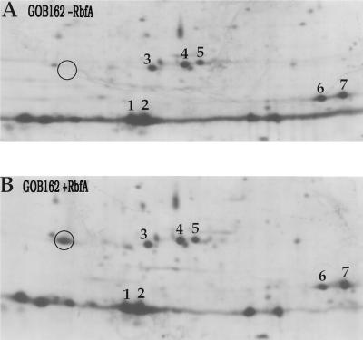
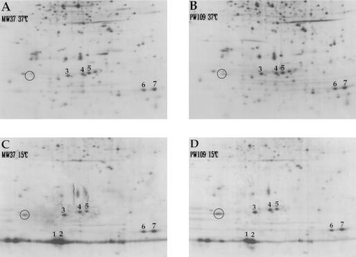
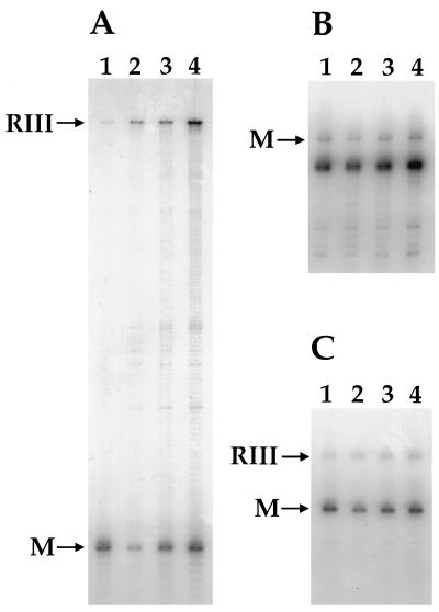
The trmD operon is located at 56.7 min on the genetic map of the Escherichia coli chromosome and contains the genes for ribosomal protein (r-protein) S16, a 21-kDa protein (RimM, formerly called 21K), the tRNA (m1G37)methyltransferase (TrmD), and r-protein L19, in that order. Previously, we have shown that strains from which the rimM gene has been deleted have a sevenfold-reduced growth rate and a reduced translational efficiency. The slow growth and translational deficiency were found to be partly suppressed by mutations in rpsM, which encodes r-protein S13. Further, the RimM protein was shown to have affinity for free ribosomal 30S subunits but not for 30S subunits in the 70S ribosomes. Here we have isolated several new suppressor mutations, most of which seem to be located close to or within the nusA operon at 68.9 min on the chromosome. For at least one of these mutations, increased expression of the ribosome binding factor RbfA is responsible for the suppression of the slow growth and translational deficiency of a deltarimM mutant. Further, the RimM and RbfA proteins were found to be essential for efficient processing of 16S rRNA.


Figures

FIG. 1
FIG. 1
Genetic organization of the trmD operon region of the chromosome of the ΔrimM-2 mutant MW37 and the congenic rimM+ strain MW38. P and T indicate the promoter and terminator, respectively, of the trmD operon. The arrows above and below the genes for Ffh (named Ffh for fifty-four homolog of the 54-kDa protein of the signal recognition particle) and a 16-kDa protein designated 16K, respectively, show the orientations of the transcripts and not their actual sizes. The Kmr gene derived from transposon Tn903 was previously inserted into the gene for the nonessential 16K protein (33).
FIG. 2
FIG. 2
Relative cell yields and growth rates for wild-type and mutant strains. The cell yields for the different strains grown in MOPS minimal medium were calculated as ΔA420/Δ(glucose concentration, expressed as a percentage). The glucose concentrations used were 0.005 to 0.1%. The growth rates are for growth in LB medium. Both the cell yield values and the growth rates have been normalized to those for strain MW38.
FIG. 3
FIG. 3
Suppression of the slow growth of a ΔrimM-2-containing mutant by plasmids that carry different parts of the nusA operon. The suppression level was judged after single-cell outstreaks on rich-medium plates. Plasmid pGOB18 conferred stronger suppression in the presence of 0.25 mM IPTG than in the absence of IPTG. IF2, translational initiation factor IF2; TruB, tRNA Ψ55 synthase; PNP, polynucleotide phosphorylase. Restriction sites used in the plasmid constructions: B, BamHI; E, EcoRI; K, KpnI; S, SalI. +, suppression; −, no suppression.
FIG. 4
FIG. 4
Northern blot analysis of nusA operon mRNA. (A) Genetic organization of the nusA operon. Abbreviations: B, C, and D, probes used in the experiments for which results are shown in panels B, C, and D, respectively; R III and R E, processing sites for RNase III (34, 35) and RNase E (25, 36), respectively; (B through D) P, promoter; T, terminator. Five micrograms of total RNA was subjected to electrophoresis in an agarose gel containing formaldehyde, transferred to a Hybond N filter, and probed with a radiolabeled PCR fragment (probe D). The probe was removed by washing, and the filter was reprobed twice (probes C and B). The exposure times in the experiments for which results are shown in panels B and C were shorter than those in the experiment for which results are shown in panel D in order to avoid overexposure of the bands in strains PW109 and GOB083. The sizes of the γ-32P-labeled ATP kinase-treated fragments of the 1-kb DNA ladder from GIBCO BRL Life Technologies Inc. (Gaithersburg, Md.) are indicated. The strains used (with the relevant genetic markers in parentheses) were MW38 (rimM+), MW37 (ΔrimM-2), PW109 (ΔrimM-2 sdr-43), PW100 (ΔrimM-2 sdr-34), GOB113 (rimM+ sdr+), and GOB083 (rimM+ sdr-43).
FIG. 5
FIG. 5
Syntheses of individual proteins at 37°C and after a shift in temperature to 15°C. Total cell extracts of strain MW38 (rimM+ rbfA+) labeled with [35S]methionine just prior to (A) and 30 min after (B) the shift to 15°C were separated on 2D gels. The indicated proteins are as follows (protein labels shown in parentheses): proteins CspG (1), CspA (2), and H-NS (3) and r-proteins S6 (4 and 5), L12 (6), and L7 (7). The identities of these proteins were obtained by comparing our gels with those of Fang et al. (11) and Jones and Inouye (14). A protein putatively identified as RbfA is indicated by a circle.
FIG. 6
FIG. 6
Identification of RbfA on 2D gels. (A) A total cell extract of strain GOB162 (rbfA::Kmr) labeled with [35S]methionine 30 min after a shift in temperature from 37 to 15°C was separated on 2D gels. (B) A [35S]methionine-labeled minicell extract of strain AA10, expressing rbfA from plasmid pGOB18, was added to the total extract. The indicated proteins are as follows (protein labels shown in parentheses): proteins CspG (1), CspA (2), and H-NS (3) and r-proteins S6 (4 and 5), L12 (6), and L7 (7). The position of RbfA is indicated by a circle.
FIG. 7
FIG. 7
Syntheses of individual proteins at 37°C and after a shift in temperature to 15°C. Strains MW37 (ΔrimM-2) and PW109 (ΔrimM-2 sdr-43) were labeled with [35S]methionine just prior to and 30 min after the shift to 15°C. (A) MW37 at 37°C; (B) PW109 at 37°C; (C) MW37 at 15°C; (D) PW109 at 15°C. The indicated proteins are as follows (protein labels shown in parentheses): proteins CspG (1), CspA (2), and H-NS (3) and r-proteins S6 (4 and 5), L12 (6), and L7 (7). The position of RbfA is indicated by a circle.
FIG. 8
FIG. 8
Primer extension analysis of the 5′ ends of 5S, 16S, and 23S rRNA in wild-type and mutant strains. Lanes 1, strain MW38 (rimM+ rbfA+); lanes 2, strain MW37 (ΔrimM-2 rbfA+); lanes 3, strain PW109 (ΔrimM-2 sdr-43 rbfA+); lanes 4, strain GOB162 (rimM+ rbfA::Kmr). The sizes of the primer extension products obtained were determined by comparing the mobilities with those of a known DNA sequencing ladder. (A) RIII indicates a primer extension product of 179 nt corresponding to pre-16S rRNA processed at the RNase III site 115 nt upstream of the 5′ end of mature 16S. M indicates a product of 64 nt corresponding to the 5′ end of mature 16S rRNA. (B) M indicates a primer extension product of 63 nt corresponding to mature 5S rRNA. (C) RIII indicates a primer extension product of 65 nt corresponding to pre-23S rRNA processed at the RNase III site 7 nt upstream of the 5′ end of mature 23S. M indicates a product of 58 nt corresponding to the 5′ end of mature 23S rRNA.

Similar articles

Cited by

References

    1. Alix J-H, Guérin M-F. Mutant DnaK chaperones cause ribosome assembly defects in Escherichia coli. Proc Natl Acad Sci USA. 1993;90:9725–9729. - PMC - PubMed
    1. Apirion D, Miczak A. RNA processing in prokaryotic cells. Bioessays. 1993;15:113–120. - PubMed
    1. Bertani G. Studies on lysogenesis. I. The mode of phage liberation by lysogenic Escherichia coli. J Bacteriol. 1951;62:293–300. - PMC - PubMed
    1. Björk G R. Modification of stable RNA. In: Neidhardt F C, Ingraham J L, Low K B, Magasanik B, Schaechter M, Umbarger H E, editors. Escherichia coli and Salmonella typhimurium: cellular and molecular biology. Vol. 2. Washington, D.C: American Society for Microbiology; 1987. pp. 719–731.
    1. Björk G R, Ericson J U, Gustafsson C E, Hagervall T G, Jönsson Y H, Wikström P M. Transfer RNA modification. Annu Rev Biochem. 1987;56:263–287. - PubMed

Publication types

MeSH terms

LinkOut - more resources