

Review
doi: 10.1136/bjo.81.9.795.
Gene therapy for inherited retinal degeneration
Affiliations
- PMID: 9422936
- PMCID: PMC1722319
- DOI: 10.1136/bjo.81.9.795
Item in Clipboard
Review
Gene therapy for inherited retinal degeneration
Br J Ophthalmol.
1997 Sep.
No abstract available
Figures
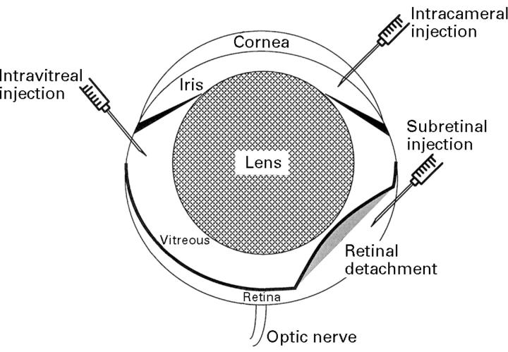
Schematic of the mouse eye. Note that the lens is proportionally much larger in mice than in humans. The routes of intraocular injections are indicated.
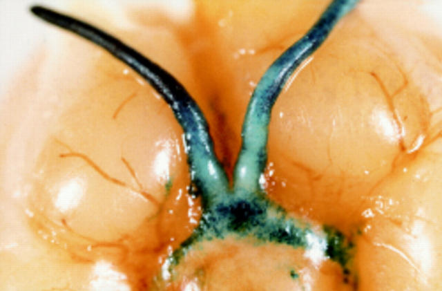
Blue X-gal staining indicates lacZ activity in the optic nerve and optic chiasma of a BALB/c mouse 3 days after subretinal injection of 2 µl suspension of replication competent HSV virus, BE8 (5 × 10⁹ pfu/ml) in which the lacZ, driven by a CMV promoter, has been inserted into the non-essential Us5 gene.57
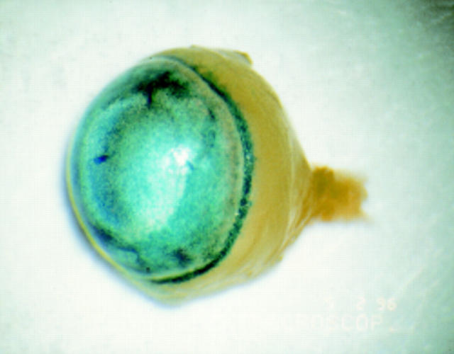
Blue X-gal staining in the anterior chamber of adult BALB/c mouse after intracameral injection of 2 µl of adenovirus carrying a lacZ gene with nuclear localisation signal driven by a CMV promoter (AV.CMV.LacZnuc) at a titre of 1 × 10⁹ pfu/ml. LacZ activity can be observed throughout the anterior segment including corneal endothelium, iris, and trabecular meshwork.
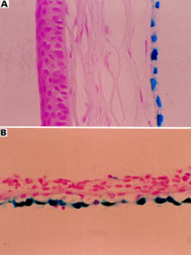
Transduced corneal endothelium (A) and iris pigment epithelium (B) in adult BALB/c mouse after intracameral injection of 2 µl of AV.CMV.LacZnuc (1 × 10⁹ pfu/ml). A 5 µm paraffin section counterstained with nuclear fast red (× 45).
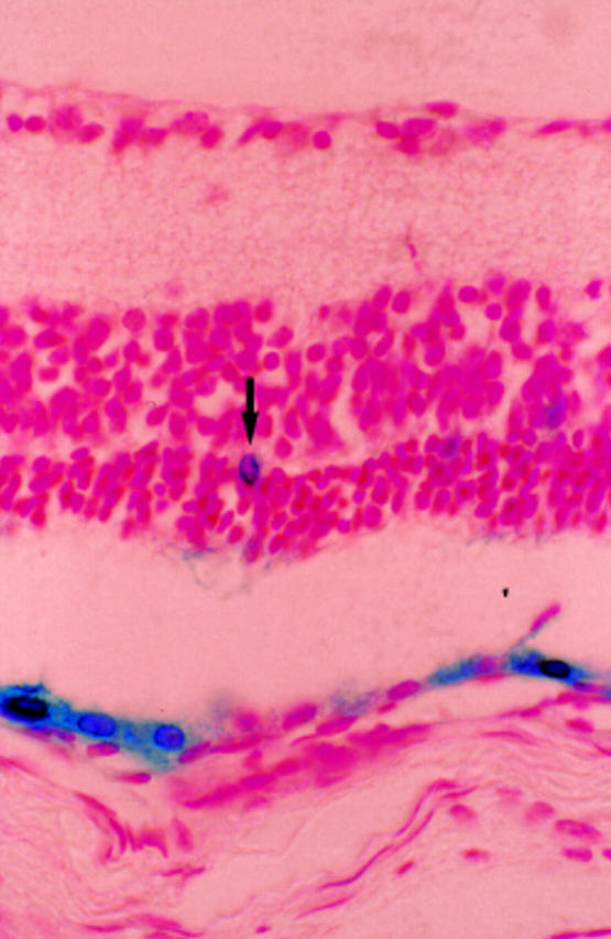
Transduced retinal pigment epithelium and occasional positive photoreceptor cell (arrow) in adult BALB/c mouse 2 weeks after subretinal injection of 2 µl of AV.CMV.LacZnuc (1 × 10⁹ pfu/ml). A 5 µm paraffin section counterstained with nuclear fast red (× 66).
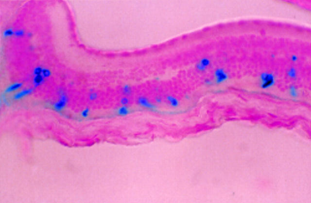
Transduced photoreceptor cells in adult nude mouse 1 month after subretinal injection of 2 µl of AAV.CMV.LacZ (1 × 10⁷ IU/ml). All the stained outer segments can be related to stained photoreceptor nuclei which is consistent with transduction of photoreceptor cells and subsequent transport of LacZ into the outer segments. A 5 µm paraffin section counterstained with nuclear fast red (×25).
References
Publication types
MeSH terms
LinkOut - more resources
Full Text Sources
Other Literature Sources
Medical